Jaime Luís Freitas Arrarte1, José Faibes Lubianca Neto2, Gilberto Bueno Fischer3
Abstract
Objective: To evaluate the effect of adenotonsillectomy on oxygen saturation measured through nocturnal pulse oximetry in children
with sleep-disordered breathing. Methods: A pre- and post-intervention study was carried out using nocturnal pulse oximetry. The study involved 31 children who were suspected of having sleep-disordered breathing and had been referred for adenotonsillectomy. Results: A total of 27 children completed the study. The mean age was 5.2 ± 1.8 years, and 18 (66.7%) of them were male. Upon physical examination, 23 (85.2%) of the children presented class III or class IV hyperplasia of the palatine tonsils. There was significant improvement in the
post-operative period over the pre-post-operative period in terms of the oxygen desaturation rate (OR = 0.65; 95% CI: 0.5–1.3 vs. OR = 1.63; 95% CI: 1.1–2.4; p < 0.001). Conclusion: Adenotonsillectomy significantly improved oxygen saturation, as measured through nocturnal pulse
oximetry, in children with sleep-disordered breathing.
Keywords: Sleep apnea obstructive; Tonsillectomy; Oximetry; Child.
* Study carried out at the Santo Antônio Children’s Hospital of the Santa Casa Hospital Complex in Porto Alegre (SP) Brazil. 1. Masters student in Pediatrics at the Federal University of Rio Grande do Sul (UFRGS, School of Medicine) – Porto Alegre (RS) Brazil.
2. Adjunct Professor of Otorhinolaryngology at the Fundação Faculdade Federal de Ciências Médicas de Porto Alegre (FFFCMPA, Federal Foundation School of Medical Sciences of Porto Alegre) – Porto Alegre (RS) Brazil.
3. Full Professor of Pediatrics at the Fundação Faculdade Federal de Ciências Médicas de Porto Alegre (FFFCMPA, Federal Foundation School of Medical Sciences of Porto Alegre) – Porto Alegre (RS) Brazil.
Correspondence to: Jaime Luís Freitas Arrarte. Rua 13 de maio, 581/803, Centro, CEP 95700-000, Bento Gonçalves, RS, Brasil. Phone 55 54 452-7670. E-mail: [email protected]
Introduction
The association between behavior/learning disorders and sleep disorders has been known for approximately 100 years.(1) In the last 30 years, several
sleep-related diseases have been defined as a result of the monitoring of vital signs during sleep. Among those, sleep-disordered breathing (SDB) encom-passes a wide spectrum of conditions presenting varying degrees of airway obstruction during sleep, ranging from primary snoring to partial or complete obstruction of the upper airways, known as obstruc-tive sleep apnea-hypopnea syndrome (OSAHS). In the general population, OSAHS is one of the most prevalent SDB conditions, affecting adults as well as children. However, OSAHS seams to be different in children than in adults in many aspects, espe-cially those related to congenital, neurological, and anatomical changes.(2,3)
SDB conditions have an important effect on the quality of life of affected children. In a study evalu-ating 61 children with OSAHS, diagnosed through the use of polysomnography, a standard quality of life questionnaire, known as the OSA-18, was used.(4)
Based on this questionnaire, the authors found the impact of OSAHS to be minimal in 20 children (33%), moderate in 19 (31%), and considerable in 22 (36%). There was a significant correlation between OSA-18 scores and the apnea-hypopnea index, even when adjusted for confounding factors in a multiple linear regression model (r = 0.5, p = 0.007). In 2003, another group of authors evaluated the impact of adenotonsillectomy using the same questionnaire in a cohort of 54 children. The size of the effect (mean of the standardized response) on the ques-tionnaire score (before and after surgery) was 1.54 (95% CI: 1.26–1.82), an effect equal to or greater than 0.8 being considered significant.(5)
Due to the positive response to the surgical treat-ment, together with the relative rarity of sleep studies in Brazil, children are diagnosed and submitted to surgery without a formal study. Therefore, the eval-uation of the effect of this surgery on peripheral oxygen saturation, as measured by nocturnal pulse oximetry (NPO), during sleep in children with SDB is relevant.
Methods
In order to evaluate the effect of adenotonsil-lectomy in children with SDB, a before and after
type cohort study was designed. The study sample was selected from among the children treated in the Pediatric Otorhinolaryngology and Pulmonology Outpatient Clinic of the Santo Antônio Children’s Hospital of the Santa Casa Hospital Complex in Porto Alegre, Brazil. All selected patients were candidates for tonsillectomy, with or without adenoidectomy (Chart 1), and had been referred for treatment by pediatricians of the Municipal Secretary of Health. The eligibility criteria of these candidates were checked by resident physicians who were unaware of the objective of the study. The inclusion criteria were being from two to ten years of age and having presented obstructive respiratory symptoms during sleep for at least three months. The exclusion criteria were as follows: presenting craniofacial, genetic or neurological abnormalities; suffering from asthma or other chronic pulmonary diseases; and having nasal polyposis.
The objectives, risks and benefits of the study were explained to the parents and legal guardians. They were then asked to observe signs and symp-toms in the children for a period of two weeks. Those who agreed to participate in the study gave written informed consent. The parents and guard-ians were then interviewed, and the children were examined. The size of the palatine tonsils seen in the nasal endoscopy was classified according to the criteria previously defined.(6) Pre-operative
labora-tory examinations (blood workup and coagulation profile) and electrocardiogram, as well as
radio-Chart 1 - Indications for tonsillectomy, with or without
adenoidectomy.
Infection Recurrent tonsillitis* Chronic nasopharyngitis Chronic/recurrent sinusitis Obstruction Hyperplasia with non-responsive
obstruction to clinical treatment, with or without obstructive apnea, severe dysphagia, and late development
Nasal obstruction with speech or orodental abnormalities
Miscellaneous Recurrent peritonsillar abscess Unilateral tonsillar hyperplasia Hemorrhagic tonsillitis Chronic tonsilloliths
logical studies of the nasopharynx and chest, were ordered.
The selected patients participated in the study by allowing themselves to be subjected to NPO during the night preceding the surgical treatment. For this study, an Ohmeda oximeter (model 3700®; Ohmeda,
Boulder, CO, USA), which has a memory for up to eight hours of study, was used at a 12 sec sampling rate. If the child was suffering from an airway infec-tion, the study and surgery were postponed. During the night, the room was monitored by two nurses. A previously trained medical student, under the super-vision of the researcher, was responsible for prepping the patient and for giving instructions to the patient and to the parent/guardian regarding the exami-nation. The sensor was attached to the finger of the patient, preferably the middle finger. For better fixation of the sensor, a splint was attached to the forearm with adhesive tape. Parents and guardians were instructed in the use of the equipment, so that they could turn it on or off in case the child had to go to the bathroom or upon ending of the examina-tion, when the child woke up. The examination was considered valid if it continued for a period of at least four hours.(7)
The following variables were studied: total number of desaturations of ≥ 4%; oxygen desatu-ration index (ODI), defined as the total number of desaturations of ≥ 4% divided by the duration of the examination in hours; percentage of time during which saturation was lower than 95%; percentage of time during which saturation was lower than 90%; mean saturation; and minimum saturation. The oximetric data collected were analyzed in oximetric data and heart rate distribution tables created with the Excel® program and using a programmed
algo-rithm to identify possible movement artifacts. All desaturations of ≥ 4% were identified and noted. Heart rate was evaluated during the 36 sec prior to desaturation, and those presenting a pulse ampli-tude modulation range of ≤ 40 were considered valid desaturations.(8)
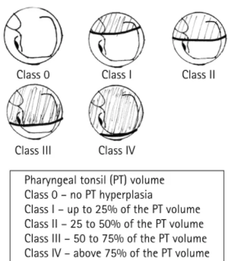
All surgical procedures were performed in the morning and always by the same surgeon. After initiation of anesthesia, the nasopharynx was evaluated. Hyperplasia of the pharyngeal tonsils (adenoids) was verified and classified using a flex-ible fiberoptic laryngoscope (Machida, Tokyo, Japan) (Figure 1). After surgery, patients remained at least four hours in the anesthesia recovery room and were
then sent to the infirmary. The volume of the pala-tine tonsils, obtained by determining the difference in liquid volume after their immersion in a graded flask containing a known volume of saline solution, was noted at the end of the procedure.
Unless there was some surgical complication, patients were discharged from the hospital on the day after the procedure. The interval between surgery and the post-operative NPO was at least four weeks. On the day of the post-operative oximetry, the questionnaire was again administered to the children. The oximetric variables were analyzed before and after surgery using the Student’s t-test or the Wilcoxon t-test, depending on the type of data distribution. In order to determine whether the pre-operative oximetry findings presented any associations with the anthropometric data or with the results of the physical examination, Spearman’s rank correlation coefficient (rs) was used. Data were tabulated and analyzed using the Excel® program
and the SPSS program, version 11.0. The anthro-pometric data were analyzed using the Nutristat®
program contained within the 2002® Epi Info™
package. The necessary sample size was estimated using the Student’s t-test to compare the difference between the mean pre-operative ODI and the mean postoperative ODI. For the calculation, a previous study was used with the same oximetry model used in a sample of patients with similar clinical characteristics.(9) The Samples® program contained
within the PEPI® statistical package was used. An
alpha error of 5% and a power of 90% were
esti-Class 0 Class I Class II
Class III Class IV
Pharyngeal tonsil (PT) volume Class 0 – no PT hyperplasia Class I – up to 25% of the PT volume Class II – 25 to 50% of the PT volume Class III – 50 to 75% of the PT volume Class IV – above 75% of the PT volume
mated. The minimum sample size calculated was 21 patients with SDB. This study was approved by the Ethics in Research Committee of the Santa Casa Hospital of Porto Alegre.
Results
Of the 35 eligible patients, 8 were excluded for not being submitted to post-operative oximetry. Therefore, the final sample consisted of 27 patients, all of whom completed the study. The mean age was 5.2 ± 1.8 years. Of those 27 patients, 18 were male (67.7%), and 22 were white (81.5%). Most parents and guardians had less than eight years of schooling (63%). The most common clinical complaints were snoring (100%), respiratory pauses during sleep (96.3%), nocturnal mouth breathing (96.3%), rest-less sleep (81.5%), and sialorrhea (74.1%) (Table 1). Most patients presented class III or class IV
pharyn-geal and palatine tonsil hyperplasia (96.3% and 85.2%, respectively) (Table 2). There were 6 children (22.2%; 3 males and 3 females) who were under the 25th percentile in terms of weight-for-age, and there
were 3 (25.9%; 2 males and 1 female) were over the 75th percentile.
There were no intra-operative or post-operative complications. The mean surgical time was 42.9 ±
13.8 min. The average palatine tonsil volume was 10.18 ± 3.11 mL, ranging between 5 mL and 18 mL.
The oximetric study results are described in Table 3. The mean interval between surgery and the postop-erative NPOs was 281.9 days. All patients reported symptom improvement after surgery, except for 2 cases in which snoring persisted. One of those 2 patients also continued to present significant desaturations during the postoperative period (ODI = 4.5).
There was a significant difference between pre-operative ODI and post-pre-operative ODI (p < 0.001), as well as between pre-operative and post-opera-tive total number of desaturations, percentage of saturation time lower than 95%, and percentage of saturation time lower than 90% (p < 0.001) (Table 3). We found no differences between the pre-operative and post-operative values for examination time, mean saturation, mean minimum values, and mean maximum values. The ODI was not found to corre-late significantly with age or weight (p = 0.526 and p = 0.496, respectively), nor with the degree of hyperplasia observed in the pharyngeal and palatine tonsils (p = 0.427 and p = 0.851, respectively).
Discussion
In this study there was a clear improvement in the principal post-operative oximetric variables in all but one of the patients studied. Prior to surgery, this patient presented class III and class IV
hyperplasia of the pharyngeal and palatine tonsils, respectively. Considering the significant clinical symptom improvement, as well as the improved oximetry findings, in children submitted to surgical treatment, it is important to emphasize that there was only a weak correlation between pre-operative ODI and the degree of hyperplasia of the pharyn-geal and palatine tonsils, which, in daily practice, is an important finding to indicate surgical treatment. This finding is similar to those of other studies.(10-12)
However, it is important to correlate the tonsil volume with the size of the pharyngeal space. In
Table 1 - Signs and symptoms of the patients studied
before and after surgery (n = 27).
Symptoms Frequency
Pre-operative Post-operative
Snoring 27 (100.0) 2 (7.4)
Respiratory pauses 26 (96.3) 0 Nocturnal mouth
breathing
26 (96.3) 5 (18.5)
Restless sleep 22 (81.5) 2 (7.4)
Sialorrhea 20 (74.1) 1 (3.7)
Daytime mouth breathing
19 (70.4) 4 (14.8)
Irritability 19 (70.4) 4 (14.8)
Rhinorrhea 14 (51.9) 3 (11.1)
Loss of appetite 12 (44.4) 3 (11.1) Nocturnal enuresis 12 (44.4) 4 (14.8)
Stunted growth 9 (33.3) 1 (3.7)
Nightmares 10 (37.0) 2 (7.4)
Excessive sleepiness 5 (18.5) 1 (3.7) Data are shown as numbers and percentages.
Table 2 - Degree of pharyngeal and palatine tonsilar
hyperplasia (n = 27).
Degree of hyperplasia Class
0 I II III IV
a narrow pharynx, even small tonsils can cause significant airway obstruction. In adults, OSAHS seams to have a multifactorial origin: genetic and neuromuscular factors can play important roles. In children, factors other than the volumetric increase in Waldeyer’s lymphoid ring can be involved.(13)
In the pediatric population, the estimated preva-lence of primary snoring ranges from 3.2% to 12.1%, compared with 0.7% to 10.3% for OSAHS.(10) In
chil-dren with SDB, the prevalence of OSAHS, diagnosed through polysomnography, has been reported to be as high as 70%.(14-17) Although polysomnography is
considered the gold standard in the investigation of SDB, NPO is an interesting investigative option due to its low cost and wide availability. However, the sensitivity of NPO is low when compared with that of polysomnography. In a transversal study, 349 children were submitted to evaluation for SDB. The authors showed that a positive NPO has a posi-tive predicposi-tive value of 97%. However, a negaposi-tive or inconclusive NPO has a negative predictive value of only 57%.(14) If we use the cut-off value
estab-lished by these authors, which is the same as that recommended by the American Thoracic Society (an ODI equal to or greater than 3), we can see that 5 (18.5%) of the children evaluated in our study presented abnormal desaturations based on the NPO results.(18) This proportion is lower than those
found in similar studies, in which the proportion of children with SDB found to present OSAHS ranged from 46% to 70%.
One hypothesis that could explain the low proportion of children with more than three desatu-rations is that an oximeter that provides an oximetric average over a prolonged period of time underesti-mates the number of short episodes of desaturation
in children with OSAHS.(19) In this study, the oximeter
was used in eight-hour memory mode. In this mode, the equipment calculates the average every 12 sec. The problem of the sampling rate in the memory of the device has been highlighted by other authors, who carried out a study involving 16 adult patients with SDB and the simultaneous use of two oxime-ters with different sampling rates. In that study, the oximeter set to use a 12-sec sampling rate registered a mean ODI of 3.2 (range, 1 to 18.3), while the other (set to a 2-sec sampling rate) registered a mean ODI of 8.34 (range, 2 to 22.8) (rs = 0.69, p < 0.001).(20)
Another possibility for the low frequency may be the fact that we have included in the sample chil-dren with indication of tonsillectomy for repeated tonsillitis, even though snoring is one of the selec-tion criteria. However, despite the above-menselec-tioned limitations of NPO, this examination can still be useful if we take into account its high specificity, its wide availability, and the limited availability of sleep laboratory polysomnography for pediatric patients.
There is still no consensus on when surgical treat-ment is indicated in children with SDB. Most authors agree that children with severe OSAHS should receive immediate treatment due to the potential risks of cardiopulmonary complications. On the other hand, the appropriate treatment for children with border-line or moderate forms of the disease is still under debate, principally because little is known regarding the natural history and late consequences of mild OSAHS in children. Another factor to be considered is that there are still no recognized criteria for clas-sifying the severity of OSAHS in children.
In the present study, most of the children selected for adenotonsillectomy in the otorhinolaryngology outpatient clinic (21-77.8%) were of pre-school
Table 3 - Comparison between the pre-operative and post-operative oximetry findings (n = 27).
Variable Pre-operative Post-operative Difference 95% CI p
ODI 1.6 (1.1 to 2.5) 0.7 (0.5 to 1.3) -1.1 -1.9 to -0.4 <0.001*
DesN 12.0 (9.0 to 18.0) 4.0 (4.0 to 7.0) -8.0 -11.0 to -3.0 <0.001* SpO2 < 95% 6.2 (4.3 to 22.8) 1.0 (0.2 to 4.6) -5.3 -16.9 to -3.5 <0.001* SpO2 < 90% 0.5 (0.0 to 1.4) 0.0 (0.0 to 0.2) -0.4 -0.8 to 0.0 <0.001*
Mean SpO2 97.2 ± 1.07 97.3 ± 1.65 -0.12 -0.9 to 0.6 0.75**
Minimum SpO2 84.5 ± 8.03 89.2 ± 5.23 -4.7 -8.5 to -0.9 0.02**
Data expressed as mean ± standard deviation or median (interquartile range, 25–75%), *Wilcoxon t-test; **Student’s t-test; 95% CI: 95% confidence interval; ODI: oxygen desaturation index; DesN: total number of desaturations; SpO2: peripheral oxygen
age (two to six years of age), of which 62.9% were between three and five years of age. These results are consistent with the findings in the literature, since children in this age bracket present a greater quan-tity of lymphoid tissue in relation to the adjacent airway, resulting in a narrower airway.(21,22) In the
present study, the prevalence of snoring was higher in boys (18–66.7%), which is different from that found in other studies, with the exception of that reported in a population-based transversal study carried out in Iceland, in which the prevalence of snoring was higher in boys than in girls (24.3% vs. 16.5%).(9,23,24) Therefore, this difference can also be
explained by the limited sample size of the present study. It is known that there is an association between growth retardation and OSAHS. One group of authors studied 41 children with SDB submitted to surgical treatment and found that 46% were in the 5th percentile in terms of weight-for-age.(25) In
contrast with these data, the proportion of children in the present study presenting weight-for-age below the 25th percentile was 22.2% (6 children),
and none presented weight-for-age below the 5th percentile.
We conclude that adenotonsillectomy has a beneficial effect on peripheral oxygen saturation measured by pulse oximetry in children presenting airway obstruction during sleep. However, children in whom the symptoms persist post-operatively should be submitted to further investigation, since oxygen desaturation can be caused by conditions other than hyperplasia of the pharyngeal lymphoid tissue.
Acknowledgements
We would like to thank the medical students of the Ana Letícia Boff Foundation for the Federal School of Medical Sciences at Porto Alegre, as well as Leonardo de Azambuja and Vinícius Richter for their dedication to the quality of the sleep studies. We are also grateful to Dr. João Lima for his sugges-tions regarding the sleep study protocols.
References
1. Lavie P. Nothing new under the moon. Historical accounts of sleep apnea syndrome. Arch Intern Med. 1984;144(10):2025-8.
2. Greene MG, Carroll JL. Consequences of sleep-disordered breathing in childhood. Curr Opin Pulm Med. 1997;3(6):456-63
3. Coleman J. Sleep studies: Current techniques and future trends. Otolaryngol Clin North Am. 1999;32(2):195-210. 4. Franco RA Jr, Rosenfeld RM, Rao M. First place--resident
clinical science award 1999. Quality of life for children with obstructive sleep apnea. Otolaryngol Head Neck Surg. 2000;123(1 Pt 1):9-16.
5. Sohn H, Rosenfeld RM. Evaluation of sleep-disordered breathing in children. Otolaryngol Head Neck Surg. 2003;128(3):344-52.
6. Brodsky L. Adenotonsillar disease in children: General considerations. In: Cotton RT, Myer CM, editors. Practical pediatric otolaryngology. Philadelphia: Lippincott Williams &Wilkins; 1999. p.15-40.
7. Smith TC, Proopos DW, Pearman K, Hutton P. Hypoxia in sleeping children: overnight studies can be reduced to 4 hours without loss of clinical significance. Clin Otolaryngol Allied Sci. 1992;17(3):243-5.
8. LaFontaine VM, Ducharme FM, Brouillette RT. Pulse oximetry: accuracy of methods of interpreting graphic summaries. Pediatr Pulmonol. 1996.;21(2):121-31. 9. Ali NJ, Pitson DJ, Stradling JR. Snoring, sleep disturbance,
and behaviour in 4-5 years olds. Arch Dis Child. 1993;68(3):360-66.
10. Schechter MS; Section on Pediatric Pulmonology, Subcommittee on Obstructive Sleep Apnea Syndrome. Technical report: diagnosis and management of childhood obstructive sleep apnea syndrome. Pediatrics. 2002;109(4): e69.
11. Ågren K, Nordlander B, Linder-Aronsson S, Zettergren-Wijk L, Svanborg E. Children with nocturnal upper airway obstruction: Postoperative orthodontic and respiratory improvement. Arch Otolaryngol. 1998;118(4):581-7. 12. Suen JS, Arnold JE, Brooks LJ. Adenotonsillectomy for
treatment of obstructive sleep apnea in children. Arch Otolaryngol Head Neck Surg. 1995;121(5):525-30. 13. Friberg D, Ansved T, Borg K, Carlsson-Nordlander B, Larsson
H, Svanborg E. Histological indications of a progressive snorers disease in an upper airway muscle.Am J Respir Crit Care Med. 1998;157(2):586-93.
14. Brouillette RT, Morielli A, Leimanis A, Walters KA, Luciano R, Ducharme FM. Nocturnal pulse oximetry as an abbreviatedNocturnal pulse oximetry as an abbreviated testing modality for pediatric obstructive sleep apnea. Pediatrics. 2000;105(2):405-12.
15. Carroll JL, McColley SA, Marcus CL, Curtis S, Loughlin GM. Inability of clinical history to distinguish primary snoring from obstructive sleep apnea syndrome in children. Chest. 1995;108(3):610-8.
16. Rosen CL. Clinical features of obstructive sleep apnea hypoventilation syndrome in otherwise healthy children. Pediatr Pulmonol. 1999;27(6):403-9.
17. Van Someren V, Burmester M, Alusi G, Lane R. Are sleep studies worth doing? Arch Dis Child. 2000;83(1):76-81. 18. Cardiorespiratory sleep studies in children. Estabilishment
of normative data and polysomnographic predictors of morbidity. American Thoracic Society. Am J Respir Crit Car Med. 1999;160(4):1381-7.
19. Nixon GM, Kermack AS, Davis GM, Manoukian JJ, Brown KA, Brouillette RT. Planning adenotonsillectomy in children with obstructive sleep apnea: the role of overnight oximetry. Pediatrics. 2004;113(1 Pt 1):e19-25.
21. Jeans WD, Fernando DC, Maw AR, Leighton BC. A longitudinalA longitudinal study of the growth of the nasopharynx and its contents in normal children. Br J Radiol. 1981;54(638):117-21. 22. Marcus CL. Sleep-disordered breathing in children. Am J
Respir Crit Care Med. 2001;164 (1):16-30.
23. Redline S, Tishler PV, Schluchter M, Aylor J, Clarck K, Graham G. Risk factors for sleep-disordered breathing in children.
Associations with obesity, race, and respiratory problems. Am J Respir Crit Care Med. 1999;159(5 Pt 1):1527-32. 24. Gislason T, Benediktsdóttir B. Snoring, apneic episodes and
nocturnal hypoxemia among children 6 Months to 6 Years Old. Chest. 1995;107(4): 963-6.