Bronchial hyperreactivity in patients with
gastroesophageal reflux disease*
MÔNICA SILVEIRA LAPA1, ROBERTO RODRIGUES JÚNIOR1, ELIE FISS2
ABSTRACT
Objective: To identify this vagal reflex using bronchial provocation tests in patients with gastroesophageal reflux disease. Methods: The study group was composed of 10 patients presenting endoscopic evidence of hiatal hernia or gastroesophageal reflux disease, and the control group consisted of 11 patients presenting no evidence of either condition. All subjects were submitted to bronchial provocation with carbachol. Results: The provocation test was positive in 5 (50%) of the study group patients and 3 (27%) of the control group patients (p = 0.64). Conclusion: The hypothesis that the airways of patients with gastroesophageal reflux disease (and no history of asthma-like respiratory symptoms) might be more responsive than those of individuals without the disease remains unproven.
Keywords: Asthma/complications; Gastroesophageal reflux/complications; Hernia hiatal; Peak expiratory flow rate;
Bronchial hyperreactivity; Forced expiratory volume; Carbachol/diagnostic use
* Study conducted at the Faculdade de Medicina do ABC (FMABC, ABC School of Medicine), Santo André (SP) Brazil. Financial support: FAPESP (grant no. 98/15363-0)
1. Attending Physician in the Pulmonology Department of the Faculdade de Medicina do ABC (FMABC, ABC School of Medicine), Santo André (SP) Brazil
2. Tenured Professor in the Pulmonology Department of the Faculdade de Medicina do ABC (FMABC, ABC School of Medicine), Santo André (SP) Brazil
Correspondence to: Mônica Silveira Lapa. Rua Ática 404, São Paulo - SP. CEP: 04634-041. Phone: 55 11 5031-7929; 55 11 9199-1639. E-mail: [email protected]
INTRODUCTION
Asthma is a disease of the upper airways that occurs in susceptible individuals and presents three main characteristics: obstruction of the upper airways that is either spontaneously reversible (although not completely in some individuals) or reversible with treatment; inflammation of the airways; and hyperreactivity of these airways to a large number of stimuli.(1)
There are a few factors that predispose to bronchial hyperreactivity (BHR): allergens, infections, gastroesophageal reflux, air pollution, inhaled irritants, physical exercise, pharmacological stimulants and emotional factors.(2)
Gastroesophageal reflux disease (GERD) may present in several forms, some including extra-esophageal symptoms. In the respiratory tract, GERD may manifest as a simple persistent cough, voice alteration or even severe clinical profiles such as chronic bronchitis, recurrent pneumonia and asthma-like manifestations.(3-5) The incidence of GERD among
asthmatic patients (ranging from 40% to 80%) exceeds the incidence in the general population. Therefore GERD constitutes an important instability factor in BHR.(6-7)
It is believed that gastroesophageal reflux may aggravate or even cause BHR in certain patients. (8-10) The possible mechanisms involved in triggering
BHR in such individuals include microaspiration of refluxed acid(8,11) and activation of a vagal reflex
from the esophagus to the lung (resulting in bronchoconstriction),(11-13) as well as sensitization of the
vagal system (acid-sensitive receptors in the esophageal wall), which can induce or accentuate BHR.(6, 14-16)
In cases of chronic cough of undefined etiology, the bronchial provocation test is used to define whether the cough is asthma-related. Upper digestive endoscopy and contrast-enhanced radiological examination of the esophagus, stomach and duodenum are used to diagnose GERD, asymptomatic or not. Although considered the gold standard exam for the diagnosis of GERD, pHmetry often presents false-negative results does not yet have a standardized value of normality. This was demonstrated in a doctoral thesis,(14) in which only 72% of the patients with
biopsy-confirmed esophagitis tested positive for GERD. Chronic cough and bronchial asthma can be caused by gastroesophageal reflux. The main consequence of such reflux in the lung is the stimulus
of an esophagobronchial and tracheal vagal reflex which could lead to diffuse bronchoconstriction. However, the intensity and the significance of this vagal reflex in individuals with GERD who do not present respiratory symptoms is unknown. Asymptomatic patients may present BHR alteration detectable only by bronchoprovocation. One published study, the only one on this issue, revealed that gastroesophageal reflux is correlated with increased BHR in patients without pulmonary symptoms.(17)
Since gastroesophageal reflux is an important causal factor of BHR, and its main mechanism of action is through an esophagobronchial vagal reflex, the objective of this study was to assess, through bronchoprovocation with carbachol, whether patients with GERD present more BHR than do normal individuals, and to reveal, through bronchoprovocation, the existence of esophagobronchial reflex in patients who do not present pulmonary disease.
METHODS
A group of 10 patients with GERD and a control group of 11 individuals without GERD were studied during the 1998-2000 period. The patients were volunteers from the Gastroenterology Outpatient Clinic and the Endoscopy Department of the Faculdade de Medicina do ABC (ABC School of Medicine).
We used the following inclusion criteria: presenting esophagitis, hiatal hernia or gastroesophageal reflux confirmed by digestive endoscopy; presenting gastritis or normal endoscopic examination (in control patients); being a nonsmoker or having quit smoking more than eight years prior; presenting pulmonary function tests within patterns of normality; having no history of respiratory symptoms.
The exclusion criteria were as follows: being over 70 years of age (since most patients in this age bracket presented considerable difficulty in performing this test); having undergone endoscopy more than three months prior to the date of the test; presenting more than two comorbidities.
between the two (FEV1/FVC) were determined. Bronchoprovocation was performed if the pulmonary function was within normality patterns, that is, if the FEV1/FVC ratio was higher than or equal to 75% or if FEV1 was higher than 80% of predicted. Bronchoprovocation was performed with carbachol at the following dilutions: 0.25; 0.50; 1.00; 2.00 and 4.00 mg/ml. Inhalation was sustained for 120 seconds, followed by new spirometric curve measurements, until the total dose or a 20% drop in FEV1 was achieved. If there was a drop in FEV1 of 20% or more, the test was considered positive. The cumulative dose that determined the test as positive was 2.5 mg.
The study was approved by the ABC School of Medicine Ethics Committee. All patients gave written informed consent.
The results obtained from the two groups were statistically compared using the paired Student's t-test. Calculations were made using the Statistical Analysis System.
RESULTS
We performed 29 pulmonary function tests and 21 bronchoprovocation tests with carbachol. Eight patients presented abnormal pulmonary function test results, which contraindicated the bronchoprovocation test.
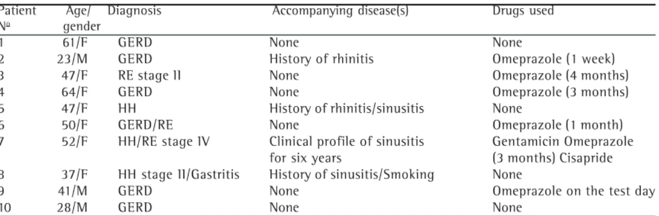
Of the 21 bronchoprovocation tests performed, 17 were performed in women and 4 in men; 10 were performed in patients with GERD, reflux esophagitis or hiatal hernia, as shown in Tables 1 and 2. We observed that 6 of these patients presented gastroesophageal reflux, 3 presented reflux
esophagitis, and 3 presented hiatal hernia (all diagnosed by digestive endoscopy). Six patients were being treated with a proton pump inhibitor. Two patients presented a history of treatment for rhinitis, and 5 of the 10 patients reacted positively to the bronchoprovocation test (drop in FEV1 > 20%).
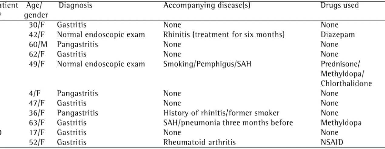
In the control group, 9 patients had gastritis and the remainder presented endoscopic exams within normality patterns. Three presented a positive bronchoprovocation test. Two patients (1 of whom was a former smoker) had a history of treatment for rhinitis, 1 had rheumatoid arthritis, and 2 had systemic arterial hypertension. Of the 2 patients with systemic arterial hypertension, one admitted being a smoker prior to the test, and the other had had pneumonia three months prior to the test (Tables 3 and 4).
We chose not to exclude the smokers from the study since they presented negative bronchoprovocation, and the final result of the study would therefore not be affected.
Ages ranged from 23 to 64 in the group with GERD and from 17 to 63 in the control group (Tables 1 and 3).
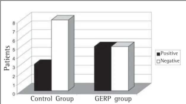
Figure 1 summarizes the results of the tests in the two groups. There were no statistical differences regarding age. Mean age in the GERD group was 45.09 (± 13.52), and mean age in the control group was 45.27 (± 14.24) (p = 0.95). The bronchoprovocation test was positive in 27% of the individuals of the control group (n = 3) and in 50% of the patients with GERD (n = 5). However, this difference was not found to be statistically significant using Fisher's exact test (p = 0.22).
TABLE 1
Patients with gastroesophageal reflux, hiatal hernia or reflux esophagitis
Patient Age/ Diagnosis Accompanying disease(s) Drugs used
No gender
1 61/F GERD None None
2 23/M GERD History of rhinitis Omeprazole (1 week)
3 47/F RE stage II None Omeprazole (4 months)
4 64/F GERD None Omeprazole (3 months)
5 47/F HH History of rhinitis/sinusitis None
6 50/F GERD/RE None Omeprazole (1 month)
7 52/F HH/RE stage IV Clinical profile of sinusitis Gentamicin Omeprazole
for six years (3 months) Cisapride
8 37/F HH stage II/Gastritis History of sinusitis/Smoking None
9 41/M GERD None Omeprazole on the test day
10 28/M GERD None None
DISCUSSION
Approximately 90% of all patients with nonallergic asthma present GERD. To date, the relationship between gastroesophageal reflux and respiratory diseases remains under debate.(7)
One group of researchers suggested that gastroesophageal reflux causes asthma symptoms but does not significantly affect the pulmonary function.(18) The authors attributed the asthma-like
symptoms to the retrosternal discomfort that increases minute ventilation and the respiratory sensation. This contradicts the findings of another study which showed a drop in FEV1 at up to 90 minutes after instillation of hydrochloric acid in the esophagus of asthmatic patients.(14)
It is currently believed that there are three mechanisms through which gastroesophageal reflux may induce or aggravate asthma: microaspirations of refluxed acid;(8,11) activation of vagal reflex from
the esophagus to the lung;(11-13) and sensitization
of the vagal reflex, which would induce or accentuate BHR.(6,14-16)
Scintigraphic studies, together with pHmetry in the esophagus and trachea, as well as double pHmetry in the esophagus, demonstrated that 20% of the patients with GERD presented microaspiration.(19) However,
other studies revealed that microaspiration is not a significant cause of gastroesophageal reflux-induced bronchoconstriction. The significance of aspiration as a causative factor of asthma remains uncertain.(15)
Some authors have demonstrated that acid infusion into the esophagus of asthmatic patients causes increased airway resistance, which was totally reversed by the use of antacids.(12) In studies
involving dogs, it has been demonstrated that acid infusion into the esophagus produced a drop in respiratory conductance, which disappeared with the bilateral section of the vagal nerve.(13)
TABLE 2
Bronchoprovocation with carbachol in patients with gastroesophageal reflux, hiatal hernia or
reflux esophagitis
Patient FEV1 FEV1 FEV1 Conclusion
No Baseline After drug (%) Carbachol dose (mg)
1 1.96 1.92 2 Negative
Carbachol 2.5
2 3.92 2.85 27 Positive
Carbachol 0.37
3 3.13 3.00 4 Negative
Carbachol 3.0
4 1.95 1.78 8 Negative
Carbachol 3.2
5 3.28 2.23 32 Positive
Carbachol 2.16
6 2.26 1.79 20 Positive
Carbachol 1.50
7 2.11 1.56 26 Positive
Carbachol 0.37
8 3.37 3.16 6 Negative
Carbachol 2.8
9 4.09 3.74 8 Negative
Carbachol 3.25
10 4.15 2.84 32 Positive
Carbachol 0.4 FEV1: forced expiratory volume in one second (in liters)
TABLE 3 Control group
Patient Age/ Diagnosis Accompanying disease(s) Drugs used
No gender
1 30/F Gastritis None None
2 42/F Normal endoscopic exam Rhinitis (treatment for six months) Diazepam
3 60/M Pangastritis None None
4 62/F Gastritis None None
5 49/F Normal endoscopic exam Smoking/Pemphigus/SAH Prednisone/
Methyldopa/ Chlorthalidone
6 4/F Pangastritis None None
7 47/F Gastritis None None
8 36/F Pangastritis History of rhinitis/former smoker None
9 63/F Gastritis SAH/pneumonia three months before Methyldopa
10 17/F Gastritis None None
11 52/F Gastritis Rheumatoid arthritis NSAID
Hyperreactivity of the airways seems to be altered by gastroesophageal reflux. Esophageal instillation of acid or acid reflux resulted in a decrease in the peak expiratory flow in asthmatics with GERD, or even in patients who presented only asthma or patients who presented only gastroesophageal reflux. This alteration was prolonged beyond the clearance of the acid in the asthmatic patients with GERD. However, the same did not occur in the other groups.(15-16) This may result from a prolonged vagal
stimulus or may be associated with the release of inflammatory mediators, which would result in bronchoconstriction.(14-16)
If the presence of acid in the esophagus causes a vagal reflex and this, in turn, results in bronchoconstriction, why is it that only certain nonasthmatic patients with GERD present this alteration? Why do some asthmatic patients with reflux present no improvement in their pulmonary clinical profile after treatment with proton pump inhibitors? The studies reviewed did not reveal how this vagal reflex could act in patients with reflux only. Would its existence depend on preexisting BHR?
In the general population, BHR seems to present a normal distribution. Approximately 20% of individuals without pulmonary disease present mild BHR. Although the significance of this BHR in patients without pulmonary disease is uncertain, studies suggest that these patients are in a latent phase of asthma.(17) In this particular study, 36%
of the patients with GERD and no respiratory symptoms presented BHR (p < 0.01), suggesting that gastroesophageal reflux is associated with increased BHR.
In the present study, 50% of the patients with GERD had BHR, whereas only 27% of the control
group tested positive. The results were not statistically relevant, making it impossible to confirm the relationship.
In patients with BHR, the effects of treating GERD with proton pump inhibitors (such as omeprazole), or even through surgical procedures, are unknown. Some studies have shown a lessening of respiratory symptoms, although with no alteration in pulmonary function. A study carried out by one group of authors is consistent with this assertion.(21) The study involved
36 asthma and chronic obstructive pulmonary disease patients with severe obstruction and BHR, as well as gastroesophageal reflux. These patients were treated with high doses of omeprazole (80 mg/day) for twelve weeks and did not present alteration in pulmonary function. The opposite was observed by other researchers who assessed patients with (mildly obstructive) asthma and GERD. The patients who used omeprazole at 20 mg/day and domperidone at 10 mg three times a day for six weeks presented significantly improved pulmonary function.(22) In our
study, there was no difference in BHR between the patients using omeprazole and the untreated patients.
Paci
en
te
Figure 1 - Results of bronchoprovocation tests carried out in the control group and in the group of patients with gastroesophageal reflux, hiatal hernia or reflux esophagitis; GERD: gastroesophageal reflux disease
TABLE 4
Bronchoprovocation with carbachol in control group patients
Patient FEV1 FEV1 FEV1 Conclusion
No Baseline After drug (%) Carbachol dose (mg)
1 2.68 1.94 27 Positive
Carbachol 2.19
2 2.22 1.54 30 Positive
Carbachol 2.09
3 2.64 2.31 12 Negative
Carbachol 2.8
4 2.23 1.83 18 Negative
Carbachol 2.5
5 1.95 1.70 12 Negative
Carbachol 2.83
6 2.25 1.99 11 Negative
Carbachol 3.0
7 2.12 1.77 16 Negative
Carbachol 2.75
8 2.94 2.93 13 Negative
Carbachol 3.0
9 1.76 1.70 21 Positive
Carbachol 0.829
10 2.45 1.96 19 Negative
Carbachol 3.20
11 2.08 1.95 6 Negative
Carbachol 3.01 FEV1: forced expiratory volume in one second (in liters)
Negative
Pa
tien
ts
Control Group GERP group
Some patients diagnosed with GERD were using omeprazole upon examination, which may increase the number of false-negative results. However, these patients were not excluded since, in the literature, there is no consensus regarding GERD treatment and decreased BHR. Of the 10 patients with GERD, omeprazole was taken by 6, 3 of whom tested positive (1 had been using the drug for a week, another for three months and the other for a month). Two patients who had been using the medication for three to four months tested negative.
One notable limitation of the study is that, due to a lack of equipment at the facility, the method used to diagnose GERD was not pHmetry, which is considered the gold standard. Had we been able to use pHmetry, this would probably have increased the number of diagnoses. However, one of the authors of the present study previously conducted a study in which only 72% of the diagnoses of GERD made through biopsy were confirmed by pHmetry,(14) demonstrating that even using the
method considered the gold standard, some diagnoses are not confirmed.
The hypothesis that the lungs of GERD patients with no history of asthma-like symptoms may be more responsive than those of patients without GERD remains unproven. However, it has been suggested that GERD constitutes a risk factor for bronchoconstrictive diseases.
This study has not provided statistical evidence that gastroesophageal reflux causes hyperreactivity in patients without respiratory symptoms. New studies on the issue are needed in order to obtain more conclusive results and to assess the importance of the use of proton pump inhibitors in patients with BHR.
REFERENCES
1. Sociedade Brasileira de Alergia e Imunopatologia. Sociedade Brasileira de Pediatria e Sociedade Brasileira de Pneumologia e Tisiologia. II Consenso Brasileiro no Manejo da Asma. J Pneumol. 1998;24(2):171-276. 2. Guidelines for the diagnosis and management of asthma.
National Heart, Lung, and Blood Institute. National Asthma Education Program. Expert Panel Report. J Allergy Clin Imunol. 1991;88(3 Pt 2):427-535. 3. Irwin RS, Corrao WM Pratter MR. Chronic persistent cough
in the adult: the spectrum and frequency of causes and successful outcome of specific therapy. Am Rev Respir
Dis. 1981;123(4 Pt 1):413-7.
4. Irwin RS, Curley FJ, French CL. Chronic cough. The spectrum and frequency of causes, key components of the diagnosis evaluation, and outcome of specific therapy. Am Rev Respir Dis. 1990;141(3):640-7. 5. Chinzon D, Moraes Filho JPP. Manifestações respiratórias
da doença do refluxo gastroesofágico. Rev Bras Med. 1998;55(4):222-5.
6. Boeree MJ, Peters FT, Postma DS, Kleibeuker JH. No effects of high-dose omeprazole in patients with severe airway hyperresponsiveness and (a)symptomatic gastroesophageal reflux. Eur Respir J. 1998;11(5):1070-4.
7. Specheler SJ, Gordon DW, Cohen J, Williford WO, Krol W. The effects of antireflux therapy on pulmonary function in patients with severe gastroesophageal reflux disease. AJG 1995,90(6):915-8.
8. Mansfield LE. Gastroesophageal reflux and respiratory disorders: a review. Ann Allergy. 1989;62(3):158-63. 9. Traube M. The spectrum of the symptoms and
presentations of gastroesophageal reflux disease. Gastroenterol Clin North Am. 1990; 19(3):609-16. 10. Levin TR, Sperling RM, McQuaid KR. Omeprazole improves
peak expiratory flow rate and quality of life in asthmatics with gastroesophageal reflux. Am J Gastroenterol. 1998;93(7):1060-3.
11. Goldman J. Gastroesophageal reflux and asthma. Motility. 1992;2:4-6.
12. Mansfield LE, Stein MR Gastroesophageal reflux and asthma: a possible reflux mechanism. Ann Allergy. 1978;41(4):224-6.
13. Mansfield LE, Hameister HH, Spaulding HS, Smith NJ, Glab N. The role of the vague nerve in airway narrowing caused by intraesophageal hydrochloric acid provocation and esophageal distention. Ann Allergy. 1981;47(6):431-4. 14. Fiss E. Estudo do refluxo esôfago-brônquico em pacientes
portadores de asma [tese]. São Paulo; Faculdade de Medicina da Universidade de São Paulo; 1993
15. Harding SM, Schan CA, Guzzo MR, Alexander RW, Bradley LA, Richter JE. Gastroesophageal reflux-induced bronchoconstriction. Is microaspiration a factor? Chest. 1995;108(5):1220-7.
16. Schan CA, Harding SM, Haile JM, Bradley LA, Richter JE. Gastroesophageal reflux induced broncoconstriction. An intraesophageal acid infusion study using state-of-art technology. Chest. 1994;106(3):731-7.
17. Bagnato GF, Gulli S, Giacobbe O, De Pasquale R, Purello D´Ambrosio F. Bronchial hyperresponsiveness in subjects with gastroesophageal reflux. Respiration. 2000;67(5):507-9. 18. Field SK. Gastroesophageal reflux and asthma: can the
paradox be explained? Can Respir J. 2000;7(2):167-76. 19. Ruth M, Carlsson S, Mansson I, Bengtsson U, Sandberg N. Scientigraphic detection of gastro-pulmonary aspiration in patients with pulmonary disorders. Clin Physiol. 1993;13(1):19-33.
20. Field SK. Omeprazole for asthma with gastro-oesophageal reflux. Eur Respir J. 1999;13(6):1495.