Anti-alpha-fodrin antibodies in patients with Sjögren’s
syndrome secondary to rheumatoid arthritis
Anticorpos antialfafodrina em pacientes com síndrome de Sjögren
secundária a artrite reumatoide
Renato Nisihara1, 2; Thelma L. Skare1; Elisa Cenci1; Denise Gabardo1; Flavia Nass3; Shirley R. R. Utiyama3
1. Faculdade Evangélica do Paraná (Fepar), Paraná, Brazil. 2. Universidade Positivo (UP), Paraná, Brazil. 3. Universidade Federal do Paraná (UFPR), Paraná, Brazil.
First submission on 03/12/15; last submission on 02/03/16; accepted for publication on 12/03/16; published on 20/06/16
ABSTRACT
Introduction: The Sjögren’s syndrome (SS) is an autoimmune disease characterized by lymphocytic iniltration. The currently most
researched antibodies for its diagnosis are anti-La and anti-Ro, which, however, have low speciicity in the case of SS secondary to
rheumatoid arthritis (RA) and systemiclupus erythematosus (SLE). The antibodies against alpha-fodrin (AF) have been proposed
to diagnose SS. Objective: In the present study, we investigated the anti-AF antibody in a group of RA patients with and without secondary
SS (sSS). Methods: Were studied 90 consecutive patients with RA (48.8% of them with SS), and samples of 45 healthy volunteers. Anti-AF immunoglobulin class G (IgG) and anti-AF immunoglobulin class A (IgA) were investigated by enzyme-linked immunosorbent assay (ELISA) and were considered positive when >15 U/ml. Demographic, clinical, and serological data were obtained from chart reviews. Results: Anti-AF IgA was positive in 46/90 (51.1%) of the RA sample and 3/45 (6.7%) of controls (p < 0.001); anti-AF IgG was found
in 21/90 (23.3%) of RA patients and none of controls (p = 0.037). Neither IgA nor IgG anti-AF antibodies showed signiicant difference in
patients with and without sSS. Conclusion: In our study, anti-AF IgA and anti-AF IgG neither alloweded diagnosis of sSS in RA patients,
nor marked any special clinical or serological inding.
Key words: Sjögren’s syndrome; autoimmunity; autoantibodies; arthritis rheumatoid.
INTRODUCTION
Sjögren syndrome (SS) is an autoimmune disease in search of its autoantibody. Although anti-Ro and anti-La have been found in
60%-90% and 30%-60% of primary cases, respectively(1), they lack
speciicity(2, 3), mainly in cases of secondary SS (sSS) associated with
rheumatoid arthritis (RA) and systemic lupus erythematosus (SLE).
Antibodies against alpha-fodrin (AF) have been proposed to ill this space. AF is an actin-binding protein found in the membrane skeleton that is involved in exocytosis and gland secretion(1). It is
cleaved in apoptosis by caspase 3 and calpain, forming a 120-kDa protein that represents a neoantigen(4). AF cleavage during apoptosis is
the main stimulus for induction of the disease in SS mouse models(4).
According to Watanabe et al. (1999)(2), anti-AF antibodies
(immunoglobulin class G [IgG] and/or class A [IgA]) are found in 95.3% of patients with primary SS (pSS) and in 62.5% of secondary
cases, have a sensitivity of 67% and a speciicity of 93% in both situations. However, these authors studied only nine patients with primary disease and 15 patients with SS secondary to SLE, comparing them with 44 SLE patients. They used the European classiication criteria for diagnosis. Similar numbers were found by Haneji et al.
(1997)(5), with a 96% prevalence of anti-AF IgG in 43 SS patients
classiied by the Japanese criteria. These autoantibodies have been associated with the intensity of lymphocytic iniltration in minor salivary glands and appear to relect the degree of disease activity(4).
Witte et al.(6) detected that anti-AF IgA provided higher
sensitivity than anti-AF IgG, but they found lower prevalence of anti-AF IgA in pSS (64%) and sSS (47%) than the previous authors. Others are even less enthusiastic. Loch et al. (2008)(7), comparing
sensitivity and speciicity of anti-AF antibodies in 321 patients with pSS, could not conirm that they had a higher performance than anti-Ro/La. Furthermore, Zandbelt et al. (2004)(8), using
the US/European classiication criteria for SS diagnosis, stated
that neither IgG nor IgA anti-AF antibodies added much to SS diagnosis. They searched for these autoantibodies in a cohort of 51 patients with rheumatic diseases that included 21 patients with pSS, six with sSS, 12 with RA, six with SLE and six with scleroderma. Turkçapar et al.(9), also looking for anti-AF IgG and IgA in patients
with pSS and sSS (classiied by the European criteria), reported low prevalence of these antibodies and found that anti-Ro and anti-La are more useful in the diagnosis of this syndrome.
A recent meta-analysis(10) encompassing 23 other studies
concluded that the anti-AF antibody has moderate accuracy for the diagnosis of SS, with high speciicity (83.1% in the IgG subtype and 82.8% in the IgA subtype) and relative low sensitivity (38% in the IgG subtype and 41.9% in the IgA subtype). These authors concluded that in order to avoid misdiagnosis of SS, anti-AF may be used in combination with anti-Ro/La antibodies.
Although common, SS disease does not have well-established classiication criteria yet, so it is dificult to compare the existing data. SS patients classiied according to the San Diego criteria, which are more stringent, seem to have higher prevalence of anti-AF IgG and anti-AF IgA than those classiied by the European criteria(4). Also, patients’ ethnic background and the sensitivity of
the different methods used to measure anti-AF have been found to contribute to the diversity of results(10).
We analyzed the presence of anti-AF IgG and anti-AF IgA in RA patients. We aimed at establishing these antibodies’ value to determine which patients had sSS in this context. Secondarily, we looked for other RA clinical and serological associations with anti-AF.
METHODS
This study was approved by the local Research Ethics Committee, and all participants signed a consent. We included sera of 90 consecutive patients who illed at least four of the American College of Rheumatology classiication criteria (1987) for RA(11) [48.8% of
them with sSS according to the American-European criteria(12)]
and sera of 45 healthy volunteers. Anti-AF IgG and anti-AF IgA were investigated by enzyme-linked immunosorbent assay (ELISA) (Orgentec Diagnostika, Mainz, Germany). In accordance with the manufacturer’s instructions, values were considered positive when > 15 U/ml, for both anti-AF IgG and anti-AF IgA.
Demographic, clinical, serological and functional(13) data
were obtained from chart review. In this sample, 20 (22%) were Afro-descendants (mulattos and Blacks), and 70/90 (78%) were Caucasians. None was Asian in origin.
Association studies were done by Fisher’s and chi-square test for
nominal data, and by unpaired t-test and Mann-Whitney test using
the software Graph Pad Prism 4.0 for numerical data. The adopted signiicance was of 5%. Data showing p < 0.05 were further analyzed
through logistic regression to assess variable independence.
RESULTS
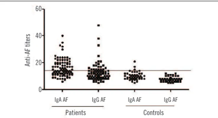
Anti-AF IgA was positive in 46/90 (51.1%) of the RA sample
and 3/16 (18.7%) of the controls (p = 0.027); anti-AF IgG was
found in 21/90 (23.3%) of RA patients and none of the controls
(p = 0.037), as show in Figure 1.
Analyzing positivity of anti-AF IgG and anti-AF IgA according to demographic, clinical and serological profiles
in RA patients, we found the data shown in Table 1. Neither
IgA nor IgG anti-AF antibodies showed significant difference in patients with and without sSS. In Figure 2, the receiver
operating characteristic (ROC) curve analysis shows the poor performance of anti-AF IgA and anti-AF IgG to distinguish RA patients with and without sSS.
Comparing titer values of anti-AF IgA and anti-AF IgG in RA patients with and without sSS, we found no statistical differences, as seen in Table 1.
The analysis of anti-AF IgA and anti-AF IgG regarding RA demographic, clinical and serological proiles is seen in Table 2.
TABLE 1 − Comparison of titer values of IgG and IgA anti-AF antibodies
in RA patients with and without sSS
Antibody RA without sSS RA with sSS p (Mann-Whitney)
Anti-AF IgA (U/ml)
6-35 Median 15 (11.5-20.5)
8-40 Median 14 (12-19)
0.97
Anti-AF IgG (U/ml)
5-38 Median 12 (8-15)
7-48 Median 11 (9-14)
0.68
IgG: immunoglobulin class G; IgA: immunoglobulin class A; AF: alpha-fodrin; RA: rheumatoid arthritis; sSS: secondary Sjögren syndrome.
FIGURE 1 −IgA and IgG anti-AF antibodies in RA patients and controls
AF: alpha-fodrin; RA: rheumatoid arthritis.
60
40
20
0
Patients Controls
Anti-AF titers
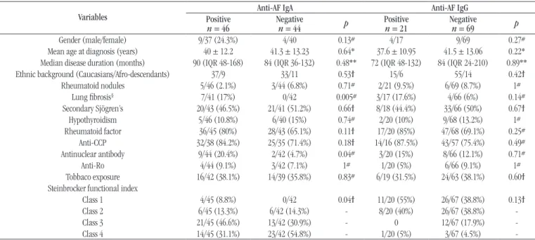
TABLE 2 − Association studies of demographic, clinical and serological data with IgA and IgG anti-AF antibodies in 90 RA patients
Variables
Anti-AF IgA Anti-AF IgG
Positive
n = 46 Negativen = 44 p Positiven = 21 Negativen = 69 p
Gender (male/female) 9/37 (24.3%) 4/40 0.13# 4/17 9/69 0.27#
Mean age at diagnosis (years) 40 ± 12.2 41.3 ± 13.23 0.64* 37.6 ± 10.95 41.5 ± 13.06 0.22* Median disease duration (months) 90 (IQR 48-168) 84 (IQR 36-132) 0.48** 72 (IQR 48-132) 84 (IQR 24-210) 0.89** Ethnic background (Caucasians/Afro-descendants) 37/9 33/11 0.53ϯ 15/6 55/14 0.42ϯ
Rheumatoid nodules 5/46 (2.1%) 3/44 (6.8%) 0.71# 2/21 (9.5%) 6/69 (8.7%) 1#
Lung ibrosis§ 7/41 (17%) 0/42 0.005# 3/17 (17.6%) 4/66 (6%) 0.14#
Secondary Sjögren’s 20/43 (46.5%) 21/41 (51.2%) 0.66ϯ 8/18 (44.4%) 33/66 (50%) 0.67ϯ Hypothyroidism 5/46 (10.8%) 6/40 (15%) 0.74# 2/20 (10%) 9/68 (13.2%) 1# Rheumatoid factor 36/45 (80%) 28/43 (65.1%) 0.11ϯ 17/20 (85%) 47/68 (69.1%) 0.25#
Anti-CCP 32/38 (84.2%) 25/35 (71.4%) 0.18ϯ 14/16 (87.5%) 43/57 (75.4%) 0.49# Antinuclear antibody 9/44 (20.4%) 2/42 (4.7%) 0.04# 3/20 (15%) 8/66 (12.1%) 0.71#
Anti-Ro 4/44 (9.1%) 3/42 (7.1%) 1# 1/20 (5%) 6/66 (9.1%) 1#
Tobbaco exposure 16/42 (38.1%) 14/39 (35.8%) 0.83# 6/19 (31.5%) 24/63 (38.1%) 0.60ϯ Steinbrocker functional index
Class 1 4/45 (8.8%) 0/42 0.04ϯ 11/20 (55%) 26/67 (38.8%) 0.13ϯ
Class 2 6/45 (13.3%) 6/42 (14.3%) - 8/20 (40%) 26/67 (38.8%)
-Class 3 21/45 (46.6%) 13/42 (30.9%) - 0 12/67 (17.9%)
-Class 4 14/45 (31.1%) 23/42 (54.8%) - 1/20 (5%) 3/67 (4.5%)
-IgA: immunoglobulin class A; IgG: immunoglobulin class G; AF: alpha-fodrin; RA: rheumatoid arthritis; IQR: interquartile range; CCP: cyclic citrullinated protein; *: Student’s t-test; **: Mann-Whitney test; #: Fisher’s exact test; ϯ: chi-square test.
As anti-AF IgA showed positive association with lung ibrosis, antinuclear antibodies (ANA) and functional classiication in univariated analysis, we further studied these variables in a logistic regression analysis where only functional class index showed association with the presence of anti-AF IgA (odds ratio [OR] = 1.8; 95% conidence interval [CI] of 1.02-3.16).
DISCUSSION
Both SS and non-Sjögren’s syndrome can show the clinical symptoms of dry eyes and dry mouth, so sicca symptoms are poor discriminators of SS(14). Secondary SS was found to be present
in 22% of RA patients in our geographical area(15). Its diagnosis,
according to the American-European criteria, requires a minor salivary gland biopsy or an objective evidence of salivary gland
involvement(12). Although salivary biopsy is a minor procedure, it
is invasive and not always well accepted by patients. The objective
measurement of salivary gland involvement by unstimulated
salivary low, parotid sialography and salivary scintilography is not accessible in daily practice. If a speciic biomarker, such as an autoantibody, were found, this problem could be avoided.
The AF antigen has been postulated to be involved in the autoimmune responses leading to destruction of exocrine glands(16).
It is associated with ion channels and pumps, and therefore it is possible that antibodies directed against AF could disturb their function(16). Thus, anti-AF seems a good candidate to mark this
FIGURE 2 −Performance of anti-AF antibodies to distinguish secondary Sjögren’s
syndrome in RA patients
A) ROC of anti-AF IgA with area = 0.502 (95% CI = 0.38-0.62); p = 0.96; B) ROC of anti-AF IgG with area = 0.52 (95% IC = 0.4-0.64); p = 0.67.
AF: alpha-fodrin; RA: rheumatoid arthritis; ROC: receiver operating characteristic; IgA: immunoglobulin class A; CI: confidence interval; IgG: immunoglobulin class G.
0 20 40 60 80
100
0 50 100 150
Specificity%
s
e
n
s
it
iv
it
y
%
FIGURE 2- rin antibo
150
100
50
0
Sensitivity (%)
Specificity (%)
A) anti-AF IgA
0 20 40 60 80 100
0 20 40 60 80
100 0
50 100 150
s
e
n
s
it
iv
it
y
%
FIGURE 2- e
150
100
50
0
Sensitivity (%)
0 20 40 60 80 100 Specificity (%)
situation. However, the present results do not favor this possibility in the case of SS secondary to RA. Although anti-AF IgA and IgG were more commonly present in RA patients than in controls, this autoantibody did not identify those with RA and sSS, neither did RA patients with sSS have a higher level of these antibodies.
There are few studies of anti-AF in RA patients with sSS; most of them are directed to primary disease, SLE and sSS. Witte et al. (2000)(6)
found that anti-AF IgA, but not IgG, was associated with sSS in RA patients, but they studied only 19 RA patients using the European criteria for sSS diagnosis. Turkçapar et al. (2006)(9), studying 10
patients with RA and sSS, found a 10% prevalence for anti-AF IgA and no positivity for anti-AF IgG, concluding that they were not useful for diagnostic purposes in this context.
We could not demonstrate that RA patients with hypothyroidism have more anti-AF antibodies than those without
it. Szanto et al. (2008)(17) published a very interesting study where
they found association of anti-AF IgG and IgA with thyroiditis. Based on the fact that AF can be associated with endocrine and exocrine secretion, they proposed that these autoantibodies could be a marker of secretory disorders.
In univariated analysis, lung ibrosis and ANA were associated with anti-AF IgA. Interstitial lung disease is an extra-articular manifestation that can be severe and a source of substantial morbidity and mortality for affected patients(18). At present, there is
no serological marker of this complication, although some authors found that it is more common in patients who are positive for anti-cyclic citrullinated protein (CCP) and rheumatoid factor (RF)(18-20).
To our knowledge, association with anti-AF has not been studied before. In addition, the presence of ANA in RA is associated with a more aggressive disease and with a higher rate for extra-articular
manifestations(18). In the logistic regression, these associations did
not remain as independent variables, but further examination of
this possibility, with a larger number of patients, could be interesting. They are, both, linked to severe RA prognosis(18).
In the present study, patients with worse functional class had higher prevalence of anti-AF IgA, but the real meaning of this inding needs additional evaluation. Functional class is a variable inluenced by many factors, such as the early institution of treatment. So, prompt access to medical care, not evaluated at present, could be a determining factor. However, it must be noted that a worse functional class, as well as lung ibrosis and presence of ANA, denotes poor prognosis.
Hu et al. (2013)(10), in their meta-analysis, demonstrated that
heterogeneity existed in anti-AF prevalence among the Chinese and Japanese populations, what may explain the different incidences of SS in different areas or nationalities. We could not ind inluence of ethnic background in the presence of either IgG or IgA anti-AF sample, but we had only patients of African and Caucasian origin, none was Asian. Also, it must be taken into account that, in our geographical area, the ethnic classiication according to external appearance may not be reliable due to a very high miscegenation rate.
A major limitation of this study is that the present RA sample had mean disease duration of seven years; so most of the patients were on glucocorticoid therapy, immunosuppressants and/or biologics. The use of these drugs could have reduced the positivity of anti-AF antibodies. Studies on newly-diagnosed patients receiving no treatment are required to avoid this interference. Another point to be considered is that we did not study RA disease activity; it would be interesting to know if a high RA disease activity would modify anti-AF results.
Concluding, in our study, IgA and IgG anti-AF antibodies neither allowed diagnosis of sSS in RA patients nor did they mark any special clinical or serological inding. Their association with functional class needs further investigation.
RESUMO
Introdução: A síndrome de Sjögren (SS) é uma doença autoimune caracterizada por infiltração linfocítica. Atualmente os anticorpos mais pesquisados
para seu diagnóstico são anti-Ro e anti-La, que, no entanto, apresentam baixa especificidade nos casos de SS secundária a artrite reumatoide (AR) e lúpus eritematoso sistêmico (LES). Os anticorpos contra alfafodrina (AF) foram propostos para diagnosticar SS. Objetivo: Investigamos o anticorpo anti-AF em um grupo de portadores de AR com e sem SS secundária (SSs). Material e métodos: Foram estudados 90 pacientes consecutivos com AR (48,8% com SS) e amostras de 45 voluntários saudáveis. Imunoglobulina da classe G (IgG) e imunoglobulina da classe A (IgA) anti-AF foram investigadas por ensaio imunossorvente ligado à enzima (ELISA), sendo consideradas positivas quando acima de 15 U/ml. Dados demográficos, clínicos e sorológicos foram obtidos a partir de revisão de prontuários. Resultados: IgA anti-AF foi positiva em 46/90 (51,1%) das amostras com AR e 3/45 (6,7%) das amostras-controle (p < 0,001); IgG anti-AF foi encontrada em 21/90 (23,3%) de pacientes com AR e nenhum dos controles (p = 0,037). Nem IgA anti-AF nem IgG anti-AF conseguiram diferenciar pacientes com e sem SSs. Conclusão: IgG anti-AF e IgA anti-AF foram investigadas e não contribuíram especificamente para diferenciar pacientes com AR que sofrem ou não de SS, além de não conseguirem estabelecer o diagnóstico de SS em pacientes com AR.
REFERENCES
1. Routsias JG, Tzioufas AG. Sjögren syndrome – study of autoantigens and autoantibodies. Clin Rev Allerg Immunol. 2007; 32: 238-51. 2. Watanabe T, Tsuchida T, Kanda N, Mori K, Hayashi Y, Tamaki K. Anti-alpha-fodrin antibodies on Sjögren syndrome and lupus erythematosus. Arch Dermatol. 1999; 135: 535-9.
3. Ruiz-Tiscar JL, Urrea FJ, Sánchez-Rámon S, et al. Prevalence of IgG anti-α-fodrin antibodies in Sjögren’s syndrome. Ann N Y Acad Sci. 2005; 1050: 210-6.
4. Ulbricht KU, Schmidt RE, Witte T. Antibodies against alpha-fodrin in Sjögren’s syndrome. Autoimmune Reviews. 2003; 2: 109-13.
5. Haneji N, Nakamura T, Takio K, et al. Identiication of alpha-fodrin as candidate autoantigen in primary Sjogren’s syndrome. Science. 1997; 276: 604-7.
6. Witte T, Matthias T, Arnett FC, et al. IgA and IgG autoantibodies against alpha-fodrin as markers of Sjögren’s syndrome. Systemic lupus erythematosus. J Rheumatol. 2000; 27: 2617-20.
7. Loch H, Pelck R, Manthorpe R. Diagnostic and prognostic signiicance of measuring antibodies to alpha-fodrin compared to Ro-52, anti-Ro-60 and anti-La in primary Sjögren’s syndrome. J Rheumatol. 2008; 35: 845-9.
8. Zanbelt MM, Vogelzangs LV, Van de Putte LB, Van Venrooij WJ, Van den Hooge FH. Anti-alpha-fodrin antibodies do not add much to diagnosis of Sjogren’s syndrome. Arthritis Res Ther. 2004; 6: 33-8.
9. Turkçapar N, Olmez U, Tutkak H, Durman M. The importance of alpha-fodrin antibodies in the diagnosis of Sjögren’s syndrome. Rheumatol Int. 2006; 26: 354-9.
10. Hu Q, Wang D, Chen W. The accuracy of the anti-α-fodrin antibody test for diagnosis of Sjögren’s syndrome: a meta-analysis. Clin Biochem. 2013; 46(15): 1372-6. doi: 10.1016/j.clinbiochem.2013.04.020. 11. Arnett FC, Edworthy SM, Bloch DA, et al. The American Rheumatology
Association 1987 revised criteria for the classiication of rheumatoid arthritis. Arthritis Rheum. 1988; 31: 315-24.
12. Vitali C, Bombardieri S, Jonsson R, et al. Classiication criteria for Sjögren’s syndrome: a revised version of the European criteria proposed by the American-European consensus group. Ann Rheum Dis. 2002; 61: 554-8.
13. Klippel J, Dieppe PA. Selected measures for outcome assessment of rheumatic diseases. In: Klippel J, Dieppe PA, editors. Rheumatology. 2nd ed, vol. 2. Mosby London; 1998. p. A1-11.
14. Chen KS, Jiang MC, Li JC, Liu OK, Tsai CSS. Discrimination between Sjögren’s and non-Sjögren’s sicca syndrome by sialoscintigraphy and antibodies against alpha-fodrin and Ro/La autoantigens. J Inter Med Res. 2009; 37: 1088-96.
15. Moura MC, Zakszewski PT, Silva MB, Skare TL. Epidemiological proile of patients with extra-articular manifestations of rheumatoid arthritis from the city of Curitiba, south of Brazil. Rev Bras Reumatol. 2012; 52: 679-94.
16. Routsias JG, Tsioufas AG. Sjögren’s syndrome – study of autoantigens and autoantibodies. Clin Rev Allerg Immunol. 2007; 32: 238-51. 17. Szanto A, Csipo I, Horvath I, Biro E, Szodoray P, Zeher M. Autoantibodies to alfa-fodrin in patients with Hashimoto thyroiditis and Sjögren’s syndrome: possible markers for a common secretory disorder. Rheumatol Int. 2008; 28: 1169-72.
18. Young A, Koduri G. Extra-articular manifestations and complications of rheumatoid arthritis. Best Prac Res Clin Rheumatol. 2007; 21: 907-27.
19. Richman NC, Yazdany J, Graf JM, Chernitskiy V, Imboden JB. Extraarticular manifestations of rheumatoid arthritis in a multiethnic cohort of predominantly Hispanic and Asian patients. Medicine (Baltimore). 2013; 82: 92-7.
20. Giles JT, Danoff SK, Sokolove J, et al. Association of ine speciicity and repertoire expansion of anticitrullinated peptide antibodies with rheumatoid arthritis associated interstitial lung disease. Ann Rheum Dis. 2014; 73(8): 1487-94.
CORRESPONDING AUTHOR
Renato Nisihara