*Correspondence: C. A. Schmidt. Departamento do Medicamento, Faculdade de Farmácia. Rua Barão do Jeremoabo, 147. Ondina, 40170-115 – Salvador - Bahia, Brasil. Phone: +55-71-3237-7635. E-mail: [email protected]; [email protected]
vol. 49, n. 4, oct./dec., 2013
A fully validated microbiological assay to evaluate the potency of
ceftriaxone sodium
Maria Luisa Manio
1, Danielle Araújo Agarrayua
1, Jaison Carlosso Machado
2,
Cleber Alberto Schmidt
3,*1Pharmacy Course, Franciscano University Center, Santa Maria, Brazil, 2Faculty of Pharmacy,
Federal University of Rio Grande do Sul, Porto Alegre, Brazil, 3Department of Medicines, Faculty of Pharmacy,
Federal University of Bahia, Salvador, Brazil
Ceftriaxone (CFTX) sodium is a third-generation, broad-spectrum cephalosporin that is resistant to beta-lactamases. An alternative bioassay for the assessment of the potency of this drug in pharmaceutical formulations has not been previously reported. Thus, this paper reports the development and full validation of a 3 x 3 agar diffusion bioassay using a cylinder-plate method to quantify CFTX sodium in pharmaceutical samples. The strain Staphylococcus aureus ATCC 6538P was used as the test microorganism, and the
results of the proposed bioassay displayed high linearity, precision, accuracy, speciicity and robustness.
All potency results were statistically analyzed using an analysis of variance (ANOVA) and were found to
be linear (r=0.99999) in the range of 16–64 μg/mL, accurate (100.5%), and precise [repeatability: relative standard deviation (RSD)=1.4%; intermediate precision: between-day RSD=2.1% and between-analyst RSD=2.5%]. The speciicity of the bioassay was determined by evaluating a degraded sample (50 ºC) at
0, 24 and 48 hours as compared against the results from the pharmacopeial liquid chromatography method for CFTX. The results validated the proposed microbiological assay, which allows reliable quantitation of CFTX in pharmaceutical samples. Moreover, it is a useful, simple and low-cost alternative method for monitoring the quality of this medicine.
Uniterms: Ceftriaxone/agar diffusion assay. Ceftriaxone/quality control. Ceftriaxone/microbiological
assay. Drugs/quality control.
A ceftriaxona sódica é uma cefalosporina de terceira geração de uso parenteral, com amplo espectro de atividade e resistente a β-lactamases. Este estudo apresenta o desenvolvimento e validação de um bioensaio por difusão em ágar usando o método de cilindros em placas para determinação da potência deste antibiótico. A validação desenvolvida apresentou bons resultados em termos de linearidade,
precisão, exatidão, especiicidade e robustez. Empregou-se o Staphylococcus aureus ATCC 6538P como micro-organismo teste. Os resultados dos ensaios foram tratados estatisticamente utilizando-se análise de variância (ANOVA). O método apresentou linearidade (r=0,99999) na faixa de doses selecionada
(16-64 µg/mL), precisão (repetibilidade: DPR=1,4%; precisão intermediária: inter-dias DPR=2,1% e inter-analistas: DPR=2,5%) e exatidão de 100,5%. A especiicidade do bioensaio foi avaliada através da análise comparativa, por cromatograia líquida de alta eiciência, de amostras degradadas a 50 ºC nos
tempos zero, 24 e 48 h. Os resultados encontrados demonstraram a validade do bioensaio proposto, o
qual permite a quantiicação coniável de ceftriaxona sódica em produtos farmacêuticos comerciais. Por
ser metodologia simples e de baixo custo constitui-se em alternativa para a análise de rotina do controle de qualidade de medicamentos.
Unitermos: Ceftriaxona/ensaio de difusão em ágar. Ceftriaxona/controle de qualidade. Ceftriaxona/
INTRODUCTION
Ceftriaxone (CFTX) sodium is a semisynthetic antibiotic that can effectively treat several types of bacterial infections. Unlike the other 3rd generation
cephalosporins, CFTX has a long plasma half-life, up to 4- to 10-times longer than the other antibiotics in this class (Neu et al., 1981; Stoeckel, 1981). This cephalosporin shows a broad spectrum against positive and Gram-negative bacteria, including enterobacteria, Haemophilus
inluenzae and Streptococcus pneumoniae (Rebuelto et al., 2002).
Regarding the official quality control of CFTX, as raw material or in pharmaceutical preparation, the pharmacopeias recommend employing reversed phase
liquid chromatography (RP-HPLC) with UV detection
and a mobile phase composed of water, phosphate buffer and acetonitrile (British Pharmacopoeia, 2009; European Pharmacopoeia, 2005; Japanese Pharmacopeia, 2006; United States Pharmacopeia, 2011).
Several alternative physicochemical methods to assay CFTX in pharmaceutical formulations are described in the literature, such as spectrophotometric methods (El-Walily et al., 2000; Al-Momani, 2001; Amim, Ragab, 2004; Sankar et al., 2006; Okoye et al.,
2007; Adegoke, Quadri, 2012), low injection analysis
(FIA) with chemiluminescence detection (Yinhuan, Jiuru, 2006), spectrofluorometry (Shah et al., 2011), reversed-phase high-performance liquid chromatography
(RP-HPLC) (Hecq et al., 2006; Jane et al., 2006; Tippa,
Singh, 2010; Akl et al., 2011), high performance thin layer
chromatography (HPTLC) (Eric-Jovanovic et al., 1998)
and differential pulse polarography (DPP) (Sengün et al., 1985).
To assess the concentration of CFTX in biological matrices such as plasma, serum, cerebrospinal fluid and bile, several physicochemical methods have been
reported, such as HPLC (Patel et al., 1981; Trautmann,
Haefelfinger, 1981; Ascalone, Dal Bò, 1983; Bowman et al., 1984; Chan et al., 1986; Granich, Krogstad, 1987; Bompadre et al., 1998; Kohlhepp et al., 1998; Tsai et al., 1999; Nemutlu et al., 2009; Mcwhinney et al., 2010), spectrofluorometric assays (Omar et al., 2009), and capillary zone electrophoresis (CZE) (Quaglia et al., 1997). Microbiological techniques are used to evaluate the
bioavailability of CFTX in biological luids (Rebuelto et
al., 2002; Ismail et al., 2005) and to study the susceptibility of several microorganisms to CFTX (Beskid et al., 1981; Eickhoff, Ehret, 1981; Baumgartner, Glauser, 1983; Emmerson et al., 1985; Dias et al., 1998).
The use of microbiological assays to evaluate the
potency of CFTX in pharmaceutical formulations is uncommon. The literature reports only one method which uses a 5 point calibration curve with paper discs in a bi-layer agar diffusion assay using Bacillus subtilis (ATCC 6633) as the test microorganism (Cantón et al., 1993). However, the original method was not validated and the
quantiication does not follow the procedure recommended
for a 5 x 1 assay (Esteban et al., 1990).
Physicochemical techniques are recognized to be fast, precise and accurate in quantifying cephalosporin antibiotics. However, some disadvantages are inherent to these methods, as the interference of the excipients, tedious extraction steps and lack of selectivity complicate the performance (Ahmed et al., 2011; Adegoke et al., 2012). Furthermore, most of these procedures are not simple in the routine analyses, they require dedicated or sophisticated equipment and expensive reagents, which are often not available in quality control laboratories (El-Walily et al., 2000; Souza et al., 2006).
Alternative methods to evaluate the potency of antibiotics, such as the fully validated microbiological agar diffusion assay, are simple and operationally inexpensive. Furthermore, such bioassays are suitable for quality control laboratories that do not have specialized and sophisticated instruments (Souza et al., 2006; Schmidt et al., 2008,2009).
Therefore, the aim of the present study was to
develop and validate a low-cost, simple, speciic, accurate
and reproducible microbiological agar diffusion assay using a cylinder-plate method and propose it as a useful alternative to the physicochemical methods described in the literature for quantitation of ceftriaxone sodium as raw
material and injectable formulation.
MATERIAL AND METHODS
Chemicals
The ceftriaxone sodium reference standard from
Brazilian Pharmacopeia (assigned purity 847.3 µg/mg)
and the sample were commercially obtained. The sample (batch 97728B) was within its shelf-life and claimed to contain 500 mg of ceftriaxone sodium sterile powder
for injection (Eurofarma, Brazil). All reagents used were
analytical grade (Difco, USA; Merck, Germany). Ultrapure and bidistilled water were used in the experiments.
Ceftriaxone sodium reference solution
Ten milligrams of the ceftriaxone sodium reference
and dissolved in sterile phosphate buffer solution pH 7
[K2HPO4 1.36% (w/v) and KH2PO4 0.4% (w/v)]. Aliquots
of this solution were diluted in the identical buffer yielding
working solutions with inal concentrations of 16, 32 and 64 μg/mL (S1, S2 and S3, respectively).
Preparation of the sample solutions
Five-hundred milligrams of the CFTX sample was
transferred to a 250 mL volumetric lask and dissolved
with sterile phosphate buffer solution pH 7. Five milliliters
of this solution were transferred to a 25 mL volumetric
flask and dissolved to obtain a final concentration of
400 µg/mL. Aliquots of this solution were further diluted
in the identical buffer solution to obtain the concentrations
of 16, 32 and 64 μg/mL (T1, T2 and T3, respectively),
which were tested against S1, S2 and S3.
Microorganism and inoculum standardization
The strain Staphylococcus aureus ATCC 6538P was selected as the test microorganism because of its susceptibility to ceftriaxone sodium, yielding sharply
deined zones of growth inhibition, which allows more
precise measurements. The culture of Staphylococcus aureus ATCC 6538P (INCQS - National Institute for Health Quality Control, Brazil), after reconstitution, were cultivated and maintained on Grove-Randall’s 1 culture medium (Difco). The microorganism standardization was prepared according to the procedure described in the Brazilian and USP Pharmacopeias (Farmacopeia Brasileira, 2010; United States Pharmacopeia, 2011). Prior to use, the microorganism was grown in a slant medium (Grove-Randall’s 1, for 24 h at 35 ± 2 °C). Using
aseptic techniques, the growth was suspended in a 0.9%
NaCl sterile solution and diluted to give a suspension
with 25 ± 2% turbidity (transmittance) at 580 nm using a 10 mm absorption cell, with 0.9% NaCl sterile solution
as blank. The Grove-Randall’s 1 culture medium at 48 °C
was inoculated with the standardized suspension at 1% (v/v) to compose the upper layer in the plate.
Agar diffusion bioassay
The bioassay followed the 3 × 3 parallel line design (3 doses of standard and 3 doses of sample in each plate),
with 6 plates/assay, in accordance with the European and
Brazilian Pharmacopoeias (European Pharmacopoeia, 2005; Farmacopeia Brasileira, 2010). For the base
layer agar, 21 mL of Grove-Randall’s 2 culture medium
(Difco) in a 100 × 20 mm Petri dish was used. After
solidifying, 5 mL of inoculated Grove-Randall’s 1
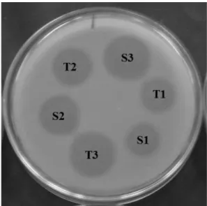
medium was poured onto the base layer. In each plate, 6 stainless steel cylinders (8 × 6 × 10 mm – external diameter x internal diameter x height) were placed on the surface of the inoculated medium. Three alternated
cylinders were illed with 150 μL of reference solutions
(S1, S2 and S3), and the other three cylinders were
illed with the concentrations of the sample solutions
(T1, T2 and T3) (Figure 1). The plates were incubated
at 35 ± 2 ºC aerobically for 16 h. The growth inhibition
zone diameters (mm) were carefully measured with an electronic caliper. All experiments were performed in a biological safety cabinet and the infected material was decontaminated before being discarded.
HPLC
In addition to the bioassay, the remaining CFTX
after hydrolysis degradation was assessed by a LC
method using the chromatographic conditions described in the USP monograph for CFTX sodium (United States
Pharmacopeia 2011). Briely, 20 µL sample volumes were injected into a LC system (Shimadzu LC-10AD, Japan)
equipped with a 270-nm detector and a C18 column (4 x 150 mm). Each sample was run in triplicate. The mobile phase (water, acetonitrile, buffer solution pH 7, buffer
solution pH 5 – 552:400:44:4) containing 0.32% of tetraheptylammonium bromide was used at a low rate of 2 mL/min.
FIGURE 1 - CFTX potency evaluation by agar diffusion assay using the cylinder-plate method with a 3 x 3 experimental design. Zones of growth inhibition observed for doses 16, 32
and 64 μg/ml of the CFTX reference substance (S1, S2, S3) and
Calculations
In all experiments, the CFTX potency was statistically calculated using the parallel-line model for a 3 x 3 assay design. The regression, parallelism and linearity of the response were evaluated by an analysis of variance (ANOVA). To complement the statistical validation, the
conidence interval (IC95) of each assay was considered
(Farmacopeia Brasileira, 2010; Hewitt, 2003; European Pharmacopoeia, 2005; ICH, 2005; Schmidt et al., 2009).
Validation of the method
All experimental conditions of the proposed method
were tested and adjusted prior to the validation to ensure
the best assay conditions. The method was validated according the International Conference on Harmonization (ICH 2005) and USP guidelines 2011). The following operational characteristics were evaluated:
Range – Assessed by the selected doses for the
calibration curve and conirmed by determination of the
accuracy, precision and linearity of the method.
Linearity – Evaluated through 12 independent assays using linear regression analysis and calculated by a least-squares method for three doses of the reference substance. Precision – Assessed through the repeatability and intermediate precision and expressed as the relative standard deviation (RSD). The repeatability was examined by assaying 6 different test solutions of the CFTX sample against the reference standard. The assays were performed by the same analyst, under identical experimental conditions in one day (intraday). The intermediate precision was evaluated by performing the analysis in the same laboratory on 2 separate days (interday) with different analysts (between-analysts).
Accuracy – The test was repeated in three consecutive
days. Three concentration levels, covering 80% to 120% of the selected range of 16, 32 and 64 μg/mL, were
tested each day. Forty mg of CFTX was transferred to
a 100 mL volumetric lask and dissolved in a phosphate
buffer solution pH 7.0 to obtain a stock solution with a
concentration of 400 μg/mL. The 3 concentration levels at 100% were prepared from aliquots of 1, 2 and 4 mL of the stock solution transferred into 50 mL volumetric lasks
and diluted with a phosphate buffer solution pH 7 to give
inal concentrations of 16, 32 and 64 μg/mL, respectively. The doses at 80% nominal concentration were prepared from aliquots of 0.8, 1.6 and 3.2 mL of the stock solution transferred into 25 mL volumetric flasks to give final concentrations of 12.8, 25.6 and 51.2 μg/mL, respectively. The doses at 120% nominal concentration were obtained
from aliquots of 1.2, 2.4 and 4.8 mL of the stock solution transferred into 25 mL volumetric lasks and diluted with phosphate buffer solution pH 7 to give solutions with inal concentrations of 19.2, 38.4 and 76.8 μg/mL, respectively.
These working solutions were assayed against the 3 concentration levels of the reference standard solution at
100% nominal concentration.
Selectivity – The ability of the proposed method to assess the content of CFTX sodium in the presence of impurities and degraded substances was tested by comparing the results obtained in the bioassay to the
results from the pharmacopeial LC method for the
identical degraded sample. A CFTX sodium commercial
sample was reconstituted with water for injection and heated to 50 ºC for 2 days. The sample was analyzed by both methods at 0, 24 and 48 h. The conditions of the LC
method were in accordance with the USP monograph for CFTX sodium (United States Pharmacopeia, 2011).
Robustness – Several method parameters were modified in assaying a CFTX sample. The considered
factors were the inoculum concentration (1.3%),
incubation temperature (32 °C), volume of the inoculated
layer (thickness – 4 mL) and the solvent used for the standard and sample dilution (sterile water for injection).
RESULTS AND DISCUSSION
The use of a suitable analytical method is fundamental in the quality control of active substances in either the pharmaceutical or raw material form. The choice of the method is normally based on several factors such as the drug source; its complexity; purity and sample quantity; and the qualitative, semi-quantitative or quantitative purpose of the method. Furthermore, the availability of equipment and reagents should be considered in the development of accessible and useful methodologies.
Taking in account that the potency of an antibiotic may be assessed through the comparison of the inhibition of growth of a susceptible microorganism induced by known concentrations of the antibiotic and its respective reference standard (European Pharmacopoeia, 2005; United States Pharmacopeia, 2011), a 3 x 3 microbiological assay was proposed for determining the CFTX sodium concentration
in injectable pharmaceutical dosage forms.
The range of the doses selected (16 to 64 μg/mL) for
the bioassay was shown to be most appropriate for this assay system for the following reasons: the susceptibility of the microorganism to lower concentrations on the curve, the linear relationship between the logarithm of the dose and the
observed response, and the signiicant slope of the curve.
6 plates/assay. The experimental average zone diameters (mm) and RSD values (%) for the standard solutions are
presented in Table I. The difference between the average
size of the inhibition zones for doses 16-32 µg/mL, and 32-64 µg/mL was approximately 3 mm, showing
good linearity in the response obtained by the method. Furthermore, the RSD values showed low variability in the response (intradose) obtained in this bioassay.
.
The logarithm of the concentrations (μg/mL) and
the mean diameter of the inhibition zones (mm) were used to calculate the calibration curve for CFTX. The method
showed good linearity for the range of 16–64 μg/mL.
The representative linear equation was y = 10.176x +
4.046. The determination coeficient (r2=0.99998) and
correlation coeficient (r=0.99999) were highly signiicant.
The bioassays were validated using an analysis of variance (ANOVA). In all experiments, the regression was highly
signiicant (P<0.05) and no deviation was found in either
the parallelism or the linearity (P>0.05). Moreover, all
bioassays gave potency results within a conidence interval of 90–115% (IC95), indicating that the experiments
performed during the validation were well executed. Simultaneously, the developed method was found to have
signiicant response differentiation between doses and signiicant sensitivity to the selected doses.
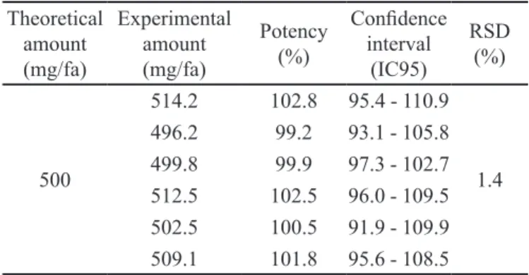
The bioassay precision, in terms of repeatability (intra-assay), was evaluated by analyzing, on identical days, six different test solutions of CFTX sodium powder
for injection with identical theoretical concentrations. The CFTX activity ranged from 99.2% to 102.8%, with an RSD value of 1.4% (Table II).
To calculate the intermediate precision, the same sample was analyzed in triplicate on 2 separate days (between-day; Table III) and by 2 different analysts (between-analysts; Table IV), yielding RSD values of
2.1% and 2.5%, respectively. These low RSD values conirmed the capacity of the method to generate, with
the same sample, reproducible results with a low response variation in independent assays.
TABLE III - Between-day precision data of the CFTX bioassay. Sample solutions at 100% theoretical concentration were tested in triplicate in two separate days
Day Potency found (%) Conidence interval
(IC95)
RSD (%)
1
100.5 91.9 - 109.9
2.1
101.8 95.6 - 108.5
102.8 95.4 - 110.9
2
102.8 93.0 – 113.8
99.6 96.0 – 103.3
97.5 93.2 – 101.9
TABLE IV - Between-analyst precision results obtained in the CFTX bioassay validation. Sample solutions at 100% theoretical concentration were tested in triplicate by two analysts
Analyst found (%)Potency Conidence interval
(IC95) RSD (%)
1
99.6 96.0 - 103.3
2.5
97.5 93.2 - 101.9
102.8 93.0 - 113.8
2
96.2 91.5 – 101.1
97.1 91.9 – 102.5
97.3 90.8 – 104.2
TABLE II – Results of the repeatability evaluation of the microbiological assay of CFTX powder for injection
Theoretical amount (mg/fa)
Experimental amount (mg/fa)
Potency (%)
Conidence interval
(IC95)
RSD (%)
500
514.2 102.8 95.4 - 110.9
1.4
496.2 99.2 93.1 - 105.8
499.8 99.9 97.3 - 102.7
512.5 102.5 96.0 - 109.5
502.5 100.5 91.9 - 109.9
509.1 101.8 95.6 - 108.5
fa – lask ampoule
TABLE I - Mean diameters of the growth inhibition zones obtained for the CFTX standard curve for 16, 32 and 64 μg/mL solutions
Concentration
(μg/mL) Mean diameter ± SD
a
(mm)
RSD (%)
16 16.3 +/- 0.3 1.9
32 19.4 +/- 0.2 1.0
64 22.4 +/- 0.5 2.0
a n= 12 independent assays with 6 Petri dishes each
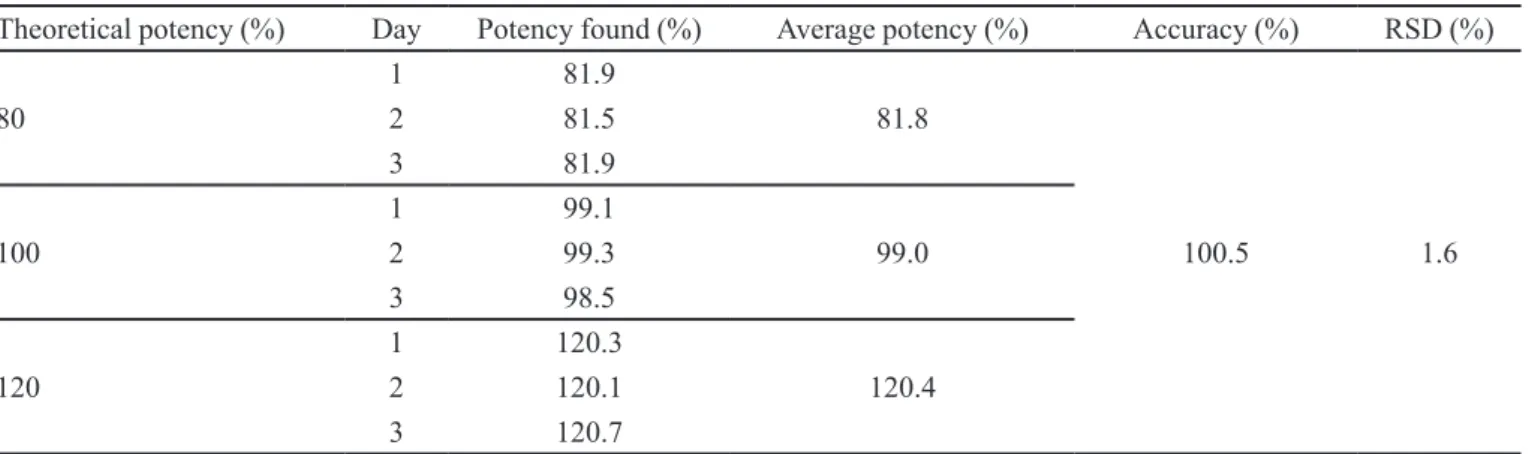
The accuracy of the method was evaluated at 80,
100 and 120% of the range selected for the bioassay (16-64 μg/mL), covering the specific range of 12.8-76.8 μg/mL. The mean accuracy was 100.5% with an RSD of 1.6% (Table V). Thus, the results obtained in
of the proposed method to accurately quantify or detect samples containing low or high concentrations of CFTX, displaying that the bioassay is able to detect samples that do not meet the assay requirements recommended by the Pharmacopeias. Generally, this essential parameter for quantitative methods is not tested in agar diffusion assay validation studies.
To evaluate the selectivity of the proposed method, a CFTX commercial sample reconstituted to
10 mg/mL was exposed to dry heat (50 ºC) for 48 h.
The concentration of the remaining CFTX in the sample
was assessed by both the LC pharmacopeial method
(United States Pharmacopeia, 2011) and the proposed bioassay at 0, 24 and 48 h. At time zero, the CFTX
potency was 103.1% ± 0.6 and 107.3% ± 0.6 in the bioassay and HPLC, respectively. The CFTX showed
low thermal stability, as shown in Figure 2, in which similar decreasing concentration curves were registered by both methods. However, the microbiological assay
presented higher sensitivity, because it showed a decrease
in potency of approximately 77%, as compared to 53% detected by HPLC for the same sample. This preliminary
result shows that the decomposition products of CFTX were not microbiologically active. Therefore, the
microbiological assay was shown to be speciic, because
the impurities and degradation products did not interfere in the ability of the method to assess the analyte.
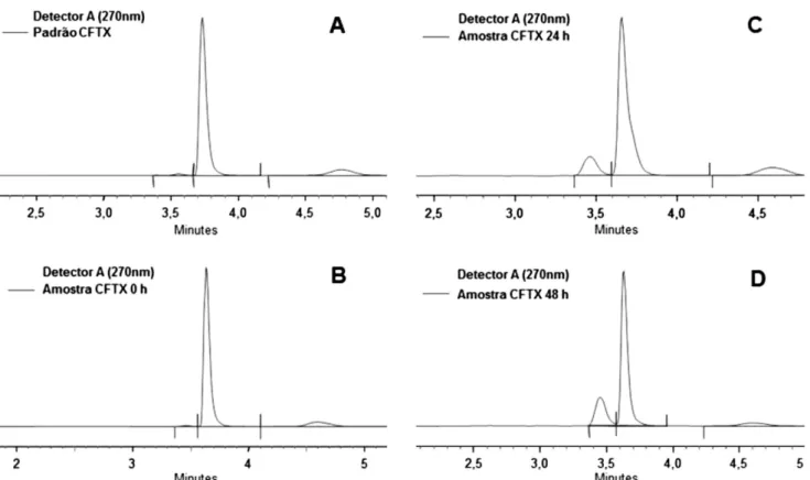
Figure 3 displays the chromatograms of the CFTX sodium reference standard (A), the freshly prepared sample (B), and after 24 h (C) and 48 h (D) of exposure to
dry heat at 50 ºC. The main peak at approximately 3.7 min
corresponds to CFTX. In 3C and 3D, the secondary peaks increase, mainly at approximately 3.5 min.
Considering the quantitation purpose of this bioassay and its inherent response variability that is characteristic of all bioassays systems (United States Pharmacopeia, 2011), it was considered essential to test the
inluence of small variations in the analytical conditions
initially proposed. Among several critical factors involved in an agar diffusion assay, 4 were selected. These are directly related to the substance diffusibility and the growth of the inoculum. Therefore, to assess the method robustness, the following parameters were modified:
inoculum concentration (1.3%), incubation temperature (32 °C), volume (thickness) of the inoculated layer (4 mL)
and the solvent used for standard and sample dilution (bidistilled water) as shown in Table VI. The results
showed no signiicant inluence on the mean potencies when an ANOVA was applied (P<0.05), supporting the
robustness of the method. The robustness results were
also close to the range of 96.2% to 102.8% obtained under normal conditions for the repeatability, between-day/ analyst precision and accuracy at 100% of the nominal
concentration.
TABLE V - Accuracy of the proposed microbiological assay assessed by the analysis, in triplicate, of CFTX sample solutions diluted to 80%, 100% and 120% of the theoretical concentration
Theoretical potency (%) Day Potency found (%) Average potency (%) Accuracy (%) RSD (%)
80
1 81.9
81.8
100.5 1.6
2 81.5
3 81.9
100
1 99.1
99.0
2 99.3
3 98.5
120
1 120.3
120.4
2 120.1
3 120.7
TABLE VI - Robustness data of the CFTX bioassay validation tested at 100% theoretical sample concentration
Condition challenged Parameter Potency found (%) Conidence interval (IC95) RSD (%)
Inoculum concentration 1.3%
99.3 99.2 100.2
93.7 - 105.4 94.2 - 104.6 93.0 - 107.2
2.3
Incubation temperature 32ºC
101.0 102.7 102.7
94.3 - 108.2 96.3 - 109.6 95.2 - 110.8
Inoculated layer 4 ml
101.4 101.2 101.4
92.3 - 111.5 92.8 - 110.4 97.1 - 105.8
Standard/sample solvent Bidistilled water
95.4 98.9 99.5
86.1 - 105.5 89.9 - 108.8 90.6 - 109.2
FIGURE 3 - Chromatograms of the CFTX reference standard (A). Freshly prepared sample (B). Sample after exposure to dry heat at 50 ºC for 24 h (C) and 48 h (D). The CFTX main peak is at 3.7 min. The chromatographic parameters were λ=270-nm, C18 column and mobile phase (water, acetonitrile, buffer solution pH 7, buffer solution pH 5 – 552:400:44:4) containing 0.32% of tetraheptylammonium bromide was used at a low rate of 2 mL/min.
CONCLUSION
Analytical methods used for the quantitative determination of active substances must generate reproducible and reliable data. Therefore, in the routine quality control of medicine, it is mandatory to use
well-characterized and fully validated analytical methods to yield reliable results that can be satisfactorily interpreted. The proposed microbiological assay for determining the potency of CFTX in pharmaceutical formulations was fully validated according to the ICH parameters and produced
precision and signiicant linearity of response. Moreover,
the bioassay produced results supporting those obtained
by the pharmacopeial LC method for CFTX. However,
the biological method has several advantages, including its simplicity and low cost, becoming increasingly
appropriate when a LC system is not available for
determining the potency of the antibiotic. Therefore, the proposed bioassay can be a useful method in the quality control of ceftriaxone sodium in pharmaceutical products and the raw material.
REFERENCES
ADEGOKE, O.A.; QUADRI, M.O. Novel spectrophotometric determinations of some cephalosporins following azo dye
formation with p-dimethylaminobenzaldehyde. Arab. J.
Chem., 2012. Available at: <http://www.sciencedirect.com/
science/article/pii/S1878535212000329>. Accessed on: 20 Feb. 2012.
AHMED, S.M.A.; ELBASHIR, A.A.; ABOUL-ENEIN, H.I. New spectrophotometric method for determination of cephalosporins in pharmaceutical formulations. Arab. J. Chem., 2011. Available at: http://dx.doi.org/10.1016/j.
arabjc.2011.08.012 Accessed on: 20 Feb. 2012.
AKL, M.A.; AHMED, M.A.; RAMADAN, A. Validation of
an HPLC-UV method for the determination of ceftriaxone sodium residues on stainless steel surface of pharmaceutical manufacturing equipments. J. Pharm. Biomed. Anal., v.55, n.2, p.247-252, 2011.
AL-MOMANI, I.F. Spectrophotometric determination of
selected cephalosporins in drug formulations using low
injection analysis. J. Pharm. Biomed. Anal., v.25, n.5-6,
p.751-757, 2001.
AMIN, A.S.; RAGABB, G.H. Spectrophotometric determination of certain cephalosporins in pure form and in pharmaceutical formulations. Spectrochim. Acta A. Mol. Biomol. Spectrosc., v.60, n.12, p.2831-2835, 2004.
ASCALONE, V.; DAL BÒ, L. Determination of ceftriaxone,
a novel cephalosporin, in plasma, urine and saliva by high performance liquid chromatography on a NH2-bonded-phase column. J. Chromatogr., v.273, n.2, p.357-366, 1983.
BAUMGARTNER, J.D.; GLAUSER, M.P. Pharmacokinetic
and microbial susceptibility studies of ceftriaxone. Eur. J. Clin. Microbiol., v.2, n.5, p.501-504, 1983.
BESKID, G.; CHRISTENSON, J.G.; CLEELAND, R.;
DELORENZO, W.; TROWN, P.W. In vivo activity
of ceftriaxone (Ro 13-9904), a new broad-spectrum
semisynthetic cephalosporin. Antimicrob. Agents
Chemother., v.20, n.2, p.159-167, 1981.
BOMPADRE, S.; FERRANTE, L.; LEONE, L. On-line
solid-phase extraction of cephalosporins. J. Chromatogr. A, v.812, n.6, p.191-196, 1998.
BOWMAN, D.B.; ARAVIND, M.K.; MICELI, J.N.; KAUFFMAN, R.E. Reversed-phase high-performance liquid-chromatographic method to determine ceftriaxone
in biological luids. J. Chromatogr., v.309, n.1, p.209-213,
1984.
BRITISH PHARMACOPOEIA. London: Her majesty’s
stationery ofice, 2009. 3841 p.
CANTÓN, E.; ESTEBAN, M.J.; RIUS, F. Factors affecting the stability of ceftriaxone sodium in solution on storage.
Int. J. Pharm., v.92, n.1-3, p.47-53, 1993.
CHAN-C., Y.; CHAN, K.; FRENCH, G.L. Rapid high
performance liquid chromatographic assay of cephalosporins
in biological luids. J. Antimicrob. Chemother., v.18, n.4,
p.537-545, 1986.
DIAS, C.A.G.; KADER, I.A.; AZEVEDO, P.; SUPERTI, S.;
ALVES, D.; OLM, G. Agar diffusion tests with cefuroxime
disks for predicting ceftriaxone susceptibility among isolates of streptococcus pneumoniae. Rev. Microbiol., v.29, n.4, p.314-316, 1998.
EICKHOFF, T.C.; EHRET, J. Comparative in vitro studies of
Ro 13-9904, a new cephalosporin derivative. Antimicrob. Agents Chemother., v.19, n.3, p.435-442, 1981.
EL-WALILY, A.F.M.; GAZY, A.A.; BELAL, S.F.; KHAMIS, E . F. Quantitative determination of some thiazole cephalosporins through complexation with palladium (II) chloride. J. Pharm. Biomed. Anal., v.22, n.2, p.385-392, 2000.
EMMERSON, A.M.; LAMPORT, P.A.; REEVES, D.S.; BYWATER, M.J.; HOLT, H.A.; WISER, R.; ANDREWS,
J.; HALL, M.J. Ceftriaxone: a three centre comparative in
E R I C - J O VA N O V I C , S .; A G B A B A , D .; Z I VA N O V-STAKIC, D.; VLADIMIROV, S. HPTLC determination
of ceftriaxone, ceixime and cefotaxime in dosage forms. J.
Pharm. Biomed. Anal., v.18, n.4-5, p.893-898, 1998.
ESTEBAN, M.J.; CANTÓN, E.; RIUS, F. Influence of temperature on degradation kinetic of Ceftriaxone in
diluted and undiluted human serum. Antimicrob.Agents
Chemother., v.34, n.6, p.1268-1270, 1990.
E U R O P E A N P H A R M A C O P O E I A . 5 . e d . E u r o p e a n directorate for the quality of medicines. Strasbourg, 2005. 3086 p.
FARMACOPÉIA BRASILEIRA. 5.ed. Brasília: Agência nacional de vigilância sanitária, 2010. Available at: <http:// www.anvisa.gov.br>. Accessed on: 14 June 2012.
G R A N I C H , G . G .; K R O G S TA D , D . J . Ion pair high-performance liquid chromatographic assay for ceftriaxone.
Antimicrob. Agents Chemother., v.31, n.3, p.385-388, 1987.
HECQ, J.D.; EVRARD, J.M.; VANBECKBERGEN, D.F.;
JAMART, J.; GALANTI, L.M. Effect of freezing, long
term storage and microwave thawing on the stability of ceftriaxone sodium in 5% dextrose infusion polyolein bags at 2-8 oC. Eur. J. Hosp. Pharm. Sci., v.12, n.3, p.52-56, 2006.
HEWITT, W. Microbiological assay for pharmaceutical
analysis: a rational approach. Boca Raton: CRC Press, Interpharm, 2003. 260 p.
ICH Harmonised tripartite guideline. Validation of analytical procedures: text and methodology - Q2(R1) - ICH steering committee. Commission of the european communities, Geneva, 2005. Available at: <http://www.ich.org/LOB/ media/MEDIA417.pdf.>. Accessed on: 01 June 2012.
ISMAIL, M.M. Pharmacokinetics, urinary and mammary
excretion of ceftriaxone in lactating goats. J. Vet. Med. A Physiol. Pathol. Clin. Med., v.52, n.7, p.354-358, 2005.
JANE, J.; SUBRAHMANYAN, E.V.S.; SATHANARAYANA,
D. HPLC analysis of ceftriaxone and ceftizoxime. Asian J.
Chem., v.18, n.4, p.3207-3209, 2006.
JAPANESE Pharmacopoeia. 15.ed. Tokyo: Yakuji Nippo LTD,
2006. 1788 p.
KOHLEPP, S.J.; GILBERT, D.N.; LEGGETT, J.E. Inluence of assay methodology on the measurement of free serum ceftriaxone concentrations. Antimicrob. Agents Chemother., v.42, n.9, p.2259-2261, 1998.
MCWHINNEY, B.C.; WALLIS, S.C.; HILLISTER, T.;
ROBERTS, J.A.; LIPMAN, J.; UNGERER, J.P.J. Analysis
of 12 beta-lactam antibiotics in human plasma by HPLC with ultraviolet detection. J. Chromatogr. B Analyt. Technol. Biomed. Life Sci., v.878, n.22, p.2039-2043, 2010.
NEMUTLU, E.; KIR, S.; KATLAN, D.; BEKSAÇ, M.S. Simultaneous multiresponse optimization of an HPLC method to separate seven cephalosporins in plasma and amniotic luid: application to validation and quantiication
of cefepime, ceixime and cefoperazone. Talanta, v.80, n.1,
p.117-126, 2009.
NEU, H.C.; MEROPOL, N.J.; KWUNG, P.F. Antibacterial
activity of Ceftriaxone (Ro 13.9904), a betalactamase stable cephalosporin. Antimicrob. Agents Chemother., v.19, n.3, p.414-423, 1981.
OKOYE, N.N.; NWOKEDI, G.I.C.; UKWUEZE, N.N.; OKOYE, F.B.C. Spectrophotometric determination of some cephalosporin antibiotics using Prussian blue reaction. Sci. Res. Essays, v.2, n.8, p.342-347, 2007.
OMAR, M.A.; ABDELMAGEED, O.H.; ATTIA, T.Z. Kinetic
spectroluorimetric determination of certain cephalosporins in human plasma. Talanta, v.77, n.4, p.1394-1404, 2009.
PATEL, I.H.; CHEN, S.; PARSONNET, M.; HACKMAN, M.R.; BROOKS, M.A.; KONIKOFF, J.; KAPLAN, S.A.
Pharmacokinetics of ceftriaxone in humans. Antimicrob.
Agents Chemother., v.20, n.5, p.634-641, 1981.
QUAGLIA, M.G.; BOSSI, E.; DELL’AQUILA, C.; GUIDOTTI, M. Determination of the binding of a fl2-blocker drug, furosemide and ceftriaxone to serum proteins by capillary zone electrophoresis. J. Pharm. Biomed. Anal., v.15, n.8, p.1033-1039, 1997.
REBUELTO, M.; ALBARELLOS, G.; AMBROS, L.; KREIL, V.; MONTOYA, L.; BONAFINE, R.; OTERO, P.; HALLU, R. Pharmacokinetics of ceftriaxone administered by the intravenous, intramuscular or subcutaneous routes to dogs.
S A N K A R , D . G . ; S U J AT H A , N . ; K U M A R , B . A . ;
MADHAVILATHA, P.V. UV-spectrophotometric
determination of valacicyclovir and ceftriaxone. Asian J. Chem., v.18, n.4, p.3244-3246, 2006.
SENGÜN, F.I.; ULAS, K.; FEDAI, I. Analytical investigations
of cephalosporins - II. Polarographic behaviour of ceftriaxone, cefuroxime, cefotaxime and ceftizoxime and assay of their formulations. J. Pharm. Biomed. Anal., v.3, n.2, p.191-199, 1985.
SCHMIDT, C.A.; AGARRAYUA, D.A.; LAPORTA, L.V.; MACHADO, J.C.; MANFIO, M.L.; BITTENCOURT, C.F. Development and validation of a microbiological agar assay for determination of cefuroxime sodium in pharmaceutical preparations. J. Microbiol. Methods, v.77, n.3, p.308-315, 2009.
SCHMIDT, C.A.; CARAZZO, M.; LAPORTA, L.; BITTENCOURT, C.F.; SANTOS, M.R.; FRIEDRICH, M. Development and validation of an agar diffusion assay for determination of ceftazidime in pharmaceutical preparations. J. AOAC Int., v.91, n.1, p.59-66, 2008.
S H A H , J . ; J A N , M . R . ; S H A H , S . ; N A E E M , M . Spectroluorimetric protocol for ceftriaxone in commercial formulation and human plasma after condensation with formaldehyde and ethyl acetoacetate. J. Fluoresc., v.21, n.6, p.2155-2163, 2011.
SOUZA, M.J.; KULMANN, R.R.; SILVA, L.M.; NOGUEIRA, D . R . ; Z I M M E R M A N N , E . S . ; S C H M I D T, C . A . Development and in-house validation of a microbiological assay for determination of cefepime in injectable preparations. J. AOAC Int., v.89, n.5, p.1367-1372, 2006.
STOECKEL, K. Pharmacokinetics of rocephin, a highly active
new cephalosporin with an exceptionally long biological half-life. Chemotherapy, v.27, n.1, p.42-46, 1981.
TIPPA, D.M.R.; SINGH, N. Reconstitution stability of ceftriaxone sodium for injection in intravenous diluents.
Pharm. Sinica, v.1, n.2, p.24-30, 2010.
TRAUTMANN, K.H.; HAEFELFINGER, P. Determination of
the cephalosporin ro 13-9904 in plasma, urine, and bile by means of ion-pair reversed-phase chromatography. J. High Resolut. Chromatogr., v.4, n.2, p.54-59, 1981.
TSAI, T.H.; CHENG, F.C.; HUNG, L.C.; CHEN, C.F. Determination of unbound ceftriaxone in rat blood by on-line microdialysis and microbore liquid chromatography.
Int. J. Pharm., v.193, n.1, p.21-26, 1999.
UNITED States Pharmacopeia 34 NF 29. Rockville: The United States Pharmacopeial Convention,2011.
YINHUAN, L.I.; JIURU, L.U. Chemiluminescence
flow-injection analysis of β-lactam antibiotics using the
luminol-permanganate reaction. Luminescence, v.21, n.4, p.251-255, 2006.
Received for publication on 17th November 2012