Arq Neuropsiquiatr 2005;63(3-A):601-604
ELECTROPHYSIOLOGIC ASSESSMENT OF
REGENERATION IN RAT SCIATIC NERVE
REPAIR USING SUTURE, FIBRIN GLUE OR
A COMBINATION OF BOTH TECHNIQUES
Roberto Sergio Martins
1, Mario Gilberto Siqueira
1, Ciro Ferreira da Silva
2,
Benedito Ortiz de Godoy
3, José Píndaro Pereira Plese
1ABSTRACT - We evaluated the repair of seccioned rat sciatic nerve by the comparison of electro p h y s i o l o g-ic parameters. The repair was effected with suture (group A), fibrin glue (group B) or a combination of both techniques (group C). The amplitude, latency and conduction velocity of the motor and nerve action potentials were assessed before the nerve section and at reoperation after 24 weeks. There was no diff e r-ence between the groups when the nerve action potential was evaluated. Rats of group B presented bet-ter results than those of group A (p<0.05) when latency and the nerve conduction velocity assessed at the reoperation, and the ratio between the conduction velocity at the reoperation and before the nerve sec-tion in the motor acsec-tion potential evaluasec-tion were measured. Animals of group C presented better re s u l t s than those of group A when the ratio between the conduction velocity of motor action potential at the reoperation and before the nerve division was considered (p<0.05). No difference between groups B and C was found. We conclude that repair using fibrin glue presented better results than suture following t r a n-section of sciatic nerve when the motor action potential was evaluated in the rat experimental model.
KEY WORDS: fibrin glue, nerve regeneration, suture, sciatic rat nerve.
Avaliação eletrofisiológica da eficácia de três tipos de re p a ro após a secção do nervo ciático do rato
RESUMO - Foram comparados os parâmetros obtidos na avaliação eletrofisiológica do potencial de ação do nervo e do potencial de ação motor antes e após 24 semanas do re p a ro no nervo ciático do rato pre v i a-mente seccionado no lado direito com a utilização de sutura (grupo A), adesivo de fibrina (grupo B) ou uma combinação das duas técnicas (grupo C). Não houve diferença entre os grupos na avaliação do poten-cial de ação do nervo. Quando consideradas a latência e a velocidade de condução mensurados na re o p e-ração e a razão entre a velocidade de condução medida na reopee-ração e o mesmo parâmetro antes da secção do nervo, durante a mensuração do potencial de ação motor, os animais do grupo B apre s e n t a r a m melhores resultados em relação aos do grupo A (p<0,05). Os animais do grupo C apresentaram melhores resultados em comparação com os do grupo A quando considerada a razão entre a velocidade de con-dução medida 24 semanas do re p a ro e antes da secção do nervo durante a avaliação do potencial de ação m o t o r. Conclui-se que os animais em que o re p a ro dos nervos foi realizado com o adesivo de fibrina apre-sentaram melhores resultados em comparação com a sutura quando considerados os parâmetros obtidos na mensuração do potencial de ação motor.
PALAVRAS-CHAVE: adesivo de fibrina, regeneração nervosa, sutura, nervo ciático do rato.
1Laboratório de Neuro c i ru rgia (LIM 45), Divisão de Neuro c i ru rgia do Hospital das Clínicas da Faculdade de Medicina da Universidade
de São Paulo, São Paulo SP, Brasil (USP);2Laboratório de Neurobiologia, Departamento de Biologia Celular e do Desenvolvimento,
Instituto de Ciências Biomédicas, USP; 3Universidade do Vale da Paraíba, SP, Brasil.
Received 15 December 2004. Accepted 12 April 2005.
Dr. Roberto S. Martins - Rua Maestro Cardim 592/1101 - 01323-001 São Paulo SP - Brasil. E-mail: [email protected]
When a nerve is transected the continuity bet-ween the two stumps may be reestablished by dif-f e rent techniques. Although direct suturing odif-f the nerve is considered the standard procedure, it can be difficult due to reduced nerve caliber and some-times can cause inflammatory reaction impairing
the axonal re g e n e r a t i o n1. The repair with fibrin glue is an alternative to the conventional suture technique, although there is no definitive exper-imental evaluation of the two techniques1-13.
602 Arq Neuropsiquiatr 2005;63(3-A)
d i ff e rent repairs techniques after the section of the rat sciatic nerve, trying to define which one allows better nerve regeneration.
METHOD
This study was approved by the local Ethics Commi-ttee. Ti rthy male Wistar rats, weighing between 260 to 355 g, were used. The whole surgical pro c e d u re was p e rf o rmed inside a Faraday's cage in order to re d u c e eventual electromagnetic interf e rences. Each rat was anesthetized intraperitoneally with diazepam and ket-amine. The animals were placed in prone position and the right sciatic nerves were exposed through a dorso-lateral incision. After the nerve exposure, we perf o rm e d an electrophysiologic evaluation with nerve action po-tential (NAP) and motor action popo-tential (MAP) meas-u rements. This first evalmeas-uation was done to verify the n e rve eletrophysiologic integrity. To reduce any possi-ble interf e rences two grounded electrodes were ins-talled. One electrode consisted of a DMF25 (25x0.30 mm - 30G - Medtronic) monopolar straight line needle which e x t remity was placed inside the muscle adjacent to the n e rve. The other electrode was a stainless steel 316L w i re with a helical extre m i t y. This electrode involving the nerve and increases the contact area. The re c o rd-ing were perf o rmed with a two channels portable elec-t romyograph (Medelec-tronic, Keypoinelec-t®p o rtable model)
with the high frequency filter regulated for five KHz and the low frequency filter regulated for two Hz. An electric monofasic stimulus were applied to the nerv e in a single square pulse of 0.04 millisecond (msec). For PAN evaluation the bipolar stimulating and re c o rd i n g e l e c t rodes were positioned under the sciatic nerve pro x-imal and distal to the repair site. The distance between the two bipolar electrodes was 2 centimeters (cm). A 1-msec supramaximal stimulus was applied to generate an action potential. From the re c o rding of this initial potential (NAP1), the latency (LATN1) and the amplitu-de (AMPN1) were measured and the conduction veloc-ity (CVN1) was calculated.
After NAP1 evaluation, the MAP (MAP1) was then m e a s u red. The re c o rding electrode was left, the gro u n d-ed electrodes and stimulus electrode were maintaind-ed in the same position as described for NAP1 re c o rd. The re c o rding electrode, a DCF25 (25 X 0.30 mm - 30G - Med-t ronic) coaxial needle, was posiMed-tioned in Med-the gasMed-tro c n e-mius muscle through a percutaneus puncture. The dis-tance between the stimulus and re c o rding electro d e s was 3 cm. From the motor potential re c o rding, the laten-cy (LATM1) and the amplitude (AMPM1) of MAP1 were m e a s u red and the conduction velocity (CVM1) was cal-culated. After this initial evaluation, with the aid of an operating microscope, the nerve was dissected from its s u rrounding tissue and transected with microscissor half way between its origin and its first division. The next step was the immediate nerve repair.
The animals were distributed into three equal gro u p s a c c o rding to the operative pro c e d u re. In group A, the n e u rotomy was re p a i red by a micro s u rgical four- s t i t c h epineural technique using monofilament 10-0 nylon. In g roup B, fibrin glue (Beriplast®, Aventis) was applied
over the anterior epineural surface of the stumps. The n e rve was gently turned to expose its posterior surf a c e w h e re the fibrin glue was again applied. The fibrin glue used in our study is a fibrinogen-based mixture with a two-component sealant. The first component consist of fibrinogen, factor XIII and bovine aprotinin. The second p o rtion contains a mixture of thrombin and calcium chloride. Before application, the components were mi-xed and the resulting compound was immediately ins-tilled in the epineurium. The nerve ends were maintai-ned close together for three minutes to allow the sta-bilization of the fibrin glue. In group C the combina-tion of the two previous techniques was performed. A single 10-0 monofilament nylon suture was associated with the application of fibrin glue over the opposite side.
The repairs were always done by the same investi-g a t o r. After the wound closurininvesti-g, each animal was main-tained in a separated cage, fed and wateredad libitum. Each animal was numbered to avoid that the examiner knew which type of repair was applied.
A second electrophysiologic evaluation was carried out 24 weeks after the initial surgical pro c e d u re. The animals were anesthetized and the sciatic nerves were again exposed. The final nerve action potential (NAP2) and final motor action potential (MAP2) were obtained. The latency (LATN2), amplitude (AMPN2) and conduc-tion velocity (CVN2) of NAP2 and the latency (LATM2), amplitude (AMPM2) and conduction velocity (CVM2) of MAP2 were evaluated. The ratio between the initial and final amplitudes of every potential in percentage (% AMPN and %AMPM) and the ration between the ini-tial and final conduction velocity of every potenini-tial in p e rcentage (%CVN and %CVM) were calculated. After e l e c t rophysiologic evaluation the animals were killed by an overdose of sodium pentobarbitol administere d intraperitoneally.
The results are presented as the means ± standard deviation. The parameters were compared by non-para-metric analysis of variance (ANOVA). The use of ANO-VA was supplemented by Tu k e y ’s or Duncan’s statistical tests when necessary. Statistical results were discussed at a significance level of 5 %.
RESULTS
Arq Neuropsiquiatr 2005;63(3-A) 603
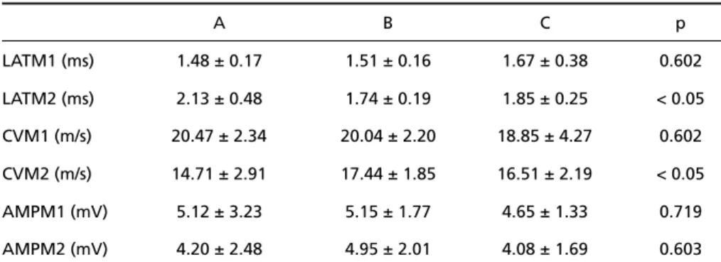
Motor action potential –Results of MAP analy-sis are shown in Table 2. The comparison of LAT M 2 and CVM2 values (Fig 2) between the groups show-ed a significant statistical difference between A g roup and the B group by Tukey method (p <0.05). In the evaluation of %CVM, was identified a
sta-tistically significant diff e rence between A gro u p and the B group and between A group and the C g roup by Duncan method (p < 0.05). The compar-ison of the other parameters presented no statis-tical significant diff e rences, included %CVM and %AMPM.
Table 1. Results of nerve action potential evaluation, including the p value.
A B C p
LATN1 (msec) 0.26 ± 0.10 0.30 ± 0.16 0.24 ± 0.08 0.508
LATN2 (msec) 0.41 ± 0.20 0.34 ± 0.15 0.33 ± 0.10 < 0.05
CVN1 (m/s) 86.07 ± 27.02 82.64 ± 35.94 92.18 ± 28.63 0.783
CVN2 (m/s) 59.55 ± 26.19 69.09 ± 28.85 66.24 ± 19.21 0.687
AMPN1 (mV) 1.29 ± 0.66 1.15 ± 0.59 1.14 ± 0.56 0.833
AMPN2 (mV) 0.37 ± 0.26 0.70 ± 0.52 0.45 ± 0.33 0.167
AMPN1, initial nerve action potentials amplitudes; AMPN2, final nerve action potentials amplitudes; LATN1, initial nerve action potentials latencies; LATN2, final nerve action potentials latencies; msec, mil-lisecond; m/s, meters per second; mV, millivolt; CVN1, initial nerve action potentials conduction velocities; CVN2, final nerve action potentials conduction velocities.
Table 2. Results of motor action potential measurement and the p value.
A B C p
LATM1 (ms) 1.48 ± 0.17 1.51 ± 0.16 1.67 ± 0.38 0.602
LATM2 (ms) 2.13 ± 0.48 1.74 ± 0.19 1.85 ± 0.25 < 0.05
CVM1 (m/s) 20.47 ± 2.34 20.04 ± 2.20 18.85 ± 4.27 0.602
CVM2 (m/s) 14.71 ± 2.91 17.44 ± 1.85 16.51 ± 2.19 < 0.05
AMPM1 (mV) 5.12 ± 3.23 5.15 ± 1.77 4.65 ± 1.33 0.719
AMPM2 (mV) 4.20 ± 2.48 4.95 ± 2.01 4.08 ± 1.69 0.603
AMPM1, initial motor action potentials amplitudes; AMPM2, final motor action potentials amplitudes; L ATM1, initial motor action potentials latencies; LATM2, final motor action potentials latencies; msec, mil-lisecond; m/s, meters per second; mV, millivolt; CVM1, initial motor action potentials conduction veloci-ties; CVM2, final motor action potentials conduction velocities.
Fig 1. Graph showing latencies values of nerve action poten -tials in the three groups of nerve repair.
604 Arq Neuropsiquiatr 2005;63(3-A)
DISCUSSION
The analysis of the alterations in the electro-physiologic parameters before and after the re p a i r showed modifications occurring in a similar way in the diff e rent groups, except for LATN2, VCM2 and %VCM. In the evaluation of these parameters a significant diff e rence was identified between g roups A and B for the three parameters and bet-ween groups A and C also for the %VCM.
These parameters were direct or indirectly re l a t-ed with the CV and the improvement pro b a b l y was the consequence of a more effective myelina-tion in the regenerated axons, mostly in the gro u p B. In a serial electrophysiologic evaluation after section and repair of rabbit sciatic nerve using fib-rin glue or suture, Moy et al.4found that CV re c o v-e ry was supv-erior to amplitudv-e rv-e c o v v-e ry at thv-e samv-e analysed period. In that study, the CV recovery in the animals whose sciatics nerves were submitted to repair with fibrin adhesive was 97 to 98 % of t h e initial CV while the amplitude re c o v e ry was 40 %.
Few studies in the literature perf o rmed an elec-t rophysiologic evaluaelec-tion proelec-tocol similar elec-to elec-the one used in this one. The results obtained in the ampli-tude and CV MAP before the nerve section were similar to the obtained by He et al.1 4. Only two stud-ies, evaluating the effectiveness of the fibrin glue used to nerve re p a i r, analysed MAP. The methodo-logy and numerical results of the one study was not i n f o rm e d5. The results of MAP latency in our study w e re similar to that obtained by Inalöz et al.1 0.
Smahel et al.1 5c o m p a red the electro p h y s i o l o g-ic parameters after rat sciatg-ic nerve repair using suture or fibrin glue. The NAP latency, show on a graph, was about three times smaller after six months in comparison to the value obtained after t h ree weeks. In the same study, the value of the NAP amplitude was about twelve to sixteen times superior after six months in comparison to the val-ue obtained after three weeks. Although the con-t rol group was conscon-ticon-tucon-ted only by four animals, the results for %CVN and %AMPN is very similar to ours.
In only two articles the exact values of the NAP parameters were informed. In the study published by Ratto et al.1 6the latency in the suture d - n e rv e g roup - 0.69 msec - was larger than our re s u l t s . This parameter was eight times larger than those obtained in our study in fibrin glue repair group. In the study of Maragh et al.7, the results were three times larger than our results.
The diff e rence observed between described PAN values in these studies and the results of our study could be justified by the used observ a t i o n period. Two months seems to be enough time for the axons to cross the repair and reach end-org a n s . H o w e v e r, the fibers maturation, which includes myelination, may needs a lengthier period to be completed. More o v e r, the presence of polyneu-ronally innervated muscular fibers, which occur-rence reduces the final efficiency of the regener-ation and consequently its pulse transmission, re-duces in a pro g ressive way after the lesion1 7. In this w a y, by the end of a prolonged observation pe-riod as we used, most remaining fibers would pre s-ent a better quality in comparison to the available fibers in shorter observation periods.
In conclusion, in our study the experimental ner-ve repair with fibrin glue presented better re s u l t s in comparison to the suture when motor action po-tential evaluation was considered. There was no d i ff e rence between the repair methods when the n e rve action potential evaluation was analyzed.
REFERENCES
1. Narakas A. The use of fibrin glue in repair of peripheral nerves. Orthop Clin N Am. 1988;19:187-199.
2. C ruz NI, Debs N, Fiol RE. Evaluation of fibrin glue in rat sciatic nerve repairs. Plast Reconstr Surg. 1986;78:369-373.
3. Feldman MD, Sataloff RT, Epstein G, Ballas SK. Autologous fibrin tis-sue adhesive for peripheral nerve anastomosis. A rch Otolaryngol Head Neck Surg 1987;113:963-967.
4. Moy LJ, Peimer CA, Konjuch MP, Howard C, Zielezny M, Katikaneni PR. Fibrin seal adhesive vs nonabsorbable micro s u t u rein peripheral nerve repair. J Hand Surg (Am). 1988;13:273-278.
5. Gilbert A. Biological glue: experimental and clinical evidence. Ann Chir Main 1989;8:302-311.
6. Nishihira S, Mc Caff rey TV. Repair of motor nerve defects: compari-son of suture and fibrin adhesive techniques. Otolaryngol Head Neck Surg 1989;100:17-21.
7. Maragh H, Meyer BS, Davenport D, et al. Morphofunctional evalua-tion of fibrin glue versus micro s u t u re nerve repairs. J Reconstr Microsurg 1990;6:331-337.
8. Zhou S. Anastomosis of peripheral nerve by fibrin glue. Zhonghua Wa i Ke Za Zhi 1990;28:682-692.
9. Povlsen B. A new fibrin seal in primary repair of peripheral nerves. J Hand Surg (Br). 1994;19:43-47.
10. Inalöz SS, Ak HE, Vayla V, et al. Comparison of microsuturing to the use of tissue adhesives in anastomosing sciatic cuts in rats. Neuro s u rg Rev 1997;20:250-258.
11. Sames M, Blahos J Jr, Rokyta R, Benes V Jr. Comparison of micro s u r-gical suture with fibrin glue connection of the sciatic nerve in rabbits. Physiol Res. 1997;46:303-306.
12. Zhang C, Gu Y, Chen L. Experimental study of fibrin glue with epineur-al anchor suture to repair peripherepineur-al nerves. Zhongguo Xiu Fu Chong Jian Wai Ke Za Zhi. 1998;12:129-132.
13. Suri A, Mehta VS, Sarkar C. Microneural anastomosis with fibrin glue: an experimental study. Neurol India. 2002;50:23-26.
14. He C, Chen Z, Chen Z. Enhancement of motor nerve regeneration by nerve growth factor. Microsurgery 1992;13:151-154.
15. Smahel J, Meyer VE, Bachem U. Glueing of peripheral nerves with fib-rin: experimental studies. J Reconstr Microsurg 1987;3:211-220. 16. Ratto S, Bignotti B, Federici A, Rossi F. Osservazioni sull´uso della
col-la di fibrina in chiru rgia nervosa esperimentale: rilievi elettro f i s i o l o g i-ci. Ort Traum Oggi 1982;2:110-113.