1D e p a rtamento de Genética Médica and;2D e p a rtamento de Neurologia, Faculdade de Ciências Médicas (FCM), Universidade Estadual de Campinas (UNICAMP), Campinas SP, Brazil.
Received 16 August 2004, received in final form 27 December 2004. Accepted 15 March 2005.
Dra. Iscia Lopes-Cendes - Departamento de Genética Médica - FCM/UNICAMP / Caixa Postal 9111 - 13084-971 Campinas SP - Brasil. E-mail: [email protected]
LABORATORIAL DIAGNOSIS OF
FRAGILE-X SYNDROME
Experience in a sample of individuals with
pervasive developmental disorders
Carlos Eduardo Steiner
1, Marilisa Mantovani Guerreiro
2,
Antonia Paula Marques-de-Faria
1, Iscia Lopes-Cendes
1ABSTRACT - Fragile X syndrome is a frequent genetic disease associated to developmental disorders, includ-ing learninclud-ing disability, mental re t a rdation, behavioral problems and pervasive developmental disord e r s (autism and related conditions). We studied a sample of 82 individuals (69 males and 13 females) pre s e n t-ing with pervasive developmental disorders ust-ing three techniques for the diagnosis of fragile X syndro m e (FXS). Cytogenetic analysis detected the fragile site in four males, but only one showed a consistent posi-tive rate. Molecular study based on the PCR technique was inconclusive for most females (92.3%), which w h e re latter submitted to Southern blotting analysis, and for one male (1.4%), excluding the FRAXA muta-tion in the remaining male individuals (98.6%). Molecular tests using the Southern blotting technique con-f i rmed only one positive case (1.2%) in a male subject. These results showed that Southern blotting analy-sis of the FRAXA mutation has the best sensitivity and specificity for the diagnoanaly-sis of FXS but also validat-ed the PCR technique as a confinable screening test.
KEY WORDS: PCR, molecular diagnosis, FRAXA, autism, mental retardation, pervasive developmental dis-orders.
Diagnóstico laboratorial da síndrome do cromossomo X frágil: experiência em uma amostra de indivíduos com distúrbios invasivos do desenvolvimento
RESUMO - A síndrome do cromossomo X frágil (SXF) é uma doença genética freqüente associada a distúr-bios do desenvolvimento neurológico, incluindo dificuldades de aprendizagem, re t a rdo mental, pro b l e-mas comportamentais e distúrbios invasivos do desenvolvimento (autismo e correlatos). Estudamos uma amostra de 82 indivíduos (69 homens e 13 mulheres) apresentando distúrbios invasivos do desenvolvimen-to, utilizando três técnicas para o diagnóstico da SXF. A análise citogenética detectou a presença do sítio frágil em quatro homens, porém apenas um deles com percentagem consistente. O estudo molecular basea-do na técnica da PCR foi inconclusivo para a maioria das mulheres (92,3%), as quais foram posteriorm e n t e submetidas a análise por Southern blotting, e para um homem (1,4%), excluindo a mutação FRAXA nos demais homens (98,6%). O teste molecular usando a técnica de Southern blotting confirmou apenas um caso positivo (1,2%) em um indivíduo do sexo masculino. Tais resultados mostraram que a técnica de S o u t h e rn blotting para análise da mutação FRAXA apresenta a melhor sensibilidade e especificidade para o diagnóstico da SXF, mas também valida a técnica da PCR como um teste confiável para seu rastreamento.
PA L AV R A S - C H AVE: PCR, diagnóstico molecular, FRAXA, autismo, re t a rdo mental, distúrbios invasivos do desenvolvimento.
Fragile X syndrome (FXS) was inittialy re p o
rt-ed in 1943 by Martin and Bell1in a large family
showing X-linked mental re t a rdation. The first lab-oratorial method for its diagnosis was described
by Lubs in 19692, who observed a fragile site [fra
o-mal region, denominated FRAXA, FRAXD, FRAXE, and FRAXF. FRAXA is the most common of these sites and it is associated with mutations in the
FMR1gene, responsible for the FXS, while FRAXE
(associated to the FMR2 gene) causes non-specif-ic mental re t a rdation and FRAXF seems to cause
no abnormal phenotype3 , 4. The molecular basis of
FXS was discovered in the early 1990´s5-8and
con-sists of a dynamic mutation characterized by ex-pansions of an unstable CGG repeat in the 5´ end
of theFMR1gene. It was found that normal
indi-viduals have 6 to 54 repeats (normal allele) where-as affected individuals present more than 200 repeats (expanded allele or full mutation). In addi-tion, phenotypically normal male and female car-riers were found to have intermediate size alleles (pre-expanded alleles or pre-mutation).
FXS has a prevalence of one per 50009, re p
re-senting one of the most common genetic disor-ders. Its clinical spectrum comprises some somatic changes, such as macro c e p h a l y, large testes, mar-phanoid build habitus, long face, and pro t ru d i n g jaw. These features are only identifiable or more p ronounced in males after pubert y. Cognitive im-p a i rment is im-present, ranging from sim-peech delay and learning disability to severe mental re t a rd a-tion. Besides, behavioral signs may be present, in-cluding hyperactivity, self-biting, poor eye contact, shyness, stereotyped movements, and other
autis-tic feature s1 0 , 1 1. This wide range of symptoms may
lead the clinician to consider the diff e rential diag-nosis of FXS in several situations, especially among individuals with diff e rent abnormalities of the neu-rological development, including the pervasive de-velopmental disorders (PDDs), a group that com-prises autism, atypical autism, and Asperger syn-drome.
We undertook the present study aiming to com-p a re cytogenetics, PCR and Southern blotting tech-niques for the laboratory diagnosis of FXS in a sam-ple of individuals with PDDs diagnosed by the DSM -IV criteria. These techniques were applied since both, chromosomal abnormalities and the FXS, are i m p o rtant etiological factors in this group of neu-ropsychiatric conditions.
METHOD
We evaluated a total of 82 patients (69 males and 13 females) with PDDs from the Genetics and Neuro l o g y Clinics of our University Hospital. Parents or legal guar-dians were invited to join the study by signing a con-sent form approved by a Research Ethics Committee. After clinical evaluation, blood samples were collected
for cytogenetic and molecular tests. All patients were also submitted to screening for inborn errors of metab-olism, TORCH sorologies, and neuroimaging studies1 2.
T h ree individuals with clinical diagnosis of Down syn-d rome were also testesyn-d for the FXS syn-due to the possibil-ity of co-occurence of both disorders, considering their high incidence in the general population.
C h romosomal analysis followed the routine cytoge-netic temporary culture in folic acid deficient medium (M-199, with addition of 5-fluoro-2’-deoxyuridine 0.0025 mg/ml). The cultures were incubated for 96 hours at 37ºC for the expression of the fra(X) site. The cell divi-sions were arrested in metaphases by adding colchicine 4 x 1 0- 5M for 30-40 minutes before harvesting the
cul-t u res. The samples were fixed in a solucul-tion of 3:1 mecul-tha- metha-nol-acetic acid, spread in slides, and submitted to G-ban-ding technique. Manual analysis in light microscope in-cluded at least 50 metaphases in male and 100 in female subjects13.
For the molecular investigation we initially used the polymerase chain reaction (PCR) technique for specific amplification of the CGG repeat in the 5´end of theF M R 1 gene, according the protocol described by Fu et al.6.
After amplification, the samples were submitted to elec-t ro p h o resis in a 1.8% agarose gel aelec-t 24 V for 15 hours together with a 250 bp molecular weight marker. Sam-ples were then transferred into Hybond N+ nylon mem-branes and hybridized with an-3 2P 3´-end labeled
(CGG)6probe.
To confirm the PCR results in all subjects and to inves-tigate the female patients, a second molecular test based on the Southern blotting technique was used. Samples w e re submitted to digestion with Pst I and Eco RI re s t r i c-tion enzymes. The resulting fragments were separated by electro p h o resis in 0.8% agarose gel at 15 V for 16 hours together with a 1 kb molecular weight marker. After that, samples were transferred into Hybond N+ nylon membranes and hybridized with the pfxa3 pro b e (Oncor®) marked with an -32P isotope14,15.
RESULTS
showing an increased length in constitutive hete-ro c h hete-romatin of the long arm of the Y chhete-ro m o s o m e ( Yqh+) and autism. All the subjects with abnorm a l cytogenetic findings were males.
Molecular studies based on the PCR technique revealed the presence of normal size alleles in all Fig 1. Fragile site at Xq27.3 (arrow) as observed in the kary -otype of individual E.C.M.
male subjects except for one patient, thus exclud-ing the presence of mutations in most individuals. The only individual with inconclusive PCR re s u l t s after repeated experiments is the same who pre-sented 17% of fra(X) cells in the chromosomal ana-lysis (Fig 2). The remaining three male individuals with low percent of fra(X) cells in the cytogenet-ic analysis showed normal CGG alleles. In the fe-males group, most patients showed only one band in the normal size range with the exception of one patient who showed two distinct bands in the mal size range. The observation of only one nor-mal size band in fenor-males may indicate that the in-dividual is homozygote for the normal allele. Ho-w e v e r, the presence of an expanded allele, Ho-which was not identified by this PCR technique, cannot be definitively excluded.
The only patient with a consistent positive fra (X) by cytogenetic evaluation was found to have
theFMR1gene mutation by Southern blot
analy-sis. We found that this patient was a mosaic for an expanded allele ranging from appro x i m a t e l y 100 to 900 CGG repeats (Fig 3).
Table 1. Results according to the three techniques used for the laboratory diagnosis of the FXS and correlation with gender and the pervasive developmental disorder (PDD) diagnosis.
PDD PCR Southern blotting
Patient (gender) diagnosis Cytogenetics CGG alleles FRAXA
T.L.B.S. (male) autism fra(X) 1% normal normal
E.C.S. (male) autism fra(X) 4% normal normal
C.R.T.S. (male) autism fra(X) 2% normal normal
E.C.M. (male) autism fra(X) 17% no PCR product expanded
E.C.C. (male) atypical autism rob(15;21) normal normal
M.C.X.A. (male) atypical autism inv(9) normal normal
D.G.M.D. (male) autism 46,Xyqh+ normal normal
J.A.B.G. (male) autism 47,XY,+21 normal normal
J.V.S.A. (male) autism 47,XY,+21 normal normal
T.V.S. (male) autism 47,XY,+21 normal normal
other 46 males autism normal normal normal
6 females autism normal normal normal
other 5 males atypical autism normal normal normal
4 females atypical autism normal most inconclusive* normal
8 males Asperger syndrome normal normal normal
3 females Asperger syndrome normal normal normal
DISCUSSION
FXS may be present in about 0.8% of persons
with autism1 6, 0.5% of pre-scholars with speech
d e l a y1 7, 0.56% of individuals with mental re t a rd
a-t i o n1 8, and 0.9% of subjects with non-specific
men-tal re t a rd a t i o n1 9. At first these frequencies may seem
too low to justify laboratory testing for FXS in these patients. However, it is important to note that these conditions are very common in the general
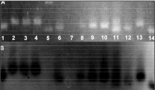
popu-lation and early intervention has a great impact in prognosis. In addition, the diagnosis of FXS is v e ry important for adequate genetic counseling of the couple and, in extension, to other maternal re l-atives. Thus, laboratory detection of FXS is indi-cated in the etiological investigation of individu-als presenting abnormalities in behavior, language, socialization or cognitive development, including the pervasive developmental disorders group. Fig 2. Molecular study based on the PCR technique showing pictures of the gel (A) and of
the X-ray film obtained after autoradiography (B). Note the absence of PCR products in ro w 7, the presence of two distinct bands in row 11, and the presence of a single band re p re s e n t ing normal alleles in the remaining rows. Row 5 corresponds to the molecular weight mark -er (250 bp).
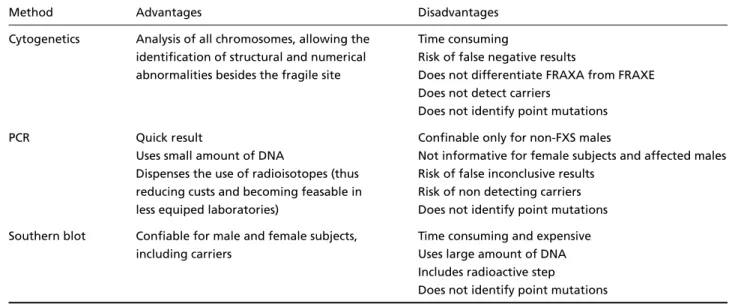
Since the discovery of the molecular basis of FXS, cytogenetic tests for its diagnosis have been less used but were not totally abandoned. Sou-t h e rnbloSou-tSou-ting Sou-technique is Sou-the mosSou-t used labora-torial method but several alternative approaches have been developed in the last decade. These in-clude PCR with radioactive probe or silver stain, reverse transcription PCR (RT-PCR), methylation-sensitive PCR (MS-PCR), and immunohistochemi-cal analyses. As for any laboratory technique a number of methodological issues are involved in the accurate diagnosis of FXS. The advantages and disadvantages of each method we used in the pre s-ent work are summarized on Table 2.
Among the three techniques, cytogenetic analy-sis showed the highest rate of altered results in t h e present sample by the detection of three individ-uals presenting structural chromosomal abnorm a l-ities that could not be diagnosed by m o l e c u l a r study of the FRAXA mutation alone. One of t h e m showed an increased length on the long arm of chromosome Y, which has been previously
repor-ted in association with autism2 0. Another patient
had a pericentric inversion of chromosome 9, situa-tion that recent literature data suggest may be in-volved in genetic susceptibility to psychiatric
disor-ders such as schizophrenia or even autism2 1. The
third one presented a roberstonian translocation 15/21 and, again, literature data suggest that ab-n o rmalities of chromosome 15 have a higher p re v
a-lence in autism than in the general population22.
Considering the technical aspects of cytogenet-ic analysis for FSX, usually a longer exposure to
colchicine causes more chromosomal condensa-tion, which turns fra(X) easier to be detected by microscopic analysis. We used a shorter exposure (30 to 40 minutes instead of 60 minutes) which is less likely to interf e re with the detection of other cytogenetic abnormalities. Although cytogenetic evaluation was sensitive and specific for the diag-nosis of FXS, the fact that it is based in a 96-hours cell culture, which can fail, may re q u i rere - t e s t i n g in several occasions. When the culture is success-ful, the study demands many hours of micro s c o p-ic analysis, especially for the female specimens. In addition, it is well known that this method may not detect all carrier females, and would miss most male carriers, since only a small pro p o rtion of their
cells will express the fra(X)10. The best advantage
of this method is that it makes it possible to detect other chromosomal abnormalities, including the other fragile X sites with one single test.
PCR is a rapid and versatile method for ampli-fying a target DNA sequence. Because it is a fast and simple method, PCR is ideally suited for muta-tion screening since it can yield results in a single day experiment. However, the optimal size range for PCR amplification is between 0.1 to 5 kb, which may limit its application. The size of the band on the gel corresponding to a fragment with norm a l
CGG repeat in theFMR1gene has approximately
250-300 bp and it will be easily amplified in the PCR experiments. However, the expanded CGG allele may be too large and fail to amplify. There-f o re, a male with a normal allele will be re a d i l y identified by PCR as having only one single band Table 2. Advantages and disadvantages of each method for the laboratorial diagnosis of the FXS.
Method Advantages Disadvantages
Cytogenetics Analysis of all chromosomes, allowing the Time consuming
identification of structural and numerical Risk of false negative results
abnormalities besides the fragile site Does not differentiate FRAXA from FRAXE Does not detect carriers
Does not identify point mutations
PCR Quick result Confinable only for non-FXS males
Uses small amount of DNA Not informative for female subjects and affected males Dispenses the use of radioisotopes (thus Risk of false inconclusive results
reducing custs and becoming feasable in Risk of non detecting carriers less equiped laboratories) Does not identify point mutations Southern blot Confiable for male and female subjects, Time consuming and expensive
including carriers Uses large amount of DNA Includes radioactive step
in the normal size range (250-300 bp). On the oth-er hand, the absence of bands could indicate that the individual has an expanded allele or that a technical problem occurred to interf e re in the opti-mal amplification of the noropti-mal allele. There f o re , the PCR technique can easily identify normal alle-les, but usually is not efficient for the detection of the expanded CGG alleles.
In our study, only one patient who had positive cytogenetic study had an altered PCR result. The t h ree individuals with low percentage of fra(X) cells in the cytogenetic study had single norm a l CGG alleles, thus excluding the FRAXA mutation which was also confirmed by the Southern blot-ting analysis.
In females, normal PCR fragments with similar sizes will result in a single band, making it impos-sible to diff e rentiate normal homozygote female f rom a carrier of the FXS mutation. In our study the PCR technique was not informative for most females, since only a single individual was het-e rozygothet-e for thhet-e normal CGG, and conshet-equhet-ently a normal non-carrier FRAXA subject. Considering
this, Weinhäusel and Haas2 3described an altern
a-tive PCR technique using fluorescence analysis called methylation-sensitive PCR (MS-PCR) that was useful for the detection of carrier females, altho-ugh distinction between pre-mutated and fully mutated females was sometimes difficult in their experience. In addition, it is important to note that, in order to confirm that the band in the gel t ruly corresponded to the CGG fragment, transfer and hybridization with specific radioactive pro b e s were performed after the gel electrophoresis. As t h e re was no other bands in our experience, we concluded that transfer and hybridization was u n n e c e s s a ry and that the analysis can be accom-plished by agarosis gel electro p h o resis stained with ethidium bromide, without the necessity of use of radioisotopes.
F i n a l l y, Southern blotting gave informative re-sults for all patients and confirm that only one in-dividual had the FRAXA mutation. It also lead to i n f o rmation in all females tested. However, this technique requires large amounts of well
preser-ved DNA (approximately 50µg) and it demands a
week of laborious laboratory work to complete all the technical steps re q u i red, including isotope manipulation.
These results are similar to those of Haddad et
a l .2 4who also studied a sample of Brazilian boys
with mental re t a rdation using the Southern
blot-ting technique and the PCR technique of Fu et al.6
modified by the use of silver staining instead of labeling with an isotopic probe. In their study, a boy with mosaicism including pre-mutated and fully mutated allele was also re p o rted and showed d i s c repant results in sequenced PCR tests, which was not seen in our work.
A final problem, common to the all three tech-niques discussed, is that point mutations in the
FMR1gene will not be detected by any of them.
F o rt u n a t e l y, this is a rare situation in FXS2 5, and
can be diagnosed only by gene sequencing. This a p p roach should be considered if the diagnosis of FXS is highly probable and all other diagnostic techniques failed.
The specific digestion followed by Southern blotting showed the best sensitivity and specifici-ty for the laboratory diagnosis of FXS and also allowed the detection of possible female carr i e r s . H o w e v e r, if only this technique was used it would have missed the six patients with chro m o s o m a l a b n o rmalities, an important group of etiologic factors of mental re t a rdation. Thus, we suggest that this method should only be the first choice when the clinical diagnosis of FXS is highly prob-able or to investigate relatives of confirmed FXS subjects (especially females) for genetic counsel-ing and prenatal diagnosis. In individuals pre s e n t-ing developmental disorders in which the diagno-sis of FXS is probable but not the main hypothe-sis, chromosomal analysis should always be perf o r-med. Considering that small structural abnorm a l-ities can be detected only in prometaphasic chro-mosomes, this type of preparation is re c o m m e n d-ed. On the other hand, this pro c e d u rewill make it more difficult to detect fra(X).
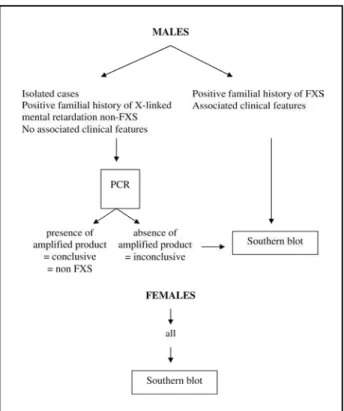
Fig. 4. Proposed algorithm for the laboratory diagnosis in Brazil of fragile X syndrome in male and female individuals with deve -lopmental disorders. These molecular tests are re c o m m e n d e d to follow cytogenetic analysis, screening for inborn errors of metabolism, TORCH sorologies, and neuroimaging as part of the complementary evaluation of individuals with disorders of neurological development.
(Fig 4). The diagnostic strategy we propose, a l-though not in accordance with other intern a t i o n-al guidelines for the laboratorin-al diagnosis of the FXS, re p resents a reasonable approach in Brazil, a c o u n t ry with limited access of families to clinical genetics services, heterogeneous distribution and conditions of laboratories, and limited govern m e n-tal re s o u rces for medical genetics serv i c e s .
Acknowledgements– The authors wish to thank all the patients and their relatives who participate of this re s e a rch, as well as the colleagues from the Labora-tório de Genética Molecular (Departamento de Genética Médica, FCM/Unicamp), especially Daniela Facchin, Te reza Cristina Lima e Silva, and Maria Eugenia Ribeiro de Camargo for valuable technical support in the PCR experiments. Thanks also to the staff from the Cytogene-tics and Molecular GeneCytogene-tics (Wo m e n ’s and Childre n ’s Hospital, Adelaide, Australia), specially Drs. John C. Mu-lley and Agi K. Gereon for the helpful discussions on the Southern blotting analysis.
REFERENCES
1. Martin JP, Bell J. A p e d i g ree of mental defect showing sex-linkage. J Neurol Neurosurg Psychiatry 1943;6:154.
2. Lubs HA. A marker X chromosome. Am J Hum Genet 1969;21:231. 3. Parrish JE, Oostra BA, Verkerk AJMH, et al. Isolation of a GCC repeat
showing expansion in FRAXF, a fragile site distal to FRAXA a n d FRAXE. Nat Genet 1994;8:229-239.
4. Ritchie RJ, Knight SJL, Hirst MC, et al. The cloning of FRAXF: trinu-cleotide repeat expansion and methylation at a third fragile site in dis-tal Xqter. Hum Mol Genet 1994;3:2115-2121.
5. Oberlé I, Rousseau F, Heitz D, et al. Instability of a 550-base pair DNA segment and abnormal methylation in fragile-X syndrome. Science 1991;252:1097.
6. Fu YH, Kuhl DPA, Pizzuti A, et al. Variation of the CGG repeat at the fragile X site results in genetic instability: resolution of the Sherman paradox. Cell 1991;67:1047-1058.
7. K remer EJ, Pritchard M, Lynch M, et al. Mapping of DNA i n s t a b i l i t y at the fragile-X to a trinucleotide repeat sequence p(CGG)n. Science 1991;252:1711.
8. Yu S, Pritchard M, Kremer E, et al. Fragile-X genotype characterized by an unstable region of DNA. Science 1991;252:1179.
9. Stoll C. Problems in the diagnosis of fragile X syndrome in young chil-dren are still present. Am J Med Genet 2001;100:110-115.
10. Mulley JC, Sutherland GR. Diagnosis of fragile X syndrome. Fetal Matern Med Rev 1994;6:1-15.
11. Félix TM, Pina-Neto JM. Fragile X syndrome: clinical and cytogenetic studies. Arq Neuropsiquiatr 1998;56:9-17.
12. Steiner CE, Guerreiro MM, Marques-de-Faria AP. Genetic and neuro-logical evaluation in a sample of individuals with pervasive develop-mental disorders. Arq Neuropsiquiatr 2003;61:176-180.
13. Jacky PB, Ahuja YR, A n y a n e - Yeboak K, et al. Guidelines for the pre p a-ration and analysis of the fragile X chromosome in lymphocytes. A m J Med Genet 1991;38:400-403.
14. Mulley JC, Yu S, Gedeon AK, et al. Experience with direct molecular diagnosis of fragile X. J Med Genet 1992;29:368-374.
15. Southern EM. Detection of specific sequences among DNA f r a g m e n t s separated by gel electrophoresis. J Mol Biol 1975;98:503-517. 16. G i l l b e rg C, Wahlström J. Chromosome abnormalities in infantile autism
and other childhood psychosis: a population study of 66 cases. Dev Med Child Neurol 1985;27:293-304.
17. Kunugi H, Lee KB, Nanko S. Cytogenetic findings in 250 schizophre n-ics: evidence confirming an excess of the X chromosome aneuploidies and pericentric inversion of chromosome 9. Schizophrenia Researc h 1999;40:43-47.
18. Bundey S, Hardy C, Vickers S, Kilpatrick MW, Corbett JA. Duplication of the 15q11-13 region in a patient with autism, epilepsy and ataxia. Dev Med Child Neurol 1994;36:736-742.
19. Klauck SM, Münstermann E, Bieber-Martig B, et al. Molecular genet-ic analysis of the FMR-1 gene in a large collection of autistgenet-ic patients. Hum Genet 1997;100:224-229.
20. Mazzoco MMM, Myers GF, Hamner JL, et al. The prevalence of the FMR1and FMR2 mutations among preschool children with language delay. J Pediatr 1998;132:795-801.
21. Millán JM, Martinez F, Candroy A, et al. Screening forFMR1m u t a t i o n s among the mentally re t a rded: prevalence of the fragile X in Spain. Clin Genet 1999;56:98-99.
22. Patsalis PC, Sismani C, Hettinger JA, et al. Molecular screening of frag-ile X (FRAXA) and FRAXE mental re t a rdation syndromes in the Hellenic population of Greece and Cyprus: incidence, genetic varia-tion, and stability. Am J Med Genet 1999;84:184-190.