Arq Neuropsiquiatr 2002;60(1):12-16
PRESERVATION OF THE OLFACTORY TRACT IN
BIFRONTAL CRANIOTOMY
Paulo H. Aguiar
1, Guilherme A. Pulici
2, Leonardo O. Lourenco
2,
Juan A.C. Flores
1, Valter A. Cescato
1ABSTRACT - The bifrontal craniotomy approach used to be associated with a high percentage of olfactory tract damage. We present our experience with this technique, that was used with excellent results in a series of 11 patients that underwent the surgical approach described in this paper. We support the idea that bilateral subfrontal craniotomy allows a wide operative exposure as well as the complete anatomic and functional preservation of the olfactory tracts bilaterally.
KEY WORDS: bifrontal craniotomy, olfactory tract preservation, anterior cranial base, olfaction, surgical technique.
Preservação do trato olfatório em craniotomias bifrontais Preservação do trato olfatório em craniotomias bifrontais Preservação do trato olfatório em craniotomias bifrontais Preservação do trato olfatório em craniotomias bifrontais Preservação do trato olfatório em craniotomias bifrontais
RESUMO - A craniotomia bifrontal costumava estar associada com alta incidência de lesão do trato olfatório. Apresentamos nossa experiência com técnica que foi usada com excelentes resultados numa série de 11 pacientes que foram submetidos à abordagem cirúrgica descrita neste estudo. Defendemos a idéia de que a craniotomia bilateral subfrontal permite uma exposição cirúrgica ampla bem como a completa preservação anatômica e funcional dos tratos olfatórios bilateralmente.
PALAVRAS-CHAVE: craniotomia bifrontal, preservação do trato olfatório, base anterior de crânio, olfato, técnica cirúrgica.
1Division of Neurosurgery of Hospital das Clinicas, University of São Paulo Medical School, São Paulo SP, Brazil. 2Neurosurgical Clinic of
Pinheiros, São Paulo SP, Brazil.
Received 23 May 2001, received in final form 10 September 2001. Accepted 26 September 2001.
Dr. Paulo Henrique Aguiar – Rua Maestro Torquato Amore 332 - Ap 12 Bl 1 - 05622-050 Sao Paulo SP - Brasil. FAX: 55 11 3082 6822. E-mail: [email protected]
The anterior cranial base and the suprasellar and
parasellar regions approach and its several methods
have been described since 1981 by Suzuki et al.
1-3.
Extended frontal approaches, however, necessitate
removal of the crista galli and sectioning of the
ol-factory rootlets with the associated risk of anosmia,
cerebrospinal fluid (CSF) leak, and the need for
com-plex reconstruction of the frontal floor
4. Bifrontal
craniotomy is the conventional approach to lesions
in these locations
5,6, but its shortcoming has been
the damage to the olfactory tract
7.
The preservation of the olfactory tract has been
the subject of many studies in extended frontal
ap-proaches. Fujiwara et al.
7and Eriksen et al.
8reported
various cases of anosmia after anterior
communi-cating artery aneurysm surgery. Spetzler et al.
4modi-fied the technique of handling the cribriform plate
to preserve the olfactory unit. Srinivasan et al.
9de-scribed the bifrontal approach that enhanced the
exposure of the suprasellar region and minimized
manipulation of the optic apparatus and the carotid
arteries.
In our study, we present our experience with the
use of bifrontal craniotomy in 11 patients with
sev-eral lesions, e.g., intracerebral schwannoma,
cranio-pharyngiomas, pituitary adenomas and Rathke Cyst.
We also report the complete preservation of
olfac-tion in these patients who underwent bifrontal
cran-iotomy.
METHOD
Operative technique
along the cranium convexity posteriorly. Both frontal si-nuses tables are accessed and the mucous membranes are removed. The dura mater is cut parallel to the base and the sagittal sinus is ligated and cut at the cecal
fora-men. Then the falx is transected and the bridging veins are preserved. The frontal poles are retracted, under mag-nification, using two self-retaining retractors adjusted stepwise (Fig 2).
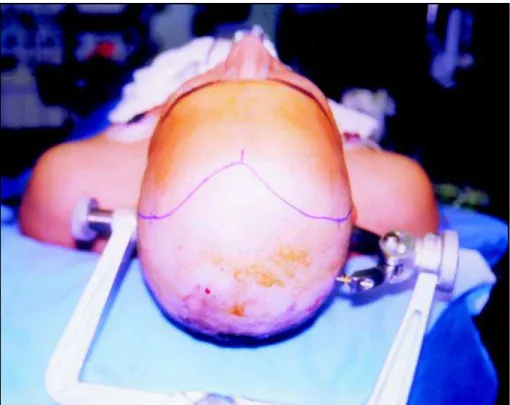
Fig 1. Case 4. Surgical view of the marked line to be incisioned.
14 Arq Neuropsiquiatr 2002;60(1)
The interhemispheric and the bilateral olfactory cis-ternae are opened in order to drain the CSF and to avoid more retraction. Both olfactory nerves should be dissected simetrically without coagulation under small bleedings.
Surgicel® is enough to stop the small bleedings mainly
near the crivous plate of ethmoidal bone.
After that, the surgical overview could better show the image of both olfactory nerves and both optic nerves composing a groove enlarged by the tumor. The pseudocapsula of the tumor could be seen through this groove and the dissection between the carotid artery and the pseudocapsula should be performed on both sides. Following this step, a debulking of the tumor should be done and a “piece meal” resection is recom-mended.
The ultrasonic aspirator could be used with extreme caution. The retraction of bilateral frontal poles should
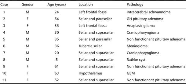
Table 1. Clinical data for 11 patients with preservation of the olfactory tract.
Case Gender Age (years) Location Pathology
1 M 24 Left frontal fossa Intracerebral schwannoma
2 F 54 Sellar and parasellar GH pituitary adenoma
3 F 35 Left frontal fossa Anaplasic glioma
4 M 30 Sellar and suprasellar Craniopharyngioma
5 M 35 Sellar and parasellar Non functionant pituitary adenoma
6 M 36 Tubercle sellar Meningioma
7 M 20 Sellar and suprasellar Craniopharyngioma
8 M 5 Sellar and suprasellar Rathke cyst
9 F 61 Sellar and suprasellar Non functionant pituitary adenoma
10 F 63 Hypothalamus GBM
11 F 52 Sellar and suprasellar Non functionant pituitary adenoma
never be strong enough to hurt the pia-mater and make damage to the brain parenchyma1.
Patient population
The surgical technique described was applied in the treatment of the 11 patients, as shown in Table 1. Of the 11 patients, 5 were female and 6 were male, with a mean age of 37.7 years (range, 5-63 yr). Histological examina-tion revealed four adenomas (Figs 3, 4, 7, 8, 9), two cranio-pharyngiomas (Figs 5, 6), one GBM, one anaplasic glioma, one meningioma, one Rathke cyst, and one Schwannoma. The symptoms at admission were typical for each patho-logical condition, and the lesions were all totally removed by using a bilateral subfrontal approach. All these patients were preoperatively evaluated with non-enhanced and enhanced magnetic resonance tomography. Prospectively, we evaluated the clinical findings at the presentation, operative treatment, and outcome of each patient.
Fig 3. Case 2. Preoperative MRI of a 54-year-old female with GH pituitary macroadenoma.
Fig 5. Case 4. Preoperative MRI of a 50-year-old male with craniopharyn-gioma.
Fig 6. Case 4. Postoperative MRI of the same 50-year-old male patient showing total resection of the tumor.
Fig 7. Case 9. Preoperative MRI of a 61-year-old female with nonfunctioning pituitary adenoma.
Fig 8. Case 9. Postoperative MRI of the same 61-year-old female patient after total resection of the tumor.
16 Arq Neuropsiquiatr 2002;60(1)
RESULTS
In our study there were no deaths or, even,
clini-cal or surgiclini-cal complications. We did not register any
case of anosmia. Neurological examination made
postoperatively did not reveal anything new in the
11 patients, except that they did have their
olfac-tory tract preserved bilaterally. No subjective or
ob-jective olfactory disturbances were noted in the
fol-low-up of those 11 patients.
DISCUSSION
In our opinion, olfaction is an extremely
impor-tant neurological function, to the point that its
dam-age can impair any individual´s quality of life.
Olfac-tory tract damage used to be regarded as the major
drawback of bifrontal craniotomy
2,10,11. Knowing that
it is fundamental to preserve the anatomy and the
function of both olfactory tracts, we strongly
rec-ommend the technique described above and first
reported by Samii et al.
1,12.
The bilateral subfrontal approach allows a good
overview of the sellar, suprasellar, presellar, parasellar
and the anterior cranial fossa region and permits
the complete preservation of olfaction. Bifrontal
cra-niotomy is the ideal approach to various midline
le-sions of those areas, as sometimes a unilateral
exten-ded pterional approach is insufficient for adequate
treatment of the lesion. We are aware that this
tech-nique requires patience and time, which, in our
im-pression is fully rewarded by its technique
advanta-ges of excellent field exposure and preservation of
the olfactory tracts.
From our point of view, we strongly agree with
the impressions and findings of Sepehrnia et al.
6.
The excellent outcome found in our series as well as
in Sepehrnia et al.
6series support the good
useful-ness of the bilateral subfrontal craniotomy with
sec-tion of the falx.
REFERENCES
1. Bini W, Sepehrnia A, Samii M. Some technical considerations regard-ing craniopharyngioma surgery: the bifrontal approach. In Samii M (ed). Surgery of the sellar region and paranasal sinuses. Berlin: Springer-Verlag 1991:381-386.
2. Suzuki J, Yoshimoto T, Mizoi K. Preservation of the olfactory tract in bifrontal craniotomy for anterior communicating artery aneurysms, and the functional prognosis. J Neurosurg 1981;54:342-345.
3. Yasui N, Nathal E, Fujiwara H, Suzuki A. The basal interhemispheric approach for acute anterior communicating artery aneurysms. Acta Neurochir (Wien) 1992;118:91-97.
4. Spetzler RF, Herman JM, Beals S, Joganic E, Milligan J. Preservation of olfac-tion in the anterior craniofacial approaches. J Neurosurg 1993;79:48-52. 5. Sekhar LN, Nanda A, Sen CN. The extended frontal approach to
tu-mors of the anterior, middle, and posterior skull base. J Neurosurg 1992;76:198-206.
6. Sepehrnia A, Knopp U . Preservation of the olfactory tract in bifrontal craniotomy for various lesions of the anterior cranial fossa. Neurosur-gery 1999;44:113-117.
7. Fujiwara H, Yasui N, Nathal-Vera E, Suzuki A. Anosmia after anterior communicating artery aneurysm surgery: comparison between the anterior interhemispheric and basal interhemispheric approaches. Neu-rosurgery 1996;38:325-328.
8. Eriksen KD, Boge-Rasmussen T, Kruse-Larsen C. Anosmia following operation for cerebral aneurysms in the anterior circulation. J Neurosurg 1990;72:864-865.
9. Srinivasan J, Dailey AT, Berger MS. The bifrontal olfactory nerve-spar-ing approach to lesions of the suprasellar region in children. Pediatr Neurosurg 1999;30:245-252.
10. Fujitsu K, Sekino T, Sakata K, Kawasaki T. Basal interfalcine approach through a frontal sinusostomy with vein and nerve preservation. J Neurosurg 1994;80:575-579.
11. Pool JL.Aneurysms of the anterior communicating artery: bifrontal cranio-tomy and routine use of temporary clips. J Neurosurg 1961;18:98-111. 12. Samii M. Olfactory nerve. In Samii M, Janetta PJ (eds). The cranial