Arq Neuropsiquiatr 2005;63(2-B):527-529
Instituto de Neurologia de Curitiba, Curitiba PR, Brazil: 1Resident of Neurosurgery; 2Postgraduation in Neurology; 3Neurosurgeon, 4Neurologist.
Received 22 June 2004, received in final form 17 November 2004. Accepted 15 January 2005.
Dra. Kelly C. Bordignon - Rua Jeremias Perretto 300 - 81210-3110 Curitiba PR - Brazil. E-mail: [email protected]
FOIX-ALAJOUANINE SYNDROME
Case report
Kelly C. Bordignon
1, María Belén Montú
2, Ricardo Ramina
3, Walter Oleschko Arruda
4ABSTRACT - In a 52-year-old woman, spinal arteriovenous malformation (AVM) has been associated with what has been known as Foix-Alajouanine syndrome. The pathophysiology of the AV fistula is probably related to increased venous pre s s u refrom the AVM plus thrombotic process.The most common initial symp-toms are sensory disturbance, pain and leg weakness. Definitive diagnosis of spinal AVMs re q u i res radiogra-phic demonstration of the vascular anomaly. Nevertheless, in this case, suggestive defects of malforma-tions could not be seen, in contrast to the MRI findings and macroscopical and anatomical-pathological lesion. These findings rise our attention, about the need to keep in mind the clinical suspicion of AVM in cases of back pain and motor deficit, and an early surgical conduct in this situation.
KEY WORDS: paraplegia, AV fistula, venous thrombosis.
Síndrome de Foix-Alajouanine: relato de caso
RESUMO - Em mulher de 52 anos, malformação artério-venosa medular (MAV) associava-se com a síndro-me de Foix-Alajouanine. A fisiopatologia da fístula artéria-venosa provavelsíndro-mente está relacionada com a p ressão venosa aumentada, a partir da MAV associada com o processo trombótico. Os sintomas iniciais mais comuns são distúrbio sensorial, dor e fraqueza do membro inferior. O diagnóstico definitivo de MAV medu-lar requer evidências radiográficas da anomalia vascumedu-lar. De qualquer modo, neste caso, defeitos sugesti-vos de malformações não foram detectados, contrastando com os achados de ressonância magnética e lesões macroscópicas e anátomo-patológicas. Estes resultados chamaram a atenção e excluíram outras causas. A alta suspeita clínica de MAV nos levou a uma conduta cirúrgica precoce em benefício do paciente. PALAVRAS-CHAVE: paraplegia, fistula AV, trombose venosa.
In 1926, Foix and Alajouanine described a suba-cute myelopathy produced by a thrombotic pro c e s s
of the spinal cord that ultimately caused death1. At
a u t o p s y, they discovered necrosis of the spinal cord and numerous thickened tortuous vessels lying on the surface of the cord. Years later, in 1931, Lherm i t t e et al., recognized this process as been associated with
a spinal arteriovenous malformation (AV M )2. It was
generally believed that the rapidly pro g re s s i v e myelopathy resulted from thrombosis of this abnor-mal vessel within the spinal cord. Consequently, this p rocesss, which came to be known as the Foix-Ala-jouanine syndrome (FAS), was felt to be irre v e r s i b l e and to carry a poor pro g n o s i s3 , 4. In most patients with
this condition, sensory symptoms, pain and leg
weak-ness are the most common initial symptoms5 - 7.
A FAS case, submitted to surgery is described, and literature is reviewed about this clinical entity.
CASE
528 Arq Neuropsiquiatr 2005;63(2-B)
A lumbar cere b rospinal fluid (CSF) was perf o rmed and showed increased protein (154 mg/dL), 144 red cells/mm3, with normal white cells (2/mm3) counting. All immunolo-gical tests were negative.
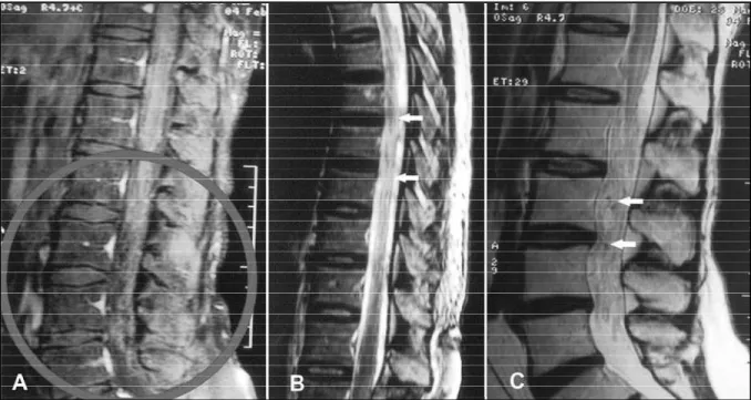
Lumbar spine MRI showed changes in keeping with multiple abnormal vascular stru c t u res at the low port i o n of the spinal cord (cone medullaris and cauda equina). Hemosiderosis could be seen at the surface of the cone region, probably due to previous hemorrhage. A hyper-intensive signal of the conus medullaris (T2-weighted ima-ges) with heterogeneous slight Gd-enhancement (T1-wei-ghted images) was observed. (Figuras 1 A,B,C) A spinal arteriography was immediately performed and did not disclose any vascular abnormalities (e.g. spinal AV fistu-la) and the Adamkievicz artery was not seen.
A laminectomy (L1 to L4) was perf o rmed and showed e n l a rged tortuous red vessels involving the spinal ro o t s . Among those abnormal vessels, a black thrombosed du-ral AV fistula was identified. The vessel was intradudu-ral- intradural-ly divided, and the dural AV fistula was coagulated and excised. Pathological analysis confirmed a thrombosed AV fistula, without evidence of vasculitis.
T h e rewas no control arteriography at the post-oper-ative period. The patient presented very mild recovery of weakness and sensibility after 3 months of follow-up. A control post-operative spinal MRI showed ischemic le-sion of the conus medullaris.
DISCUSSION
Rosemblum et al.8compared dural AV fistulas
and intradural AVM in 81 patients. He described that the clinical and physiopathological aspects in both conditions are different. In intradural AVM, 50% present in an acutely form, and hemorrhage commonly accompanies this acute clinical deteri-oration. The high flow of these intradural malfor-mations and the arterial flow steal from eloquent a reas complete the pathological mechanism. Acute n e u rological deterioration may be also due to s p o n-taneous hemorrhage from AVM. On the other hand, dural AV fistulas typically cause progressive myelopathy and only 10-15% present an acute p re-sentation. They are low-flow fistulas, draining in-tradurally into the coronal venous plexus, which becomes tortuous and elongated with time. A pa-thophysiological event - venous congestion of spinal cord - results from absence of valves between coronal venous plexus and intramedullary veins, resulting in venous medullary outflow impairm e n t . Venous congestion is also reflected by a delayed venous phase on angiography. There f o re, the low-flow aspect plus venous congestion predisposes to venous thrombosis as final event.
Arq Neuropsiquiatr 2005;63(2-B) 529
ty but a complication of spinal AVM that was cau-sed by thrombosis within the abnormal vessels of the spinal cord. This is the end-stage of a sub-acu-te myelopathy due to venous congestion of the
spi-nal cord. Criscuolo et al.6re a s s e rted this mechanism
based on their previous case re p o rts. The symptoms of subacute myelopathy are paraparesia, pare s t h e-sias, spasticity, and urinary hesitancy. These symptoms can be enhanced or exacerbated by Va l s a l v a -like manoeuvers. Recently, a case re p o rt buttre s s e d the association between spinal AVM and acute e p
i-sodes of paraplegia caused by singing9. The
diag-nosis of dural AV fistulas can be made by MRI that, t y p i c a l l y, shows swelling of medullary conus and s l i-ght central spinal enhancement. Our patient pre-sented similar image alterations with additional ser-pentine vessels involving the spinal roots.
The suspected diagnosis of spinal dural AV fis-tulas must be confirmed by angiogram. Despite its high sensibility, arteriography may be inconclus i v e .
van Dijk et al.7described two patients whose
angio-grams did not reveal a spinal AV fistula, although the patients had classic signs and symptoms, both
clinically and on MRI. Criscuolo et al.6described two
patients with diagnosis of Foix-Alajouanine syndro-me and a negative spinal art e r i o g r a p h y. Those p re-operative arteriographies were inconclusive, the same what happened with our patient. Likewise, t h e rewas no control exam at postoperative period.
When a patient presents a subacute clinical pic-t u re, correlapic-ted pic-to venous congespic-tion phase, pic-the remission of the symptoms can be placed by intra-durally surgical division of the shunting vein to the venous plexus. However, the potential for a re v e r-sal of this process of vascular lesion, including sur-gical stripping of the dorsal veins of the spinal cord has been advocated. At these cases, the surg e ry o f-ten led to clinical deterioration due to total lack of spinal cord normal venous drainage. At our case re-p o rt, slight re-pro g ressive recure-peration on re-postore-per
a-tive period corroborated to the arterializated v e n o u s plexus origin, and proper surgical fistula division.
Criscuolo et al.6and Wi rth et al.4emphasized t h e
futility of treating patients with thrombosed AV f i s-tula, as they considered it an end-stage process wi-thout hope of useful recovery of function. This is an argument against our case re p o rt. Some authors describe similar postoperative results achieved by endovascular embolization by liquid adhesive em-bolics. There f o re, despite of some controversies, the s u rgical pro c e d u reis the standard treatment, pre-venting clinical deterioration. Improvements are o f-ten possible to some exf-tent.
In conclusion, spinal arteriovenous malform a t i-on has been associated with what has been known as Foix-Alajouaine syndrome. The clinical picture is a subacute pro g ressive myelopathy. The MAV clini-cal high suspicion must led us to an early surgiclini-cal conduct to patient benefit. Arteriography and MRI are sensitive diagnostics tools to vascular altera-tions, but can be inconclusive at some point.
REFERENCES
1. Foix C, Alajouanine T. La myélite nécrotique subaigue. Rev Neurol (Paris) 1926;2:1-42.
2. Lhermitte J, Friboury-Blanc A, Kyriaco N. La gliose angéio-hyperthro-phique de la moelle épinière (myélite nécrotique de Foix-Alajouanine). Rev Neurol (Paris) 1931;2:37-53.
3. Pia HW, Vogelsang H.Diagnose und Therapie spinaler Angiome. Dtsch Z Nervenheilkd 1965;187:74-96.
4. Wirth FP Jr, Post KD, Di Chiro G, Doppman JL, Ommaya AK. Foix-Alajouanine disease. Spontaneous thrombosis of a spinal cord arteriove-nous malformation: a case report. Neurology 1970;20:1114-1118. 5. Tobin WD, Layton DD. The diagnosis and natural history of spinal cord
arteriovenous malformations. Mayo Clin Proc 1976; 51:637-646. 6. Criscuolo GR, Oldfield EH, Doppman JL. Reversible acute and
subacu-te myelopathy in patients with dural arsubacu-teriovenous fistulas: Foix-Ala-jouanine syndrome reconsidered. J Neurosurg 1989;70:354-359. 7. Van Dijk JMC, Te r B rugge KG, Willinsky RA, et al. Multidisciplinary
man-agement of spinal dural arterovenous fistulas: clinical pre s e n t a t i o n and long-term follow-up in 49 patients. Stroke 2002;33:1578-1583. 8. Rosemblum N, Oldfield EH, Doppman JL, et al. Spinal arteriovenous
malformations: a comparison of dural arteriovenous fistulas and intra-dural AVM’s in 81 patients. J Neurosurg 1987;67:795-802.