The success of an endodontic treatment relies on a correct working length determination. The canal terminus must be detected accurately and a precise control of the working length during the endodontic treatment must be maintained. The aim of this study was to investigate the working length determination in vivo with the Quill Apex Locator® for apical limit established at 1 mm short from the apical foramen (AF). Patients scheduled to dental extraction due to orthodontic or periodontal reasons were selected for this study, resulting in a sample of 24 root canals. Written informed consent was obtained from each patient before the treatment. After the administration of local anesthesia each tooth had its pulp cavity accessed. Next, the reading corresponding to the AF showed on the device’s display was recorded and the file was removed. With the use of a digital caliper, 1 mm was subtracted from that first measurement. The file with the new measurement was introduced into to the root canal again then fixed with light curing flow composite and the tooth was extracted. Next, one of the apical third walls was worn out to visualize the instrument’s point and the AF. The distance from the file tip to the AF was measured by scanning electron microscopy. The average of the measurements was 1.089±0.437 mm. The Bicaudal t-test showed no significant difference (p=0.338) between the experimental values and the hypothetical value tested at 1 mm. The present investigation confirmed that the Quill Apex Locator® was able to determine the working length with good reliability for the endodontic treatment, established at 1 mm short from the AF.
An
In Vivo
Study of Working Length
Determination with a New Apex Locator
Key Fabiano Souza Pereira1, Pedro Gregol da Silva1, Fabio Simões de Vicente1, Fabio Nakao Arashiro1, Carmem Regina Coldebella1, Carlos Alberto Spironelli Ramos2
1Dental School, UFMS - Federal
University of Mato Grosso do Sul, Campo Grande, MS, Brazil
2Dental School, UEL - University
of Londrina, Londrina, PR, Brazil
Correspondence: Prof. Dr. Key Fabiano Souza Pereira, Avenida Senador Filinto Muller, Cidade Universitária, s/n, 79070-900 Campo Grande, MS, Brasil. Tel: +55-67-3345-7383. e-mail: [email protected]
Key Words: electronic apex locators, endodontics, working length.
Introduction
The determination of the root canal length must be accurate and reliable since it is one of the most important conditions that influence the success of root canal treatments (1). The majority of researches understand that root canal instrumentation and filling should be at the level of the apical constriction. The best histological conditions occur when those steps are performed at the apical constriction (1,2). The apical constriction’s location and its shape are variable and not detectable by radiographs. Its location is generally 1.2 mm previous to the apical foramen (AF) (3) and its position may vary from 1 to 3 mm (1).
The most popular techniques for working length determination are defined by radiographic methods. However, such methods can provide only a 2D image of a 3D object, which might result in subjective interpretation (4,5). Moreover, many studies have shown that the AF is not always located at the anatomical apex (6,7). Such factors increase the inaccuracy of a radiographic canal length determination. Electronic apex locators (EALs) have been developed with the purpose to overcome those (8).
Since the first studies of Suzuki (9) and Sunada (10), electronic working length determination has presented
a great technological advancement improving the initial problems, especially regarding to the lack of capacity of the first devices in proving reliable and accurate readings in root canals containing irrigants, which conduct electric currents. Current devices can locate the AF accurately (11,12) and are regularly employed as a standard auxiliary tool in the endodontic therapy.
Although several studies (11-15) have shown the reliability of new-generation EALs, some models have not been assessed in vivo yet. This is the specific case of the Quill Apex Locator® (Ultradent Products, South Jordan, UT, USA), a third-generation EAL. The aim of this study was to investigate electronic working length determination with Quill Apex Locator® (QAL®) in vivo .
Material and Methods
18
K
.F
.S
. P
ereira et al.
referred to extraction due to orthodontic or periodontal reasons: 4 maxillary first premolars, 3 maxillary second premolars, 6 maxillary lateral incisors, 6 mandibular central incisors, 1 mandibular canine and 1 mandibular first molar. After local anesthesia, each tooth was isolated with rubber dam and the pulp cavity was accessed with a tungsten carbide bur in a high-speed handpiece under abundant water spray. Any metal restoration was completely removed. Next, the coronal and middle root canal thirds were preflared with Orifice Shaper 1,2 (Dentsply Mailleffer, Ballaigues, Switzerland) under continuous irrigation with 1% NaOCl. QAL® was used for working length determination according to the manufacturer’s instructions. The lip clip was attached to the patient’s lip and the electrode of the apex locator was connected to a stainless stell file inside the root canal. The file was introduced as far as the display of the QAL® indicate the AF. The next step was to remove the file and subtract 1 mm from that first measurement using a digital caliper. The instrument with the new measurement was introduced into the root canal again, fixed with a lightcuring flowable composite (FGM, Joinville, SC, Brazil) and the tooth was extracted, cleaned and stored in saline.
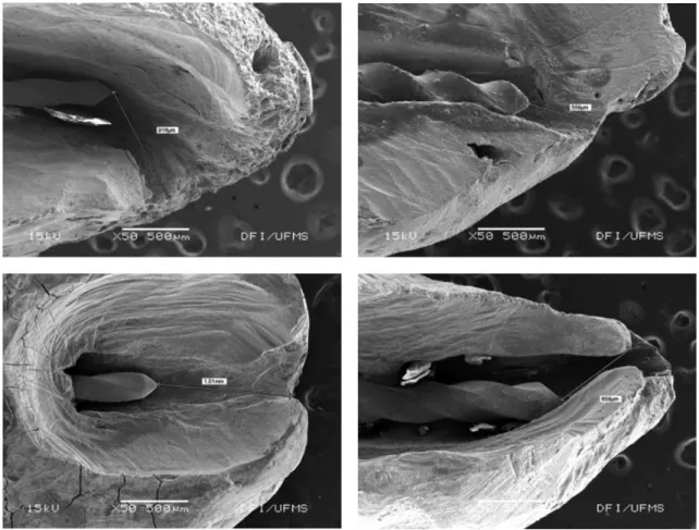
After visual identification of the root apex ending, the apical 4 mm was gently resected with diamonds burs 1013 and/or 2200 (KG Sorensen, Cotia, SP, Brazil) at high speed and under abundant water spray coolant under. When a thin dentin layer was reached between the surface and the file’s tip, their remains were removed using a scalpel blade to permit visualization of the file’s tip and its way as far as the AF. The samples were then prepared for analysis in a scanning electron microscope (JSM; 6380LV; Jeol, Tokyo, Japan) at 40× or 50× magnification was used to measure the distance from the file tip to an imaginary tangent line over the AF (Fig. 1). The measures obtained were compared with a hypothetical value of 1 mm by statistical analysis (D’Agostino-Pearson test of normality and Pearson’s correlation test, and the Bicaudal t-test).
Results
The distances from the file tip to an imaginary tangent line over the AF (I-AF), the diameter of the reading instrument (RI) and the initial apical instrument (AII), as well as for the pulp condition are in Table 1. Figure 2 shows the distances in mm from the file tip to an imaginary tangent
19
In vivo study of a new apex locator
line over the AF.
The preliminary data analysis (D’Agostino-Pearson test of normality and Pearson’s correlation test) presented a Gaussian sample distribution (p=0.608), and thus, a parametric test could be applied. The Bicaudal t-test revealed that there were no statistically significant differences (p=0.338) between the experimental values and the hypothetical value at 1 mm.
Discussion
The radiographic method is usually employed to determine the working length of the root canal. However,
studies have shown that radiographs without distortion are difficult to obtain (16,17). Abbot (18) has highlighted that radiographs can be inaccurate because of the morphological variations of root canal systems. He also pointed out that the AF not even corresponded to the anatomical root apex, so some errors may occur in the operator’s radiographic interpretation. The time elapsed for radiographic processing is another possible cause, as well as the risk for patients’ and dentists’ health due to exposure to ionizing radiation. Giving support to the problem of the variations in the apical morphology, studies (6,19,20) show a substantial inconstancy in the measurements corresponding to the distance from the AF to the anatomical root apex, which values were ranging from 0 to 3.80 mm. Being the anatomical root apex the structure seen in the radiograph in two dimensions it could be affirmed that the radiograph exam is not able to indicate nor even suggest the exact point that corresponds to the AF.
Several in vivo studies (2,11-14) were carried out to
evaluate the accuracy and the reliability of the electronic method. The investigations selected patients who had been referred to tooth extraction due to periodontal, orthodontic or prothetic reasons. This specific kind of methodology seems to be the one that offers effective results of what happens clinically as it allows the direct visualization of the apical limit determined by the electronic method in relation to the real location of the AF.
In the present study the methodology used was similar to that most of studies that evaluated the accuracy of EALs
in vivo. The adopted resources to measure those distances
are usually a digital caliper or image softwares from 7× to 100× magnification, through stereoscopic magnifiers (22). SEM was because it has a great magnification power and provides high-quality images, allowing high sharpness and focus deepness (three-dimensional image). Additionally, SEM has specific accuracy software to Table 1. The teeth, reading instrument (RI), apical initial instrument (AII),
Pulp condition and the distance file’s tip to the apical foramen (I-FA).
Tooth RI AII Pulp condition I-AF distance
(mm)
35 25 30 Vital pulp 0.559
24 - B 15 20 Vital pulp 0.906
24 - L 10 15 Vital pulp 0.898
14 - B 10 15 Vital pulp 0.724
24 - B 25 30 Vital pulp 1.280
24 - L 20 30 Vital pulp 1.520
32 15 20 Vital pulp 0.916
35 25 30 Vital pulp 0.658
32 15 20 Vital pulp 1.090
14 - L 15 20 Vital pulp 0.404
42 15 20 Vital pulp 2.010
41 15 20 Vital pulp 0.602
42 15 20 Vital pulp 0.451
43 20 25 Vital pulp 1.460
31 15 20 Vital pulp 1.470
32 10 20 Vital pulp 1.010
45 15 25 Necrosis 1.200
41 10 20 Necrosis 1.140
31 15 25 Necrosis 1.630
41 15 20 Necrosis 0.622
42 15 20 Necrosis 1.080
31 15 25 Necrosis 1.060
36 - D 15 20 Vital pulp 1.530
36 - MB 15 20 Vital pulp 1.920
Mean 1.089 +0.437
B = buccal; L = lingual; MB = mesiobuccal; D = distal.
20
K
.F
.S
. P
ereira et al.
calibrate measurement units. When small distances are being measured to evaluate the accuracy of a device that operates at a tenth of a millimeter, it seems reasonable to select a tool that represents a high standard quality in measurement, providing reliability to the the research’s outcome (13,21).
All measurements were performed under the conditions and clinical variables of a conventional endodontic treatment. Different morphological groups of teeth were employed (n=24), resulting in a higher diversification of possible anatomical situations during the experiment. Corroborating, we found that most studies on this subject used approximately the same sample numbers (2,11-13).
Preflaring was performed prior to the electronic measurement. According to Ibarrola et al. (22), who evaluated the Root ZX apex locator, the values obtained with this EAL using a crown-down instrumentation technique were more precise than those obtaiend with conventional measurement. This fact probably occured because the instrument was allowed to touch the apical walls more precisely, consequently reading the area’s impedance.
In this study, measurings we carried out up to the AF limit. Then, a digital caliper was used to measure the reading file and subtracted 1 mm from it. In the sequence, the endodontic file that better fitted into the root canal walls at that limit was fixed. Several important researchers ensure that AF represents an anatomic structure more reliable to carry out the eletronical measurements (11,12,15,21,23).
According to Lee et al. (12), more important than the anatomic site where the reading is carried out is how the measurements can be consistently reproduced. No matter where the device indicates, if the indication is reproducible, if we know where we are or if we know the average distance betwen the file tip and the true dentin-cementum junction (CDJ) then we can obtain an accurate length by subtracting the average distance from the reading. The authors found out measurements of 97% for the AF and showed that measurements from the major foramen were more consistent than from the CDJ. Corroborating with this case, Shabahang et al. (11) showed that Root-ZX, the EAL gold standard in the scientific literature - was able to localize the AF within a range of 0.5 mm in 96.2%.
Rambo et al. (15) evaluated the “ratio method” in vivo.
For such assessment a modified version of QAL® prototype was used. A total of 21 root canals were analyzed in vivo
and the results demonstrated the ability of the apex locators which are based on the “ratio method” to accurately localize the AF position, this fact is of significant clinical value. The results also explained and demonstrated why the apex locators based on the “ratio method” are not able to accurately determine the file tip position inside the root canals. The reason it happens is because the ratio of
impedance (or amplitudes) does not significantly change in such region. These readings that are between -3 and -0.5 mm from the AF can only be used by the dentists as a reference to know that the file tip is getting closer to the AF.
It is important to highlight that the procedure of subtracting 1 mm from the AF measurement was carried out to allow the clear visualization of the anatomic apex area, identifying accurately the real foramen opening. If the endodontic file had been in this area, this visualization would have been proved difficult.
There were no statistically differences between QAL® readings (mean=1.089 mm; SD=±0.437 mm) and the hypothetical measurement of 1 mm. Considering the biological pulp conditions as well as the average location of the apical constriction, the device presented an acceptable clinical limit of working length determination.
The interval of confidence supplied by the statistic test, calculated at 99.99% was comprehended between 0.682 and 1.496 mm. What became clear that there is only one possibility of 0.01% that the device can provide measurements out of this interval, which proves the reliability of the EAL. The calculated “r” coefficient was of 0.996 (maximum value of such parameter is of one). This value indicates the degree of accuracy between QAL® measurements and the hypothetical measurement of 1 mm. Therefore, according to that coefficient it is possible to state that the device is very accurate.
The variation of the measurement values generated by QAL® can be explained by the complex anatomy of the root canal in its apical region. In cases where there are large lateral canals the measurements can be influenced indicating a shorter working length (24). This statement is consistent with the results of Ardeshna et al. (25), who investigated the relationship between root canal impedance and apical anatomy in human teeth and found that the impedance values of single-foramen root canals were significantly higher than the complex-anatomy root canals (several foramina). In view of this these authors state that the device interprets the increase of root canal apical region capacitance, generating shorter readings.
There should be emphasized that even in the specimens that presented extreme measurements, the root canal preparation and the obturation would be performed at acceptable endodontic treatment limits.
In conclusion, the results of the present investigation confirmed that QAL® was able to determine working length with good reliability for the endodontic treatment, established at 1 mm shorter from the AF.
Resumo
21
In vivo study of a new apex locator
precisão e o controle do comprimento de trabalho durante o tratamento deve ser mantido. A proposta dessa investigação foi avaliar a capacidade do localizador foraminal Quill® determinar o comprimento de trabalho, a partir da localização do forame apical, estabelecido no presente estudo em 1 mm aquém do forame apical. Pacientes com indicação para extração dental por motivos ortodônticos ou periodontais foram selecionados, resultando em uma amostra de 24 canais. O termo de consentimento livre e esclarecido foi obtido de cada paciente antes dos tratamentos. Após a administração de anestesia local, a cavidade pulpar foi acessada. Em sequência, as leituras correspondentes ao forame apical mostradas no display do aparelho foram registradas e a lima removida. Com o uso de paquímetro digital, 1 mm foi subtraído da primeira medida. A lima com a nova medida foi introduzida no canal radicular novamente, fixada com resina flow e então o dente foi extraído. Após, uma das paredes do terço apical foi desgastada para vizualização da ponta do instrumento e forame apical. A distância da ponta da lima ao forame apical foi mensurada no MEV. A média das medidas foi de 1,089mm (±0,437mm). O teste t Bicaudal mostrou não haver diferenças significantes (p=0,338) entre os valores experimentais e um valor hipotético testado de 1 mm. O localizador foraminal Quill® foi capaz de determinar um satisfatório comprimento de trabalho para o tratamento endodôntico, estabelecido em 1 mm aquém do forame apical.
References
1. Ricucci D, Langeland L. Apical limit of root canal instrumentation and obturation: Part 2 - A histologic study. Int Endod J 1998;31:394-409. 2. Dunlap C, Remeikis NA, Begole EA, Rauschenberger CR. An in vivo
evaluation of an electronic apex locator that uses the ratio method in vital and necrotic canals. J Endod 1998;24:48-50.
3. Hassanien EE, Hashem A, Chalfin H. Histomorphometric study of the root apex of mandibular premolar teeth: An attempt to correlate working length measured with electronic and radiograph methods to various anatomic positions in the apical portion of the canal. J Endod 2008;34:408-412.
4. Lambrianidis T. Observer variations in radiographic evaluation of endodontic therapy. Endod Dent Traumatol 1985;1:235-241. 5. Martínez-Lozano MA, Forner-Navarro L, Sánchez-Cortés JL, Llena-Puy
C. Methodological considerations in the determination of working length. Int Endod J 2001;34:371-376.
6. Dummer PMH, Mcginn JH, Rees DG. The position and topography of the apical constriction and apical foramen. Int Endod J 1984;17:192-198. 7. Wu MK, Wesselink PR, Walton RE. Apical terminus location of root
canal treatment procedures. Oral Surg Oral Med Oral Pathol Oral Radiol Endod 2000;89:99-103.
8. Pratten DH, McDonald NJ. Comparison of radiographic and electronic working lengths. J Endod 1996;22:173-176.
9. Sunada I. New method for measuring the length of the root canal. Journal of Dental Research 1962;41:375-387.
10. Suzuki K. Experimental study on iontophoresis. Journal of the Japanese Stomatological Society 1942;16:411–417.
11. Shabahang S, Goon WWY, Gluskin AH. An in vivo evaluation of Root ZX eletronic apex locator. J Endod 1996;22:616-618.
12. Lee SJ, Nam KC, Kim YJ, Kim DW. Clinical accuracy of a new apex locator with an automatic compensation circuit. J Endod 2002;28:706-709. 13. Pagavino G, Pace R, Baccetti T. A SEM study of in vivo” accuracy of the
Root ZX electronic apex locator. J Endod 1998;24:438-441. 14. Wrbas KT, Ziegler AA, Altenburger MJ, Schirrmeister JF. In vivo
comparison of working length determination with two electronic apex locators. Int Endod J 2007; 40:133-138.
15. Rambo MV, Gamba HR, Borba GB, Maia JM, Ramos CA. In vivo
assessment of the impedance ratio method used in electronic foramen locators. Biomed Eng Online. 2010;9:46.
16. Olson AK, Goerig AC, Cavataio RE, Luciano J. The ability of the radiograph to determine the location of the apical forâmen. Int Endod J 1991;24:28-35.
17. Williams CB, Joyce AP, Roberts SA. Comparison between in vivo
radiographic working length determination and measurement after extraction. J Endod 2006;32: 624-627.
18. Abbott P. Clinical evaluation of an electronic root canal measuring device. Aust Dent J 1987;32:17-21.
19. Morfis A, Sylaras S N, Georgopoulou M, Kernani M, Prountzos F. Study of the apices of human permanent teeth with the use of a scanning electron microscope. Oral Surg Oral Med Oral Pathol 1994;77:172-176. 20. Gutierrez JH, Aguayo P. Apical foraminal openings in human teeth –
number and location. Oral Surg Oral Med Oral Pathol 1995;79:769-777. 21. Chita JJ, Silva PG, Pereira KFS, Onoda HK, Borba JC, Ramos CAS.
Accuracy and reliability of a new apex locator. Pesq Bras Odontoped Clin Integr 2012 12:457-463.
22. Ibarrola JL, Chapman BL, Howard JH, Knowles KI, Ludlow MO. Effect of preflaring on Root ZX apex locators J Endod 1999;25:625–626. 23. Cunha D’assunção FL, Albuquerque DS, Ferreira LCQ. The ability of two
apex locators to locate the apical foramen: an in vitro study. J Endod
2006;32:560-562.
24. Kobayashi C. Electronic canal length measurement. Oral Surg Oral Med Oral Pathol 1995;79:226-231.
25. Ardeshna SM, Flanagan M, Ng YL. Gulabivala K. An ex vivo investigation of the relationship between apical root impedance and canal anatomy. Int Endod J 2011;44:525-533.