FRONTOPARIETAL CORTICAL ATROPHY WITH GLIOSIS
IN THE GRAY MATTER OF CEREBRAL CORTEX
Case report
Paulo Roberto de Brito-Marques
1, Roberto Vieira de Mello
1,2, Luciano Montenegro
1,2ABSTRACT - The case of a patient who suffered from progressive amnesia, depressive humor, language and visuospatial disturbances, and hallucination episodies with interference at the daily living activities is reported. She had moderate neuropsichological diffuse deficits at the first examination, especially at the executive and visuo-constructive functions. Her cerebrospinal fluid test presented high total protein. Magnetic resonance image showed slight white matter increase in periventricular, semi-oval center bilateral and left external capsule regions, besides light frontal and parietal lobe atrophy, bilaterally. Brain single photon emission computerized tomography revealed both a bilateral moderate frontal and a severe parietal lobe hypoperfusion, especially on the left side. Macroscopic examination showed cortical atrophy, severe on the frontal, moderate on the parietal and mild on the posterior third temporal lobes, bilaterally. There was a slight atrophy on the neostriatum in the basal ganglia. The histopathological findings of the autopsy showed severe neuronal loss with intensive gemioscytic gliosis and variable degrees of status spongiosus in cortical layer. Hematoxylin-eosin and Bielschowsky staining did not show neuronal swelling (balooned cell), argyrophilic inclusion (Pick’s bodies), neurofibrillary tangles nor senile plaques. Immunohistochemical staining for anti-ubiquitin, anti-tau, anti-β-amyloide, and anti-prion protein were tested negative.
KEY WORDS: presenile dementia, cortical gliosis, diffuse neuronal loss, spongiosis, inespecific cortical atrophy. Atrofia cortical frontoparietal com gliose na substância cinzenta do córtex cerebral: relato de caso Atrofia cortical frontoparietal com gliose na substância cinzenta do córtex cerebral: relato de caso Atrofia cortical frontoparietal com gliose na substância cinzenta do córtex cerebral: relato de caso Atrofia cortical frontoparietal com gliose na substância cinzenta do córtex cerebral: relato de caso Atrofia cortical frontoparietal com gliose na substância cinzenta do córtex cerebral: relato de caso RESUMO – É descrito o caso de uma paciente que apresentava amnésia, humor deprimido, distúrbio de linguagem e visoespacial, e alucinação visual com evolução progressiva, interferindo nas atividades de vida diária. Na primeira avaliação neuropsicológica havia déficit difuso de intensidade moderada, especialmente nas funções executivas e viso-construtivas. O exame de líquido céfalo-raqueano mostrou a taxa de proteína elevada. Ressonância magnética evidenciou leve hiperintensidade de sinal na região periventricular, centro semi-oval bilateral, e alta hiperintensidade de sinal na região da cápsula interna esquerda, além de leve atrofia bilateral nos lobos frontoparietais. Tomografia cerebral por emissão de fóton único revelou hipoperfusão de intensidade moderada nos lobos frontais e severa nos parietais, especialmente à esquerda. Os achados de necrópsia evidenciaram atrofia cortical, sendo severa nos lobos frontais, moderada nos parietais e leve no terço posterior dos temporais. Havia também leve atrofia no neostriado. Do ponto de vista histopatológico, existia na camada cortical severa perda neuronal com intensa gliose gemioscítica e grau variável de status spongiosus. As colorações por hematoxilina-eosina e Bielschowsky não revelaram células baloniformes (células de Pick) e corpúsculos argirofílicos (corpos de Pick), degeneração neurofibrilar ou placa senil. As reações imuno-histoquímicas foram negativas para anti-ubiquitina, anti-tau, anti-β amilóide e proteína anti-prion. PALAVRAS-CHAVE: demência pré-senil, gliose cortical, perda neuronal difusa, espongiose, atrofia cortical inespecífica.
There are several progressive degenerative
di-seases of the central nervous system which are
lo-osely classified in the literature as “presenile
demen-tia”. These several variants or subgroups are usually
clinically indistinguishable
1, despite some
neuropsy-chological differences of right and left
frontotem-poral dementia compared to Alzheimer´s disease
(AD)
2. The most common of these disorders is AD
3.
However, there are several presenile frontotemporal
dementia types, which are much less common than
1Behavioral Neurology Unit, Department of Neurology, Faculty of Medical Sciences, University of Pernambuco, Brazil; 2Department of
Pathology, Health Sciences Center, Federal University of Pernambuco, Brazil.
Received 24 September 2001, received in final form 16 January 2002. Accepted 29 January 2002.
AD, like amyotrofic lateral sclerosis (ALS)
4, classic
Pick’s disease (PD)
5, Creutzfeld-Jakob disease and the
Heidenhain variant
1, prion disease resembling
fron-totemporal dementia and parkinsonism linked to
chromosome 17
6, presenile dementia with diffuse
calcified neurofibrillary tangles
7, dementia with ALS
features and diffuse Pick body-like inclusions
8, and
non-specific familial presenile dementia
9. Most of
these diseases are characterized by an insidious but
invariably progressive deterioration of mental and
physical capacities, culminating on a purely
vegeta-tive existence in which all patients need must be
cared for
1. In 1974, Constantinidis et al.
10after
study-ing 32 cases of lobar atrophy, classify PD into three
variants. Type A, which is classic PD; type B, the
atro-phic and gliosed cortex contained neuronal
swell-ings but no argyrophilic inclusions and a number of
those within type B that would probably be
diag-nosed as cases of corticobasal degeneration
11; and
type C. In type C the cases have neither Pick’s bodies
nor bollooned neurons, only gliosis and neuronal
loss. There are 12 cases: eight cases with temporal
atrophy (C
1) and a strong gliosis found in the white
axis of atrophied convolutions, and the other four
cases with frontal lobe atrophy (C
2) and gliosis with
extension to the precentral gyrus. This process
spre-ads to the cingular, orbital, insular and even
tempo-ral areas. The neostriatum, pallidum, thalamus and
locus niger are also severely involved and the lateral
ventricles are strongly dilated. The symptomatology
of the temporal group includes: moria, bipolar
hu-mor, and fixation amnesia, in some cases aphasic
signs and transient logorrhoea. The symptoms of the
frontal group include: apragmatism and depressive
humor, frontal type dysmnesia, boulimia, gluttony,
palilalia, echolalia, stereotypic activities and in some
cases mutism.
The histological hallmarks of PD are not present
in all cases that show typical clinical and macroscopic
pathological features of the disease. Then, a further
question related to the definition of PD is whether
the condition should be expanded still further so that
cases with a similar clinical presentation, but
lack-ing gross lobar atrophy as well as Pick cells and Pick
bodies, should be included. The consensus on these
cases is that they should not be classified as cases of
Pick’s disease but as cases of non-specific frontal lobe
dementia
12. On the other hand, the so-called Pick’s
complex, contains the clinical variants of PD
includ-ing a substantive overlap between the clinical
syn-dromes of frontal lobe dementia, frontotemporal
de-mentia, primary progressive aphasia, cortico-basal
degeneration picture, an ALS associated with severe
cortical degeneration and subcortical gliosis of
tem-poral lobes
13.
In keeping with this study, we have found
simi-larities with our case with C
2type of PD, but some
differences still exist
10. The report of this case
involv-ing the frontal and parietal and posterior third of
temporal lobes shows the clinical and
morphologi-cal aspects of a rare case of inespecific cortimorphologi-cal
atro-phy.
METHOD
A diagnosis of progressive cortical frontoparietal gliosis associated with presenile dementia was made in a female patient according to Neary et al.14. Neuropsychological
as-sessment was made on the basis of Mini-Mental State Exa-mination (MMSE)15, Clinical Dementia Rating (CDR)16. She
was also submitted to cerebrospinal fluid (CSF), and neuro-imaging studies as magnetic resonance (MR) and to brain single photon emission computed tomography (SPECT). The neurophatological examination was performed accord-ing to European Concerted Action on PD17. The brain was
fixed in 10% formalin for 4 weeks and cut into coronal sections. We examined 6-µm-thick paraffin-embedded sections from the middle frontal, upper temporal, middle temporal, parietal lobes, hippocampus and parahippocam-pus gyri, amygdaloid and Meynert nuclei of the brain. They were routinely processed and stained with hematoxilin-eosin, Masson, Wölcke’s myelin and Bielschowsky meth-ods. Immunohystochemical reactions with antibodies against tau, ubiquitin, β-amyloid and prion proteins were carried out.
CASE
home. At the age of 50, moderate anterograde persistent amnesia appeared, she was not aware of her forgetful-ness. Questions were repetead again and again, she for-got what had just been discussed. She tended to neglect the care of herself as well as that of the house, and her memory deficits had increased, involving also autobio-graphical memory. She confabulated about past events and tended to get lost in her rambling discourse. Her spon-taneous speech disappeared with progressive reduction, stereotype of speech (repetition of limited repertoire of words, or small phrases). Under these conditions her solo excursions were sharply limited. The patient had no his-tory of hypertension, diabetes and other vascular risk fac-tors. Her family history disclosed no dementing diseases. No psychiatric abnormalities emerged from her medical history.
The findings of the neurological examination 10 years after the onset of symptoms revealed a right-handed wo-man. She was limited by poor schooling. The MMSE15,
scor-ing 06/30 and CDR,216. She was fully desoriented in time
and space, but she had collaborated adequately during the test sessions. The pattern of her memory impairment manifested itself as extreme difficulty in tasks with attentional requests. Memory in everyday life seemed very disturbed. The language in informal conversation was very poor in content, she resorted to ceremonial expressions and got lost in circumlocutions demonstrating very poor verbal planning abilities; she also presented many anomias in spontaneous speech. Comprehension seemed also very impaired. As a whole, verbal communication appeared very
disturbed, even if there were no clear aphasic deficits. She named almost all common objects correctly. At this state, the simplest of geometric forms and patterns could not be copied. Her copy of a overlap pentagones was very altered.
Routine biochemical, haematological and sorological investigations were normal. The CSF from basal cistern test showed an increase in total protein content (60mg/ dl). It also included the research of virus, and electropho-retic methods. Her MRI performed when she was 50 years old revealed a slight white matter increase in the periven-tricular, semi-oval center bilateral and left external cap-sule regions. There were also a slight bilateral frontal and parietal lobes atrophy. Six months after initial evaluation, a SPECT using 99mTc-hexamethyl propyleneamine oxime
(99mTc HMPAO) showed moderate bilateral frontal and
parietal lobes hypoperfusion, especially on the left side. Her cerebral blood flow ranged from 35 to 42ml/100g tissue/min (normal 65-85) in the visual and temporal cor-tex (Fig 1).
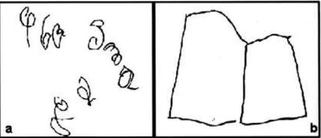
During the use of anticholinesterasic drug by 18 months her copy of overlap pentagones (Fig 2) and some daily living activities had improved. For about three years prece-eding her death, she had started to develop a progressive verbal inertia, and an stereotyped expression appeared, reduced communication and comprehension to a mini-mum, almost as a global aphasia. Despite all that she still answered with automatic language. She evolved into a clinical picture of palilalia, echolalia and mutism. The pa-tient forgot how to use common objects and tools while
Fig1. SPECT using 99mTc-hexamethyl propyleneamine oxime (99mTc HMPAO) showed moderate bilateral frontal and
retaining the necessary motor power and coordination for these activities. She had presented a poorly organized paranoid delusional state, sometimes with visual halluci-nations and also generalized tonic-clonic seizures. She took some phenobarbital until her death. Eventually, the pa-tient lost the ability to stand and walk, being forced to lie inert on the chair or in bed. During the last year of her life, she remained bedridden with arms in flexion and legs extended (decorticate rigidity) and all personal needs were cared for by her familiy in bed. She died at the age 54, after a total clinical course of 14 years. She died in August
1999 due to respiratory infection problems. The clinical diagnosis was presenile dementia Alzheimer type.
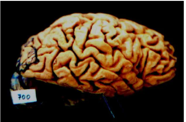
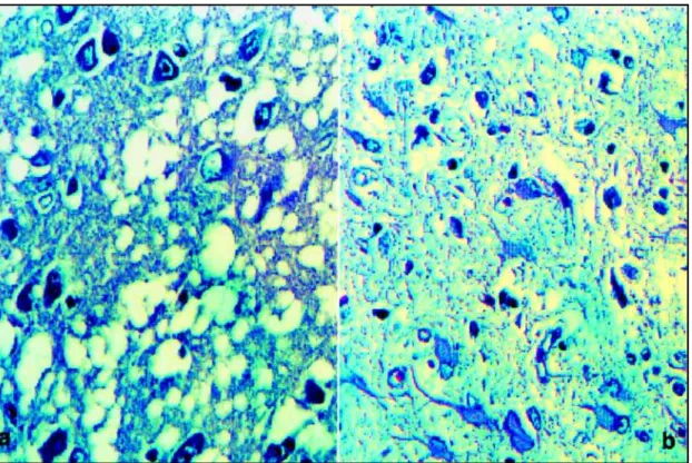
She was approximately 150 centimeters tall and weighted 50Kg. The brain was small, weighting 700 gram-mes before fixation. Macroscopic examination revealed cortical atrophy, especially severe on the frontal and mod-erate on the parietal and mild on the posterior third tempo-ral lobes (Fig 3 and 4). The diagnostic confirmation was made by the histopathological findings of the autopsy that showed severe neuronal loss with status spongiosus (mi-crovacuolation) and intensive gemistocytic gliosis in
cor-Fig 2 a,b. The overlap pentagones copy was very disturbed. The overlap pentagones copy was less disturbed during treatment.
tical structures (Fig 5). Hematoxylin-eosin and silver stains did not show neuronal swelling (balooned cell or Pick’s cells), argyrophilic inclusion (Pick’s bodies), neurofibrillary tangles, and senile plaques. Immunohistochemical stain-ing for antibodies against ubiquitin, tau, β-amyloide, and prion proteins were tested negative. There was important absence of neurons. The residual neurons were sometimes normal, sometimes hypotrophic. No focal white matter or vascular lesions were observed.
DISCUSSION
This case presented a clinical picture suggestive
of probable presenile AD in a patient who was not
used to handling complex problems due to her poor
schooling. Clinical patterns similar to those found in
AD patients occur also in non-AD patients,
highligh-ting the difficulty of making a definitive diagnosis
solely on clinical grounds
18. Then, some data were
not expected from the AD such as: a high protein
content (60mg/dl) was found in the CSF from a basal
cistern sample, in opposition to the normal CSF
usu-ally seen in AD. The MRI showed slight bilateral
fron-tal and pariefron-tal lobe atrophy without temporal lobe
involvement. In cases of AD is expected at least a
mild parietal and especially temporal lobe atrophies
on the MRI. Her brain
99mTcHMPAO SPECT showed
moderate bilateral frontal and parietal lobe
hypo-perfusion, especially on the left side. In this last test
we observed that the remaining grey matter
hypo-perfusion was ranged from 35 to 42 ml/100g/tissue/
min (normal 65-85)
19. However, it is also needed the
involvement of both temporal lobes to be
charac-terized as AD, according to what was described by
Holman et al.
20. Their criteria to the probability of
AD are the highest in patients with bilateral
tem-poroparietal defects (82%) and nearly as high (77%)
in patients with bilateral temporal and parietal
de-fects plus other dede-fects. We believed the probable
AD was the clinical diagnosis of this case because
the symptoms had been characteristic, for instance:
when she was about 41 years, she presented some
features of episodic memory deficits - she went to a
farm with her boss to spend a week, and when the
photos arrived she denied that had gone there. The
most of the episodic memory deficits presented by
her had been of the autonoetic awareness type. The
right frontal lobe, in particular, is thought to be
rel-evant for episodic memory that conects the recall of
personal past, and the related emocional
associa-tions, with plans for the future
21. Also, when she
started getting lost in well-known places, familiar
to her, she asked for help to come back home. Some
time later, she delayed to arrive at home as a result
of getting lost in common streets. These two late
facts suggest the involvement of parietal, frontal and
temporal lobes. However, it was seen in her MRI and
brain SPECT only impairment on the parietal and
frontal lobes. Brain SPECT in AD is expected both in
parietal and in temporal lobes to be involved. It is
possible that some episodic memory deficits could
have suffered influence of her poor schooling, but
the fact that she got lost in places where she went
shopping daily, we believed, could not be related to
any poor schooling.
From the neuropathological point of view, there
were a frontoparietal cortical atrophy related with
spongiosis and a diffuse cortical gliosis (DCG). These
changes were associated with neuronal loss without
specific neuronal changes, and no remarkable myelin
lesion. There was mild temporal lobe atrophy noted
on its posterior region. The immunohistochemical
reactions against tau,
β
-amyloid, ubiquitin and prion
proteins in all the cerebral lobes were negative in
tests. The clinical diagnosis of AD was not confirmed
at the autopsy, because there were neither NTs nor
SPs or immunohistochemical stainings against tau,
β
-amyloid and ubiquitin were tested negative.
Reac-tion against prion-protein was performed by the
spongiosis and intensive diffuse cortical gliosis, but
it was also tested negative. The neuropathological
findings showed compatible changes with cortical
diffuse gliosis involving the gray matter of cerebral
cortex, especially on the frontal and parietal lobes.
We observed a resemblance between our case
and the C
2type cases of Constantinidis et al.
10.
How-ever, their C
2type presents symptoms of frontal
de-mentia type associated with circumscribed frontal
cortical atrophy. The histological process spreads to
the cingular, orbital, insular, and even temporal
ar-eas, but there is not extension to the parietal lobes.
Besides, there is a varying involvement with the deep
gray matter and strong gliosis is found in the white
axis of atrophied convolutions. There were two
dif-ferent changes between the Constantinidis’ et al.
10cases and this one. First, the macroscopic atrophy
on the parietal lobes and the strong gliosis
involv-ing the cerebral cortex. This change presented some
difference in the concept of Pick’s lobar atrophy
10.
In the PD, the frontotemporal cortex is impaired for
it always spares the posterior third of the superior
temporal gyrus. In this case, the atrophy was severe
on the frontal and mild on the posterior third on the
temporal lobes. The parietal cortex is seldom
invol-ved, being moderately impaired in our case. The
oc-cipital cortex is always spared
22. The association
be-tween the three lobes is possible, but only in very
advanced disease. Acordding to Tissot et al. (apud
Adams and Victor
23) the frontal, temporal, and
parie-tal lobes are all affected in 75 percent of patients on
time disease terminates, even so they did not
scribe isolated frontal and parietal atrophy. Second,
there were not a very strong gliosis of the white
matter and neostriatum, pallidum, thalamus and
locus niger besides strongly dilated lateral ventricules
as is described in the C
2type cases
10. These are the
two great differences found among our case and
the described in the C2 type cases
10. Another study
by Neumann and Cohn
1design a new entity as
“pro-gressive subcortical gliosis” (PSG) associated with a
clinical picture of behavior and personality changes
and presenile psychosis, but Neumann´s cases are
of frontal and temporal atrophy associated with PSG
and they were different from ours.
In conclusion, nowadays, it seems impossible to
distinguish clinically primary degenerative diseases
without histological confirmation. The knowledge
of the pathological features of this case, associated
with the clinical picture of presenile dementia, show
a rare case of inespecific cortical atrophy: 1) a severe
frontal and moderate parietal and a mild posterior
third temporal lobe atrophies; 2) the presence of
strong gliosis in the gray matter of cerebral cortex
without important involvement of white matter axis
of atrophied convolutions; 3) the absence or a little
gliosis in the subcortical structures, and the lateral
dilated ventricules; 4) the absence of neuronal
swell-ings and argyrophilic inclusions, besides senile
pla-ques, neurofibrillary tangles, and prion protein were
stained negative. Several other cases will be
identi-fied and further continuous and long-term
clinico-pathological studies are clearly needed.
REFERENCES
1. Neumann MA, Cohn R. Progressive subcortical gliosis: a rare form of presenile dementia. Brain 1967;90:405-418.
2. Razani J, Boone KB, Miller B, et al. Neuropsichological performance of right and left frontotemporal dementia compared to Alzheimer´s disease. J Int Neuropsichol Soc 2001;7:468-480.
3. McKhann G, Drachman D, Folstein M, et al. Clinical diagnosis of Alzheimer’s disease: report of the NINCDS-ADRDA work group un-der the auspices of Department of Health and Human Services Task Force on Alzheimer’s disease. Neurology 1984;34:939-944.
4. Brito-Marques PR, Mello RV, Montenegro L. Amyotrophic lateral scle-rosis with dementia. Arq Neuropsiquiatr 1999;57:277-283.
5. Brito-Marques PR, Mello RV, Montenegro L. Classic Pick’s disease type with ubiquitin-positive and tau-negative inclusions. Arq Neuropsi-quiatr 2000;59:128-133.
6. Nitrini R, Silva LST, Rosemberg S, et al. Prion disease resembling fronto-temporal dementia and parkinsonism linked to cromosome 17. Arq Neuropsiquiatr 2001;59:161-164.
7. Kosaka K. Diffuse neurofibrillary tangles with calcification: a new pre-senile dementia. J Neurol Neurosurg Psychiat 1994;57:594-596. 8. Hamada K, Fukazawa T, Yanagihara T, et al. Dementia with ALS
fea-tures and diffuse Pick body-like inclusions (atypical Pick’s disease?). Clin Neuropathol 1995;14:1-6.
9. Schaumburg HH, Suzuki K. Non-specific familial presenile dementia. J Neurol Neurosurg Psychiat 1968;31:479-486.
10. Constantinidis J, Richard J, Tissot R. Pick’s disease: histogical and clini-cal correlations. Eur Neurol 1974;11:208-217.
11. Rossor MN. Pick’s disease: a clinical overview. Neurology 2001; 56(Suppl 4):S3-S5.
12. Brun A, Englund B, Gustafson L, et al. Clinical and neoropathological criteria for frontotemporal dementia. J Neurol Neurosurg Psychiat 1994;57:416-418.
13. Kertesz A, Davidson W, Munoz DG. Clinical and pathological overlap between frontotemporal dementia, primary progressive aphasia and corticobasal degeneration: the Pick complex. Dement Geriatr Cogn Disord 1999;10 (Suppl.):46-49.
14. Neary D, Snowden JS, Northen B, et al. Dementia of forntal lobe type. J Neurol Neurosurg Psychiat 1988;58:353-361.
15. Folstein MF, Folstein SE, McHugh PR. Mini-mental state: a pratical method for grading the cognitive state of patients for the clinician. J Psychiat Res 1975; 12:189-198.
16. Hughes CP, Berg L, Danziger WL, et al. A New clinical scale for the staining of dementia. Br J Psychiatry 1982;140:566-572.
17. European Concerted Action on Pick’s Disease (ECAPD) Consortium: Provisional clinical and neurophatological criteria for the diagnosis of Pick’s disease Eur Neurol 1998;5:519-520.
18. Neary D, Snowden JS, Bowen DM, et al. Neuropsychological syndromes in presenile dementia due to cerebral atrophy. J Neurol Neurosurg Psychiatry 1986; 49:163-174.
19. Ingvar DH, Cronqvist S, Ekbert R, et al. Normal values of regional ce-rebral blood flow in man, including flow and weight estimates of gray and matter. Acta Neurol Scand (Suppl.) 1965;14:72-78.
20. Holman BL, Johnson KA, Gerada B, et al. The scintigraphic appearence of Alzheimer’s disease: a prospective study using technetium-99m-HMPAO SPECT. J Nucl Med 1992;33:181-185.
21. Wheeler MA, Stuss DT, Tulving E. Toward a theory of episodic memory: the frontal lobes and autonoetic consciousness. Psychol Bull 1997; 121:331-354.
22. Escourolle R, Poirier J. Manual of basic neuropathology. 2.Ed. Phila-delphia: Saunders. 1978:242.