Arq Neuropsiquiatr 2002;60(2-B):475-477
SPINAL CORD TUMOR IN A PATIENT
WITH MULTIPLE SCLEROSIS
Case report
Mario Augusto Taricco
1, Andre Machado
2, Dagoberto Callegaro
3, Raul Marino Jr
4ABSTRACT - The association between multiple (MS) sclerosis and cerebral gliomas has been sporadically reported in the literature, causing a long lasting discussion if these lesions occur coincidentally or if MS plaques may actually lead to the genesis of gliomas. We report a 36 year old man who developed a rapid onset of right side weakness and loss of vision, having established a diagnosis of MS which was confirmed by CSF analysis and MRI. Nine years later he developed progressive tetraparesis, leading initially to suspicion of illness relapse and a demyelinating plaque in the spinal cord. However, after MRI investigation, a spinal cord tumor was diagnosed. The patient underwent cervical spine laminotomy for microsurgical removal of the spinal cord tumor diagnosed as ependimoma. The neurological deficits improved significantly.
KEY WORDS: multiple sclerosis, gliomas, spinal cord tumors.
TTTTTumor de medula espinal em paciente com esclerose múltipla: relato de casoumor de medula espinal em paciente com esclerose múltipla: relato de casoumor de medula espinal em paciente com esclerose múltipla: relato de casoumor de medula espinal em paciente com esclerose múltipla: relato de casoumor de medula espinal em paciente com esclerose múltipla: relato de caso
RESUMO - A associação entre esclerose múltipla (EM) e gliomas cerebrais foi relatada esporadicamente na literatura, levando a longa discussão quanto à possibilidade das placas de esclerose estarem envolvidas na etiologia dos gliomas ou dessas lesões ocorrerem coincidentemente. Relatamos um paciente de 36 anos que desenvolveu hemiparesia direita rapidamente progressiva e perda visual, sendo estabelecido o diagnóstico de EM após análise do LCR e imagens de RM de encéfalo. Após nove anos o paciente desenvolveu tetraparesia lentamente progressiva, levantando inicialmente a hipótese de atividade da doença e aparecimento de placa de EM na medula espinal. Contudo, após investigação com RM de coluna, um tumor medular foi diagnosticado. Foi então submetido a laminectomia cervical para ressecção microcirúrgica do tumor, que foi diagnosticado como ependimoma. Os déficits neurológicos melhoraram significativamente.
PALAVRAS-CHAVE: esclerose múltipla, gliomas, tumores medulares.
Multiple sclerosis (MS) is a demyelinating disease of the central nervous system (CNS) classically des-cribed as a relapsing-remitting disorder that affects multiple white-matter tracts, with usual onset in young adults and displaying marked clinical hetero-geneity. The varied clinical features reflect the mul-tifocal areas of CNS myelin destruction. The diagno-sis of MS was traditionally based on clinical history, neurological examination and analysis of cerebrospi-nal fluid (CSF). Evoked potential examination and magnetic resonance imaging (MRI) have brought new aids to the diagnosis1. Frequently, a pattern of
disseminated encephalomyelitis occurs, when sev-eral plaques of demyelinization can be observed throughout the neuroaxis. The plaques are typically surrounded by scar tissue, in which gliosis and glial
cell division are observed. The differential diagnosis of MS is quite limited in the setting of a young adult with two or more clinically distinct episodes of CNS dysfunction. Difficulty arises when atypical presen-tations occur, such as monophasic episodes on a pro-gressive illness. Great care must be taken to exclude treatable etiologies (compressive spinal cord lesions, arteriovenous malformations, cavernous angiomas, Chiari malformation).
The concomitance of MS and primary CNS tu-mors could be explained by simple coincidence based on the statistics of incidence and prevalence of both diseases. However, since few reports have shown the coexistenceof these conditions and it is not known how many patients actually carry them, such affir-mation cannot yet be done. In the same way, it is
Division of Neurosurgery, Hospital das Clínicas, University of São Paulo Medical School, São Paulo SP, Brazil:1Assistant physician; 2Resident
physician; 3Assistant physician, Department of Neurology; 4Professor and Chairman, Division of Neurosurgery.
Received 11 October 2001, received in final form 4 January 2002. Accepted 24 January 2002.
476 Arq Neuropsiquiatr 2002;60(2-B)
not possible to demonstrate that the gliosis around the MS plaques should be implicated in the genesis of the CNS tumors. Nevertheless, one could expect such transformation to be plausible, thinking from a strict pathophysiological point of view. There is a greater chance for a tumor to be initiated in a region of frequent mitosis than in another of infrequent mitosis. This risky area would be only for the initiation of gliomas, ruling out from this theory all other CNS tumors. Previous reports, including a review from Currie and Urich2 have shown patients with clinical
diagnosis of MS that developed cerebral gliomas, mostly malignant. We report on a case of a 36 year old man with a clinical diagnosis of MS who later on developed a spinal cord glioma.
CASE
A 36 year old man, business administrator, presented with a sudden onset of weakness and loss of sensitivity on the right side and loss of vision of the right visual field. It lasted for approximately four weeks and spontaneously improved to a lower than normal status. He underwent encephalic MRI which revealed white matter lesions sug-gestive of multiple sclerosis and a cerebrospinal fluid (CSF) tap was performed, exhibiting 1 leukocyte per ml and 37 mg of protein per 100 ml. The protein electrophoresis sho-wed a peak of 17.3 % of gammaglobulin. Diagnosis of
MS was established and he received initially prednisone therapy (40 mg daily) which was later changed to immu-nosuppressive treatment with azathioprine (100 mg daily) which was maintained for a total of seven years with a single one year interval.
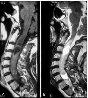
Nine years after the first episode (when the patient was 45) he presented progressive spastic tetraparesis des-pite the use of regular immunosuppressive therapy. The medication was discontinued and a new MRI was perfor-med, exhibiting a large spinal cord lesion with iso-intense-signal on T1 weighed MR (Fig 1) between the second and seventh cervical vertebrae, with a cystic nodule at the C3 level. At this time an encephalic T2 weighed MR exhibited small lesions suggestive of demyelinization plaques (Fig 2). He was submitted to a C2-C7 laminotomy for microsur-gical removal of the tumor. The patholomicrosur-gical examination revealed an ependimoma. He recovered well, with gradual improvement of muscular strength. He now presents grade 3 to 4 strength on the left upper limb and grade 4 to 5 on the right. The lower limbs present grade 5 strength with exacerbated deep-tendon reflexes. The post operative T1 weighed spinal MR confirmed the complete resection and did not exhibit any surgical complication (Fig 3).
DISCUSSION
The concomitance of MS and cerebral gliomas has been reported by few authors in the literature. There has been questioning of a possible pathological
re-Fig 1a. T1 weighed sagittal MR with gadolinium contrast of the cervi-cal spine exhibiting a large iso-signal-intensity mass lesion between C3 and C7, with a cyst in the upper level. Fig 1b. T2 weighed sagittal MR exhibiting a hyper-signal-intensity lesion and cyst.
A B
Arq Neuropsiquiatr 2002;60(2-B) 477
lationship between the two diseases, without con-clusive evidence yet. Aarli et al.3 have also proposed
the possibility of the immunosuppressive treatment for MSfacilitating the initiation of primary CNS tu-mors. However, none of the cases reported in the literature received full immunosuppressive treat-ment, including that in Aarli’s report. In the current report , the patient received a prolonged immuno-suppressive therapy. Although there is no proof that lacking imunological defenses may have facilitated tumoral initiation, it is an important consideration to make when any tumor arises.
The possible tumor initiation from a MS plaque has been discussed mainly on a pathological basis. In the Currie and Ulrich2 review, the main evidence
of initiation comes from whether there is or not a plaque contiguous to the glioma on the microscopic examination of the specimen. It is questionable if the tumor was actually induced by the contiguous plaque or if it initiated elsewhere and extended itself to meet a “false” contiguous plaque. The same is valid for
Brihaye et al.4 and Lahl5 cases. Nahser et al.6 reported
two cases in which no contiguity was observed. If the tumors developed from a plaque, it was possibly obliterated by the growing mass, making microscopic observation of contiguity very difficult. Russel and Rubinstein7also presented three cases with this
association, one of which did not present any contiguity between the tumor and plaques, supposedly because the tumor cells destroyed the plaque as well. Thus, whether gliomas occur in MS patients by mere coincidence or whether they are induced by the demyelinization plaques cannot yet be determi-ned. A larger number of cases will need to be re-ported before MS becomes a known risk factor for the initiation of gliomas.Early investigation of MS patients with MRI may help in the future to deter-mine if diagnosed plaques will give rise to tumors.
We present a report which is distinct from the others in two aspects. It is the first case of concomi-tant MS and primary spinal cord tumor, all others represented by the association of primary brain tu-mor and MS. Second, the diagnosis and report has been done with the patient alive, based on clinical and CNS imaging diagnosis. The small amount of spinal cord glial tissue (in comparison to the brain) and the rare occurrence of spinal cord plaques and tumors may explain why these lesions were not known to occur in concomitance.
REFERENCES
1. Francis GS, Duquette P. Inflammatory demyelinating diseases of the central nervous system. In Bradley GW, Daroff RB, Fenichel GM, Marsden CD (eds). Neurology in Clinical practice. New York: Butterworth-Heinemann, 1996:1307-1343.
2. Currie S, Urich H. Concurrence of multiple sclerosis and glioma. J Neurol Neurosurg Psychiatry 1974;37:598-605.
3. Aarli JA, Mφrk SJ, Myrseth E, Larsen JL. Glioblastoma associated with multiple sclerosis: coincidence or induction? Eur Neurol 1989;29:312-316.
4. Brihaye J, Périer O, Sténuit J. Multiple sclerosis associated with a cere-bral glioma. J Neuropathol Exp Neurol 1963;22:127-137.
5. Lahl R. Combination of multiple sclerosis and cerebral glioblastoma. Eur Neurol 1980;19:192-197.
6. Nahser HC, Vieregge P, Nau HE, Reinhardt V. Coincidence of mul-tiple sclerosis and glioma: clinical and radiological remarks on two cases. Surg Neurol 1986;26:45-51.
7. Russel DS, Rubinstein LJ. Pathology of tumors of the central nervous system. 4.ed : London: Edward Arnald, 1971:179.