Studies on the genetics of artemisinin
resistance in malaria
Ana Júlia Pinto Fonseca Sieuve Afonso Rodrigues
Universidade Nova de Lisboa
Instituto de Higiene e Medicina Tropical
Centro de Malária e outras Doenças Tropicais – Laboratório Associado
Ana Júlia Pinto Fonseca Sieuve Afonso Rodrigues
Studies on the genetics of artemisinin resistance in malaria
Dissertação de candidatura ao grau de Doutor em Parasitologia Médica submetida ao Instituto de Higiene e Medicina Tropical Universidade Nova de Lisboa
Ana Júlia Pinto Fonseca Sieuve Afonso Rodrigues foi bolseira da Fundação para a Ciência e Tecnologia do Ministério da Ciência Tecnologia e Ensino Superior
I declare that unless stated otherwise, the work presented here is my own. A number of figures and tables have been kindly made available by other scientists or
developed in partnership with other scientists.
Ítaca
Se um dia partires rumo a Ítaca, reza para que o caminho seja longo, cheio de aventura e de conhecimento.
Não temas monstros como os Ciclopes ou o zangado Poseidon: Nunca os encontrarás no teu caminho
enquanto mantiveres o teu espírito elevado,
enquanto uma rara excitação agitar o teu espírito e o teu corpo. Nunca encontrarás os Ciclopes ou outros monstros
a não ser que os tragas contigo dentro da tua alma, a não ser que a tua alma os crie em frente a ti. Deseja que o caminho seja bem longo
para que haja muitas manhãs de Verão em que, com quanto prazer, com tanta alegria,
entres em portos que vês pela primeira vez;
Para que possas parar em postos de comércio fenícios aí comprar coisas finas, madrepérola, coral e âmbar, e perfumes sensuais de todos os tipos
tantos quantos puderes encontrar;
e para que possas visitar muitas cidades egípcias
e aprender e continuar sempre a aprender com os seus escolares. Tem sempre Ítaca na tua mente.
Chegar lá é o teu destino. Mas não te apresses na viagem. Será melhor que ela dure muitos anos para que sejas velho quando chegares à ilha, rico com tudo o que encontraste no caminho, sem esperares que Ítaca te traga riquezas. Ítaca deu-te a tua bela viagem.
Sem ela não terias sequer partido. Não tem mais nada a dar-te. E, sábio como te terás tornado, tão cheio de sabedoria e experiência,
já terás percebido, à chegada, o que significa uma Ítaca.
Ithaca
When you set out on your journey to Ithaca, pray that the road is long,
full of adventure, full of knowledge. The Lestrygonians and the Cyclops, the angry Poseidon -- do not fear them: You will never find such as these on your path, if your thoughts remain lofty, if a fine
emotion touches your spirit and your body. The Lestrygonians and the Cyclops,
the fierce Poseidon you will never encounter, if you do not carry them within your soul, if your soul does not set them up before you. Pray that the road is long.
That the summer mornings are many, when, with such pleasure, with such joy
you will enter ports seen for the first time; stop at Phoenician markets,
and purchase fine merchandise,
mother-of-pearl and coral, amber and ebony, and sensual perfumes of all kinds,
as many sensual perfumes as you can; visit many Egyptian cities,
to learn and learn from scholars. Always keep Ithaca in your mind. To arrive there is your ultimate goal. But do not hurry the voyage at all. It is better to let it last for many years; and to anchor at the island when you are old, rich with all you have gained on the way, not expecting that Ithaca will offer you riches. Ithaca has given you the beautiful voyage.
Without her you would have never set out on the road. She has nothing more to give you.
And if you find her poor, Ithaca has not deceived you. Wise as you have become, with so much experience, you must already have understood what Ithacas mean.
Este projecto foi desenvolvido no Instituto de Higiene e Medicina Tropical (IHMT), Centro de Malária e Outras Doenças Tropicais-Laboratório Associado (CMDT-LA) e na
University of Edinburgh, School of Biological Sciences, Institute for Immunology and Infection Research.
Este trabalho foi financiado pelos projectos: ”Busca de novos marcadores de resistência a antimaláricos” (PRAXIS/P/SAU/14070/98) e “RESMALSHIP – Development of a malaria resistance DNA chip as a public health tool for managment of Plasmodium falciparum
malaria drug resistance” (QLK2-CT-2002-01503) e pelo financiamento
SFRH/BD/8913/2002, da Fundação para a Ciência e Tecnologia.
Ao Professor Doutor Virgílio Estólio do Rosário, Director da UEI Malária, membro do CMDT-LA e Professor Catedrático do IHMT, pela orientação e supervisão deste trabalho, assim como pelo apoio e disponibilidade que sempre demonstrou em todas as fases da realização deste trabalho. Acima, de tudo e a um nível pessoal, gostaria de agradecer a confiança e amizade sempre transmitidas e o tempo dispensado.
Ao Professor Doutor Pedro Vítor Lemos Cravo, membro do CMDT-LA e Professor Auxiliar do IHMT, pela orientação e supervisão deste trabalho. Agradeço-lhe todo o apoio, disponibilidade e confiança. Mesmo nas fases menos positivas do trabalho. A sua confiança fez toda a diferença. A nível pessoal, agradeço toda a amizade, paciência e toda a ciência conversada logo desde o café da manhã. Muito obrigada pela sua ajuda sem a qual nada disto teria sido possível.
To Dr Paul Hunt from the University of Edinburgh for all the supervision during this project. For all the science I have learned and for all the knowledge that I have received. More importantly, from a personal point of view, for all the friendship that has grown, during this “journey”. His friendship made the whole process immensely enjoyable.
To Dr Richard Culleton from the University of Edinburgh (now at Laboratory of Malariology, International Research Centre of Infectious Diseases, Osaka University, Osaka, Japan) for the work done and help during the Linkage Group Selection. His perseverance, his knowledge on the genetic of the parasite and above all, his way to improvise, made all the difference.
Ao Professor Doutor Celso Cunha, Director da UEI Biologia Molecular, membro do CMDT-LA e Professor Auxilar Convidado do IHMT pela disponibilidade enquanto membro da minha comissão tutorial.
To Professor Richard Carter from the University of Edinburgh for taking more than time to discuss this project.
To Dra Sandie Cheesman from the University of Edinburgh for the time spent discussing Real-Time PCR.
To Dra. Alison Creaseyfrom the University of Edinburgh for being a friend. For making my life in Edinburgh much more enjoyable. For all the science discussed late at night or over many cups of tea. For being as she is.
To Mr. Richard Fawcett and Mr. Les Steven from the University of Edinburgh for all the technical help during my time in Edinburgh
To Mr. Sittiporn Pattiradilokrat from the University of Edinburgh for his assistance with AFLP.
To Dr. Axel Martinelli for his friendship and for making my life in Edinburgh more complete. Thank you.
À Mestre Ana Catarina Alves pela ajuda técnica nas primeiras etapas deste trabalho. Sob o ponto de vista pessoal pela amizade demonstrada ao longo de todo este tempo.
Às amigas Doutora Dinora Lopes Ferreira e à Mestre Fátima Nogueira. Foi um privilégio dividir o gabinete de trabalho convosco. Pelas conversas, pelo que aprendi e pelo que foi partilhado o meu obrigada.
À Dra. Isabel Ferreira, outra “vitima da artemisinina” por ter partilhado quase simultaneamente as mesmas etapas desta jornada e pelo que aprendi com o seu método de trabalho, o meu obrigada.
À D. Encarnação Horta por toda a assistência durante este trabalho. À D. Celeste Figueiredo e à Dra. Catarina Costa pela disponibilidade na resolução pratica de todos os problemas burocráticos insolúveis. O meu sincero obrigado.
A toda a equipa do CMDT-LA, em especial à Dra. Patrícia Abrantes por toda a amizade e ajuda.
À Professora Doutora Maria de Sousa do Instituto de Ciências Biomédicas Abel Salazar da Universidade do Porto pela amizade e exemplo científico e pessoal.
À Professora Doutora Ana Tomás do Instituto de Ciências Biomédicas Abel Salazar da Universidade do Porto pela amizade e pela ajuda no pensar para a frente.
À Dra. Esmeralda Delgado da Faculdade de Veterinária da Universidade Técnica de Lisboa pelos ensinamentos transmitidos acerca da esplenectomização de ratos.
A todos os meus professores que de um modo ou outro não me espartilharam a vontade de descobrir.
A todos os meus amigos por terem compensado de um modo que eu não merecia as minhas ausências com as suas presenças constantes. Pelo que aprendi, pelo que me deram, por sempre acreditarem que era possível o meu muito obrigada.
A todas as “meninas do Norte” em especial à Doutora Fátima Macedo, Dra. Isabel Carvalho e Dra. Susana Almeida pela ajuda prática mas acima de tudo por toda a amizade À Dra. Natália Freitas porque se fosse só para a ter conhecido já valia a pena ter começado este projecto.
À Dra. Cláudia Gaspar pela amizade e pelo exemplo cientifico.
À Caia pela amizade que vem de longe e vai até sempre. Pelo exemplo de preserverança obrigada.
Ao Rui à Xana e mais que tudo à Mafalda pela amizade, pelo ombro sempre presente. Obrigada por existirem.
À Marta, pilar mestre de como eu sou, exemplo de como há coisas além da ciência, por acreditares sempre em mim o meu obrigada agora e sempre.
À minha mãe, por me ter feito como sou, porque está lá sempre porque me dá mais que tudo… Dá-me o seu exemplo. Obrigada.
Ao meu marido António porque este projecto não é definitivamente só meu é completamente nosso, porque esteve e está sempre comigo, porque sempre me deu o amor e a paz indispensáveis para a minha vida. Porque acreditou e acredita em mim. Mil vezes obrigado. À Dorothy pela companhia durante a escrita desta tese.
Page
Acknowledgements / Agradecimentos 11
Table of Contents 17
List of Figures 23
List of Tables 27
List of Abbreviations 31
Abstract 35
Resumo 39
Chapter I – Introduction 43
1.1 Malaria: general features 45
1.2 Malaria today 47
1.3 The parasite and its life-cycle 50
1.4 The genetics of malaria parasites 53
1.5 Antimalarial drugs and targets 57
1.6 Artemisinin and its derivatives 58
1.6.1 Artemisinin mode of action 63
1.7 Drug Resistance 67
1.7.1 Resistance to artemisinin 71
1.8 Rodent malaria parasites 73
1.8.1 The genetics of rodent malaria parasites 78
1.9 Linkage Group Selection 80
1.9.1. Amplified Fragment Length Polymorphism 83
1.10 Aims of the project 84
Chapter II – Materials and Methods 85
2.1. Mice 87
2.2. Mosquitoes 87
2.3. Parasites 87
2.4 Preparation of 107 standard parasite inocula 88
2.5 Cloning 88
2.6 Preparation and administration of artemisinin and artesunate 88
2.7 Drug tests 89
2.8 Artemisinin and artesunate selection experiments: general procedure 89 2.9 Tests to evaluate the stability of drug-resistance 90 2.10. Transmission through Anopheles stephensi 90
2.11. DNA extraction 91
2.12. Identification of the P. chabauditctp and atp6 genes 91
2.13. Amplification and sequencing of the mdr1, cg10, tctp and atp6 genes of P. chabaudi 92
2.14. Estimation of copy numbers of the pcmdr1, pctctp and pcatp6 genes 92
2.15 Production of cross progeny - Overview of procedure 93
2.15.1 Mosquito feeds 94
2.15.2 Infection of mice from sporozoites 95 2.15.3 Preparation of artemisinin and artesunate 95
2.16 Linkage Group Selection 95
2.16.1 Selection of cross progeny 95
2.16.2 Amplified Fragment Length Polymorphism (AFLP) analysis 96 2.16.2.1 Preparation of parasite DNA from experimental groups 96
2.16.3 AFLP analysis 97
2.16.4 Measurement and comparison of the intensity of AFLP markers 101 2.16.5 Assignment of AFLP markers to locations in a P. chabaudi genetic linkage map. 102
2.16.6 Sequencing of AFLP markers, and their location on the Plasmodium falciparum
genome 103
2.16.7 AFLP band sequencing and purification of PCR products 103 2.17 Experiments with genetic crosses of AS-ART and AJ or AS-ATN and AJ 105 2.17.1 Blood collection for Proportional sequencing analyses 105 2.17.2 Extraction of parasite DNA from blood or sporozoites 106 2.17.3 Determination of the proportions of clones in genetic crosses by proportional
sequencing 106
2.17.3.3 Proportional sequencing – Sequencing and purification of PCR
products 110
2.17.3.4 Proportional sequencing – Analyses of sequencing results 110 2.18. Amplification and sequencing of the ubp-1 gene of P. chabaudi 112
Results 113
Chapter III – Experiments for drug selection of artemisinin and artesunate resistance
117
3.1 Introduction 119
3.2 Artemisinin drug selection 120
3.3 Artesunate drug selection 124
3.4 Cloning of resistant parasites 128
3.5 Drug resistance stability tests 130
3.5.1. Resistance stability after liquid nitrogen preservation (deep-freezing) 130 3.5.2. Resistance stability after blood passages in the absence of drug pressure 131 3.5.3. Resistance stability after cyclical transmission through mosquitoes 131 3.6 Test for cross-resistance between artemisinin and artesunate clones 133
3.7 Discussion 134
Chapter IV – Analysis of the putative genetic modulators for artemisinin and artesunate resistance
137 4.1. Sequencing of P. chabaudi mdr1, cg10, tctp and atp6 genes, in the selected mutant
clones. 139
4.1.1. Isolation of the mdr1, cg10, tctp and atp6 P.chabaudi orthologues 139
4.2.2. Sequnece comparions of P. chabaudi mdr1, cg10, tctp and atp6 in the
selected mutant clones and their sensitive progenitors. 143 4.2. Estimation of gene copy numbers of the pcmdr1, pctctp and pcatp6 genes, in the
selected mutant clones.
144
4.3. Discussion 146
4.3.1. Summary 146
4.3.2. General discussion 147
Chapter V – Experiments using Linkage Group Selection as an attempt to identify the locusor lociinvolved in artemisinin and artesunate resistance
149
5.1. Introduction 151
5.2 Production of cross progeny 152
5.3 Selection of cross progeny 153
5.3.1 General procedure 153
5.3.2 Results 154
5.3.2.1 Selection of the AS-ART x AJ cross 155 5.3.2.2 Selection of the AS-ATN x AJ cross 158
5.4 AFLP analysis 160
5.4.1 General procedure 160
5.4. 2 Results 161
5.4.2.1 AFLP analysis for the AS-ART x AJ 162 5.4.2.2 AFLP analysis for the AS-ATN x AJ 165 5.4.2.3 AFLP analysis comparison between pooled crosses AS-ART x AJ
and AS-ATN x AJ 169
5.5 Sequencing and mapping of AFLP bands under selection 170
5.5.1 General procedure 170
5.5.2 Results 171
5.6 Further investigation of markers under selection on chromosome 2 173 5.6.1 Identification of ubp-1 gene mutation on P. chabaudi chromosome 2 175
5.7 Discussion 178
Chapter VI – General conclusions 181
Supplement 185
Appendix 1 -Solutions and buffers 187
Appendix 2 – Gene and primer sequence for the genes pcmdr1, cg10, pctctp and pcatp6 189 Appendix 3 – Primer sequence and PCR amplification conditions for determining gene
copy number of pcmdr1, pctctp and pcatp6 genes
193
Appendix 4 –Gene and primer sequence for the gene ubp-1 194 Appendix 5 –The relative intensity and comparative intensities of all AFLP markers
analysed in the LGS experiments described in Chapter V
Glossary 221
References 225
Online references 239
Publications that arose from this project 243
HuntP, AfonsoA, CreaseyA, CulletonR, Sidhu A, LoganJ, ValderramosS, McNaeI, CheesmanS, do Rosario V, Carter R, Fidock D and CravoP. Gene encoding a de-ubiquitinating enzyme is mutated in artemisinin- and chloroquine-resistant rodent malaria parasites. In press at the Molecular Microbiology
Cheesman S, Creasey A, Degnan K, Kooij T, Afonso A, Cravo P, Carter R, Hunt P. Validation of Pyrosequencing for accurate and high throughput estimation of allele frequencies in malaria parasites Mol Biochem Parasitol. 2007 Apr; 152 (2): 213-219.
Cravo P, Culleton R, Afonso A, Ferreira ID, do Rosário VE Mechanisms of Drug Resistance in Malaria: Current and New Challenges. Anti-Infective Agents in Medical Chemistry 2006. 5: 63-73.
Page Cover - Artemisia annua flowers and leaves.
Source: The University of York (www.york.ac.uk)
Figure 1 - Illustration drawn by Laveran of various stages of malaria parasites as seen on fresh blood. 46
Figure 2 – World geographic distribution of malaria, data from 2003. 49
Figure 3 – Malaria transmission areas and P. falciparum drug resistance distribution data from World
Heath Organization data from 2004 49
Figure 4 - Life cycle of malaria parasite. 52
Figure 5 – Crossing and chromosomal events in Plasmodium. 55
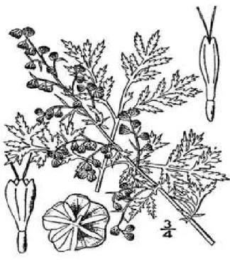
Figure 6- Schematic representation of Artemisia annua L. 58
Figure 7 – A - Chemical structure of (1) Qinghaosu or artemisinin and some of its derivatives (2) dihydroartemisinin, (3) artemether; (4) arteether and (5) artesunic acid or artesunate. 60
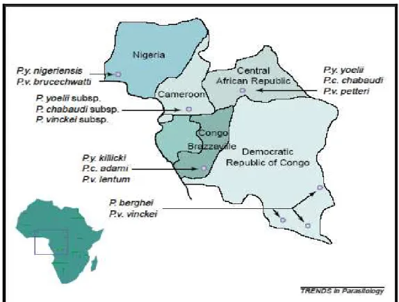
Figure 8 The geographic origins of the rodent malaria parasites. 73
Figure 9 Plasmodium chabaudi parasites (trophozoite stage) in mouse peripheral blood. 75
Figure 10 – Rodent malaria genomes synteny map to P. falciparum. 79
Figure 11 – Schematic representation of the Linkage Group Selection protocol. 82
Figure 12 - An example of an AFLP gel. 100
Figure 13- The four polymorphisms on the pcdhps gene that allowed differentiating between strains
AS and AJ. 108
Figure 14 – Example of an electropherogram for proportional sequence analysis of two polymorphisms on the pcdhps gene.
111
Figure 15 – A schematic representation of the artemisinin selection procedure. AS-ART* is uncloned. 120
Figure 16 – The increase of the artemisinin dose (mg/kg/day), tolerated by AS-30CQ clone, during 15
blood passages. 121
Figure 17 – The mean day of recrudescence on each passage under selection and artemisinin dose
(mg/kg/day). 122
Figure18 – A schematic representation of the artesunate selection procedure. AS-ATN* is uncloned. 124
Figure 19 – The increase of the artesunate dose (mg/kg/day), tolerated by AS-15CQ clone, during 14
blood passages. 125
Figure 20 – The mean day of recrudescence under selection and artesunate dose (mg/kg/day). 126
Figure 21 – A schematic representation of the clones and parasite lines of Plasmodium chabaudi used
in this project.
128
Figure 22 – A schematic representation of the artemisinin and artesunate selection procedure. 132
Figure 23 – Extent of sequence analysed for the gene pcmdr1 for the clones AS-15CQ, AS-30CQ,
AS-ART and AS-ATN. 141
Figure 24 – Extent of sequence analysed for the gene pccg10 for the clones 15CQ, 30CQ,
AS-ART and AS-ATN. 141
Figure 25 – Extent of sequence analysed for the gene pctctp for the clones 15CQ, 30CQ,
AS-ART and AS-ATN. 142
Figure 26 – Extent of sequence analysed for the gene pcatp6 for the clones 15CQ, 30CQ,
AS-ART and AS-ATN
142
Figure 27 – Relative differences (N-fold) in gene copy number between artemisinin (AS-ART) and artesunate (AS-ATN) resistant parasites and their sensitive progenitors, AS (30CQ) and AS (15CQ) respectively.
145
Figure 28 - Schematic representation of the selection of the pooled cross progeny (LGS) experiment using previously generated individual crosses between AJ and AS-ART
153
Figure 29 - Parasitaemia curves for the “untreated” and “artemisinin treated” uncloned cross progeny
of the AS-ART x AJ cross. 155
Figure 30 – Detail of the pcdhps gene sequence with indication of polymorphisms 1-4 between AS
and AJ. For entire sequence of the gene please check figure 13. 156
Figure 31 - Parasitaemia curves for the “untreated” and “artesunate treated” uncloned cross progeny
of the AS-ATN x AJ cross. 158
Figure 32 - Comparative Intensities (CI) of AJ (sensitive parent) specific AFLP markers in the ART treated group compared to the untreated group, for the AS-ART x AJ cross. 163
Figure 33 - Comparative Intensities (CI) of AS (resistant parent) specific AFLP markers in the ART treated group compared to the untreated group, for the AS-ART x AJ cross 164
Figure 34 - Comparative Intensities (CI) of AJ (sensitive parent) specific AFLP markers in the ATN treated group compared to the untreated group, for the AS-ATN x AJ cross. 167
Figure 35 - Comparative Intensities (CI) of AS (resistant parent) specific AFLP markers in the ATN treated group compared to the untreated group, for the AS-ATN x AJ cross. 168
1 and P. falciparum chromosome 7.
Figure 37 – Part of the ubp-1 gene alignments where the different mutations are represented in blue
Page Table 1 – Classification of antimalarial agents according to their stage of action. 57
Table 2 – Qinghaosu chemical information. 59
Table 3 – Some pharmacokinetic data of artemisinin and some of its derivatives. 61
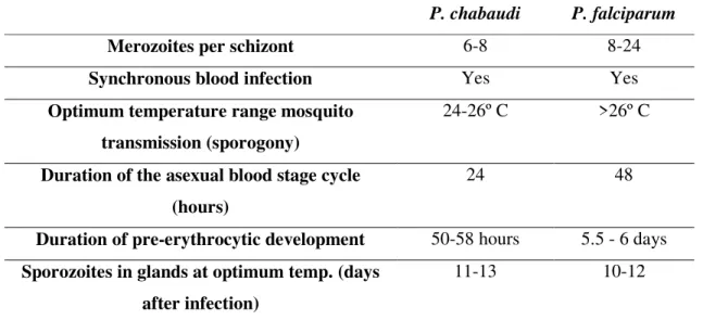
Table 4 Some biological similarities and differences between P. chabaudi and the human malaria
parasite, P. falciparum.
74
Table 5- Clones of Plasmodium chabaudi available for this project. 77
Table 6 - Parasite clones and lines used in the present work. 87
Table 7 – Maximum doses tolerated by the progenitor parasite lines used to select for resistance. 119
Table 8 – N-fold resistance, using the equation above and calculated for each passage under
artemisinin pressure. 123
Table 9 – N-fold resistance calculated for each passage under artesunate pressure. 127
Table 10 - N-fold resistance of P. chabaudi AS-ATN and AS-ART. The absolute and relative
(N-fold) drug sensitivities of AS-ATN and AS-ART after blood passage in the absence of treatment, freeze/thaw and mosquito transmission are given.
133
Table 11 – Proportional sequencing results for each group of the selection procedure for the genetic cross AS-ART x AJ (“unpassaged”, “untreated” and “artemisinin treated”) for all the dhps gene analysed..
157
Table 12 – Proportional sequencing results for each group of the selection procedure for the genetic cross AS-ATN x AJ (“unpassaged”, “untreated” and “artesunate treated”) for all the polymorphisms of the dhps gene analysed.
159
Table 13 - CI of AJ specific AFLP markers under selection in the ART treated group, compared to the untreated group for the AS-ART x AJ genetic cross, and their P. chabaudi chromosomal locations
according to the genetic linkage map.
162
Table 14 - CI of AJ specific AFLP markers under selection in the ATN treated group, compared to the untreated group for the AS-ATN x AJ genetic cross, and their P. chabaudi chromosomal locations according to the genetic linkage map.
166
Table 15 - Physical and genetic mapping of the AFLP markers with low CI. 172
Table 16 – A summary of the ubp-1 gene mutations and their correspondence on ubp-1 transcript for
each of the P. chabaudi clones and parasite lines analysed.
176
Table A3-1 - Polymerase Chain Reactions for gene quantification of pcmdr1, pctctp and pcatp6
genes in comparison to pcmsp1.
3D7 P. falciparum culture, sensitive
6-PGD - 6-Phosphogluconate dehydrogenase
A - Adenine (in DNA context)
A Alanine (Ala) (in protein context)
a.a. - Amino acid
ACT - Artemisinin combination therapy
AFLP - Amplified fragment length polymorphism
ART - Artemisinin
AJ - Plasmodium chabaudi clone, totally drug sensitive. Genetically different from AS line.
AS-ART - Plasmodium chabaudi clone, pyrimethamine-, high chloroquine- resistant, obtained from
AS-30CQ by artemisinin drug pressure (clone obtained during this project)
AS-ART* - Plasmodium chabaudi parasite line, pyrimethamine-, high chloroquine- resistant, obtained
from AS-30CQ by artemisinin drug pressure (parasite line obtained during this project), this parasite line was then cloned by limiting dilution
AS-ATN - Plasmodium chabaudi clone, pyrimethamine-, intermediate chloroquine- resistant, obtained
from AS-15CQ by artesunate drug pressure (clone obtained during this project)
AS-ATN* - Plasmodium chabaudi parasite line, pyrimethamine-, intermediate chloroquine- resistant,
obtained from AS-15CQ by artesunate drug pressure (parasite line obtained during this project), this parasite line was then cloned by limiting dilution
AS-SENS - Plasmodium chabaudi clone, totally drug sensitive. Genetically different from AJ.
AS-PYR - Plasmodium chabaudi clone, pyrimethamine-resistant obtained from AS-SENS by
pyrimethamine drug pressure
AS-3CQ - Plasmodium chabaudi clone, pyrimethamine and low chloroquine resistant obtained from
AS-PYR by chloroquine drug pressure
AS-15CQ - Plasmodium chabaudi parasite isolate, pyrimethamine and intermediate chloroquine
resistant, obtained from the AS-3CQ by cloroquine drug pressure
AS-15MEF - Plasmodium chabaudi parasite clone, pyrimethamine, intermediate chloroquine resistant
and mefloquine resistant, obtained from the AS-15CQ by mefloquine drug pressure
AS-30CQ - Plasmodium chabaudi parasite clone, pyrimethamine and high chloroquine resistant,
obtained from the AS-15CQ by cloroquine drug pressure
ARMD - Accelerated resistant to multiple drugs phenotype
ama-1 - Apical membrane antigen-1
ATN - Artesunate
atp6 - Encoding the Sarcoplasmic and Endoplasmic Reticulum Ca2+ atpase gene
C - Cysteine (Cys)
CDC - National Center for Disease Control
cDNA - Complementary DNA
cg10 - Gene coding for a putative protein transporter, P. chabaudi orthologue of the P. falciparum crt.
CI - Comparative intensity
CQ - Chloroquine
crt - Chloroquine resistance transporter gene
CSP - Circumsporozoite protein
D - Aspartic acid (Asp)
Dd2 P. falciparum culture, from Indochina, pyrimethamine, mefloquine and chloroquine
resistant
DDT - Dichloro-diphenyl-trichloroethane
DHA - Dihydroartemisinin
DHFR Dihydrofolate-reductase enzyme
DMSO - Dimethyl sulfoxide
DNA - Deoxyribonucleic acid
F - Phenylalanine (Phe)
gDNA - Genomic DNA
HB3 - P. falciparum culture, from Honduras, pyrimethamine, mefloquine and chloroquine
resistant
IC50 - The drug dose necessary to eliminate 50% of the parasites
II Intensity index
i. p. - Intraperitoneally
iRBC - Infected red blood cells (erythrocytes)
L Leucine (Leu)
LDH - Lactate dehydrogenase
LGS - Linkage group selection
Mb - Mega base
MCD - Minimum curative dose
mdr1 - Multi drug resistance 1 gene
msp-1 Merozoite surface protein-1
msp-2 - Merozoite surface protein-2
N - Asparagine (Asn)
NADH - Hydrogen nicotinamide adenine dinucleotide
NCBI/NIH - National Institute of Health
nM - Nanomolar
PABA - Paraminobenzoic acid
pcdhps - Gene of Plasmodium chabaudi codifying for enzyme dihydropteroate synthetase
PCR - Polymerase chain reaction
Phe - Phenylalanine
PYR - Pyrimethamine
RBC - Red blood cells (erythrocytes)
RIIs - Relative intensity indices
RMP - Rodent malaria parasite
RNA - Ribonucleic acid
rRNA - Ribosomal RNA
RTQ-PCR - Real time quantitative – PCR
S - Serine (Ser)
s. c. - Subcutaneous
SDS - Sodium dodecyl sulfate
sRNA - Small RNA
SP - Sulfadoxine-pyrimethamine
T - Tymine
tctp - Translationally controlled tumour protein gene
tRNA - Transfer RNA
Tyr - Tyrosine
ubp-1 - De-ubiquitinating enzyme, ubiquitin carboxyl-terminal hydrolase, putative, 1
ubp-1 De-ubiquitinating enzyme, ubiquitin carboxyl-terminal hydrolase, putative, 1 gene
V - Valine (Val)
WHO - World Heath Organization
Y - Tyrosine (Tyr)
Resistance of Plasmodium falciparum to multiple drugs including chloroquine (CQ) and
sulfadoxine-pyrimethamine (SP) is a major problem in malaria control. New drugs, such as artemisinin (ART) derivatives, particularly in combination with other drugs, are thus increasingly used to treat malaria. Although stable resistance to ART has yet to be reported from laboratory or field studies, its emergence would be disastrous because of the lack of alternative treatments.
The work presented in this thesis describes the selection of parasites with stable resistance to ART and artesunate (ATN), and their genetic analysis. This work was carried out using the rodent malaria parasite Plasmodium chabaudi chabaudi (Plasmodium chabaudi).
Two different rodent malaria parasite lines AS-15CQ and AS-30CQ were continually passaged in the presence of increasing concentrations of ATN or ART, respectively. After selection, these lines, named AS-ATN and AS-ART, showed 6-fold and 15-fold increased resistance to ATN and ART respectively. Resistance remained stable after cloning, freeze/thawing, blood passage in the absence of drug pressure and transmission through mosquitoes. The nucleotide sequences and the gene copy number of the possible genetic modulators of ART resistance mdr1, cg10, tctp and atp6; were compared between sensitive
and resistant parasites. No mutations or changes in the gene copy number of these genes were found.
Linkage Group Selection (LGS) was used to investigate the genetic basis of ART resistance. Genetic crosses between AS-ART or AS-ATN and the ART-sensitive clone AJ were analysed before and after drug treatment. Using quantitative markers, a genetic locus on chromosome 2 was found to be under strong selection. Loci on chromosomes 1, 8 and 14 of P. chabaudi also
A existência de estirpes do parasita, Plasmodium falciparum resistentes a multiplos fármacos
tais como; cloroquina (CQ) e sulfadoxina-pirimetamina (SP) é um dos problemas mais graves no controlo da malária.
Novos fármacos, como a artemisinina (ART) e seus derivados, particularmente em combinação com outros fármacos, são cada vez mais utilizados no tratamento da malaria. Embora até ao momento a fármaco-resistência estável à ART quer in vitro quer in vivo não
tenha sido registada, o seu surgimento seria desastroso devido á falta de alternativas.
O trabalho apresentado nesta tese descreve a selecção de resistência estável a ART e ao artesunato (ATN). Este trabalho foi realizado usando o modelo roedor de malária Plasmodium chabaudi chabaudi (Plasmodium chabaudi).
Duas linhas parasitáricas diferentes, AS-15CQ e AS-30CQ, foram feitas crescer na presença de concentrações crescentes de ATN e ART, e que no final apresentavam uma resistência de 6 e 15 vezes superior ao ATN e à ART, respectivamente (estas novas linhas obtidas foram nomeadas AS-ATN e AS-ART).
A resistência é estável mesmo após clonagem, congelamento/descongelamento, passagem sanguínea na ausência de pressão de fármaco e transmissão através do mosquito vector. A sequência nucleotídica e o número de cópias dos genes descritos como moduladores putativos de resistência à ART: mdr1, cg10, tctp e atp6; foi comparada entre parasitas
resistentes e sensíveis. Não tendo sido encontradas alterações na sequência ou no número de cópias destes genes.
Numa tentativa de identificar os genes encolvidos na resistância à ART e ao ATN a técnica de Linkage Group Selection (LGS) foi utilizada e dois cruzamentos genéticos entre os clones fármaco-resistentes; AS-ART e AS-ATN e o clone geneticamente distinto dos anteriores e sensível aos fármacos em estudos; AJ; foram realizados. Foram encontrados sobre selecção em ambos os cruzamentos genéticos quatro loci; cromossomas de P. chabaudi 1, 2, 6 e 8.
1.1 Malaria: general features
Malaria parasites are micro-organisms that belong to the genus Plasmodium. There are more
than 100 species of Plasmodium, which can infect many animal species such as reptiles, birds,
and various mammals. Only four species of Plasmodium infect humans; Plasmodium falciparum, Plasmodium vivax, Plasmodium malariae and Plasmodium ovale [Reviewed by:
Aikawa M 1971; Collins WE et al. 2005; Cowman et al. 2006; Gauthier C et al. 2005;
Mackinnon MJ et al 2004].
Although the symptoms of malaria were known since ancient times, the discovery of the causative agent of the disease had to wait until the end of the nineteenth century. Charles Louis Alphonse Laveran, a French army surgeon stationed in Constantine, Algeria, was the first to notice parasites in the blood of a patient suffering from malaria (see Figure 1). For his discovery, Laveran was awarded the Nobel Prize in 1907 [Anderson WK et al. 1927; Celli A
Figure 1 - Illustration drawn by Laveran of various stages of malaria parasites as seen on fresh blood. Dark pigment granules are present in most stages. The bottom row shows an exflagellating male gametocyte.
1.2 Malaria today
Although more than 100 years have gone by since the pioneering malariologists uncovered the causes of the disease, malaria is today the world’s most important parasitic infection, ranking among the major health and developmental challenges for the poor countries of the world [Sachs J et al. 2002]. Although four parasite species of the genus Plasmodium infect
human beings nearly all malaria deaths and the larger proportion of morbidity are caused by
Plasmodium falciparum.
More than a third of the world’s population (about 2 billion people) live in malaria endemic areas and 1 billion people are estimated to carry parasites at any one time (See Figure 2). In Africa alone, there are an estimated 200-450 million cases of fever in children infected with malaria each year [Breman JG et al. 2001]. Estimates for annual malaria mortality range from
0.5 to 3 million people [Marsh K 1998], although malaria related mortality is particularly difficult to measure because the symptoms of the disease are non-specific and most deaths occur at home. Although the use of ineffective antimalarials will inevitably result in an increase in mortality [Trape JF 2001], the real effects of antimalarial drug resistance on malaria morbidity and mortality tend to be under-estimated [White NJ 1999].
After World War II, the widespread use of DDT coupled with the covering and draining of breeding grounds resulted in a substantial reduction in mosquito populations. This, together with effective treatment, eradicated malaria in Southern Europe, Russia and some parts of Asia. Substantial successes were achieved in subtropical regions but control of malaria in the tropics proved far more challenging. The effectiveness of the control effort was undermined through a combination of difficulties with access to health facilities, the lack of health infrastructures, and the gradual development of insecticide resistance. As a consequence, plans for eradication of malaria through mosquito vector control had to be abandoned in the late 1960s.
Nowadays, prompt and effective drug treatment is probably the most cost-effective element of malaria control [Goodman CA et al. 1999]. The majority of antimalarial therapy worldwide is
oral drugs for uncomplicated P. falciparum malaria. Oral treatment prevents progression to
P. falciparum has become resistant to almost all drug classes except the artemisinin
derivatives. Nowadays chloroquine-resistant P. falciparum occurs across all malaria endemic
areas. The effectiveness of sulfadoxine-pyrimethamine has rapidly declined in all regions where it has been introduced due to resistance, and multidrug resistance is now established in Southeast Asia, South America and Africa (See Figure 3) [Collins WJ et al. 2006; Green MD
2006; Kshirsagar NA 2006; Linares GE et al. 2007].
Drug resistance is most likely to emerge when background immunity is weak, parasite numbers in an individual are high, transmission is low and drug pressure is intense or very intense [Hastings IM et al. 2000].
With an increase in insecticide and antimalarial-drug resistance, the development of a malaria vaccine and above all new drugs or new drug combinations, using drugs already in use, carries huge expectations [Chatterjee S et al. 2006; Girard MP et al. 2007; Greenwood B et al.
Figure 2 – World geographic distribution of malaria, data from 2003.
Source: National Center for Disease Control (CDC) with kind permission of CDC.
Figure 3 – Malaria transmission areas and P. falciparum drug resistance distribution data from World Heath
Organization data from 2004.
1.3 The parasite and its life-cycle
In nature, malaria parasites spread by infecting successively two types of hosts: humans and female Anopheles mosquitoes.
Malaria is transmitted among humans by female mosquitoes of the genus Anopheles. Female
mosquitoes require blood meals in order to carry out egg production, and such blood meals are the link between the human and the mosquito hosts in the parasite life cycle. Of the approximately 430 known species of Anopheles, only 30-50 transmit malaria in nature
(“vectors”). The successful development of the malaria parasite in the mosquito (from the “gametocyte” stage to the “sporozoite” stage – See Figure 4) depends on several factors. The most important is ambient temperature and humidity and whether the Anopheles survives long
enough to allow the parasite to complete its cycle in the mosquito host (“sporogonic” or “extrinsic” cycle, duration 10 to 18 days). Malaria’s life cycle is comprised of both the sexual and asexual forms (See Figure 4). The sexual cycle occurs mainly in the mosquito; while the asexual cycle takes place in the human host after the parasites have entered the host’s blood stream when the mosquito bites for a blood meal. During a blood meal, a malaria infected female Anopheles mosquito inoculates sporozoites into the human host. Though the salivary
glands of an infected mosquito contain thousands of sporozoites, less than 100 of these are transmitted in any one bite [Rosenberg R et al. 1990]. Within 30-45 minutes of the parasite’s
sporozoites entering the bloodstream, they enter parenchymal cells of the liver; this is achieved by the binding of the thrombospondin domains of the circumsporozoite and thrombospondin-related adhesive proteins (csp and trap respectively) to the heparin sulphate proteoglygan on the hepatocytes [Frevert U et al. 1993]. This phase is called the
pre-erythrocytic stage lasting 5-15 days in which the parasite undergoes asexual reproduction (schizogony): the end products of this are the merozoites. In P. vivax and P. ovale a dormant
stage called hypnozoites, can persist in the liver and can cause relapses by invading erythrocytes weeks or years later [Durante Mangoni E et al. 2003].
“asexual cycle”. Some parasites undergo gametocytogenesis within the erythrocyte, producing male or female micro and macrogametocytes respectively. These remain in the blood circulation where they are available for ingestion by a feeding mosquito. The asexual reproductive stage, occurring in the blood of the vertebrate host (human, primate or rodent), is the target of most antimalarial drugs, including artemisinin and its derivatives. Inside the mosquito mid-gut, female and male gametocytes undergo gametogenesis, in which the female macrogametocyte escapes from the erythrocyte membrane and the male microgametocyte undergoes the process of exflagellation which produces 8 motile microgametes. The micro and macro-gametes fuse to form a zygote that in turn becomes an ookinete, (the only diploid stage of the parasite). The ookinete crosses the gut wall and encysts on the outer wall of the gut beneath the basal lamella forming an oocyst or sporocyst. Division and multiplication of the sporocyst takes place to produce many haploid sporozoites. When the sporocyst bursts the sporozoites are released and then migrate to the salivary gland, waiting to re-infect again once the mosquito takes another blood-meal [Barnwell JW et al. 1998; Beier JC et al. 1998; Sinden
RE 1997].
Figure 4 - Life cycle of malaria parasite.
Most of the biological work presented here occurs in the erythrocytic cycle of the parasite (presented in the Figure as B). The drug resistance selection process occurs on this part of the parasite life cycle.
Briefly: the malaria parasite life cycle involves two hosts. During a blood meal, a malaria-infected female
Anopheles mosquito inoculates sporozoites into the human host . Sporozoites infect liver cells and mature
1.4 The genetics of malaria parasites
Malaria parasites, as all members of the phylum Apicomplexa, are haploid for almost their entire life cycle (exo-erythrocytic and erythrocytic blood stages, sporogony and microgametogenesis), and in the haploid phase of the parasite life cycle they multiply by mitosis.
The only phase of the parasite life cycle where the parasite genome is diploid is the zygote stage (ookinetes), prior to the meiotic division that results in the production of sporozoites. Malaria parasites have three individual genomes; an extra-chromosomal mitochondrial genome, a 35kb circular genome and a large nuclear genome.
The mitochondrial genome, also known as the 6kb element contains genes encoding two truncated ribosomal sRNA and three proteins components involved in the electron transport system; cytochrome c oxidase subunits I and II and cytochrome b [Funes S et al 2004]. The
inheritance of the 6 kb element appears to follow the same pattern as other mitochondrial genomes in eukaryotes meaning it is inherited from the female parent only [Creasey AM et al.
1993].
The 35kb circular genome associated with the apicoplast encodes 30 proteins, mainly rRNA, tRNA, which are primarily involved in gene expression [Funes S et al. 2004, Gardner MJ et al. 2002]. The exact role of the apicoplast remains unclear, but it is known to be involved in
the anabolic synthesis of fatty acids, isoprenoids and haem [Gardner MJ et al. 2002].
The haploid nuclear genome of P. falciparum is where most parasite genes reside. It consists
of 14 chromosomes and encodes approximately 5,300 genes with a total genome size of 22.8 Mb. The parasite chromosomes have a central domain that contain conserved coding regions and chromosome ends that consist of telomeric repeat sequences and subtelomeric repeat regions, containing polymorphic gene families (for example pfemp1, stevors and rifins)
[Lanzer M et al. 1994].
The P. falciparum nuclear genome is very (A+T)-rich, with an overall (A+T) content of 81%,
rising to 90% in intronic and intergenic regions [Gardner MJ et al. 2002]. There is
considerable chromosomal size polymorphism between strains of parasites [Corcoran LM et al. 1986], which could be due to unequal crossing-over of homologous chromosomes during
meiosis, or non-meiotic chromosome breaking and healing events [Babiker HA et al. 1994,
Gardner MJ et al. 2002, Hernandez-Rivas R et al. 1996, Scherf A et al. 1992].
Various genetic polymorphisms can be observed when comparing different strains even within the same Plasmodium species. This diversity observed to the genotype level has its
development and also through genetic recombination occurring in the mosquito vector stage. Genetic recombination which the parasites undergo in the mosquito midgut can then result in independent assortment of genes on different chromosomes (See Figure 5) [Walliker D et al.
1998].
A B C D
Figure 5 – Crossing and chromosomal events in Plasmodium.
Clone 1 and clone 2 are gametes (haploid stage) from two genetically distinct parasites, after the blood meal zygotes are formed. The formation of these zygotes can result from selfing (equal to the progenitor gametes) or from crossing (cross between different clones and are therefore heterozygous). Through meiosis four genetically distinct haploid daughter cells are produced, which are called the recombinant progeny. A and D are the result of selfing on the other hand B and C are the product of recombination.
From: Walliker D 2000, with kind permission of Professor David Walliker.
which differed in their response to the anti-malarial drug pyrimethamine and in the electrophoretic patterns of two enzymes (6-phosphogluconate dehydrogenase (6-PGD) and lactate dehydrogenase (LDH)) were mixed in mosquitoes and the resulting progeny were cloned and characterized for their enzyme type and their phenotypic response to pyrimethamine. It was found that not only had the two enzyme isoforms recombined, but that pyrimethamine susceptibility segregated independently [Walliker D et al. 1975], which
showed that recombination between the parental characters had occurred.
In P. falciparum the production of heterozygotes (in the oocyst) between two heterologous
malaria parasites has also been demonstrated experimentally, by dissecting individual oocysts from mosquitoes that had fed on a mixed P. falciparum blood infection of clones 3D7 and
HB3. After performing genetic typing of alleles of msp-1 and msp-2 genes, it was found that
some oocysts contained alleles exclusively from HB3, some contained alleles only from 3D7, and the remainder of the oocysts contained alleles from both parents, and were therefore hybrids, meaning, the products of fertilization between the two different parental strains. The proportion of the homozygous and heterozygous forms was consistent with random fertilization between parents [Ranford-Cartwright L et al. 1993].
With the objective of sequencing the genome of the human malaria parasite Plasmodium falciparum (clone 3D7), an International Malaria Genome Sequencing Consortium was
formed in 1996. The genome was sequenced by three groups: The Institute for Genomic Research and the Malaria Program of the Naval Medical Research Center (chromosomes 2, 10, 11 and 14), The Wellcome Trust Sanger Institute (chromosomes 1, 3-9, 13) and Stanford University (chromosome 12).
In 2002, the complete genome of Plasmodium falciparum was published, triggering the
1.5 Antimalarial drugs and targets
Antimalarial drugs are one of the most important measures to control the disease. The drug of choice depends on the parasite species and local conditions, drug resistance prevalence and specificity. Traditionally, antimalarial agents are classified as blood schizontocides, tissue schizonticides, gametocides and sporontocides, depending on the stages of the malaria life cycle which are targeted by the drug [Tracey J et al. 1996]. For details see Table 1.
Blood schizontocides are drugs acting on asexual intraerythrocytic stages of malarial parasites. They suppress the proliferation of plasmodia in the erythrocytes.
Tissue schizontocides prevent the development of hepatic schizonts. They are causally prophylactic because they affect the early developmental stages of the protozoa and prevent the invasion of the erythrocytes.
A hypnozoiticide acts on persistent intrahepatic stages of P. vivax and P. ovale in the liver.
Gametocides destroy the intraerythrocytic sexual forms (gametes) of the protozoa and the prevent transmission from human to another mosquito. Antimalarials are rarely used clinically just for their gametocidal action
Table 1 – Classification of antimalarial agents according to their stage of action.
Stage of Action Antimalarial Tissue
schizontocides
Primaquine, pyrimethamine, sulfonamides (and other 8-aminoquinolines and other folate inhibitors)
Hypnozoiticides Primaquine, tafenoquine
Blood schizontocides Type 1, quick onset: Chloroquine, mefloquine, quinine,
halofantrine, artemisinin
Type 2, slow onset: Pyrimethamine, sulfonamides, sulfones, other
antibiotics, atovaquone
Gametocides Primaquine for P. falciparum
Quinine for P. vivax, P. malariae and P. ovale
1.6 Artemisinin and its derivatives
Artemisinin, known in Chinese as Quinghaosu, is the active principle extract of the medicinal herb, know in Chinese as Qinghao (Artemisia annua L. also know as Sweet Wormwood,
Annual Wormwood, Sweet Annie or Chinese Wormwood), and has been used in traditional medicine in China for about 2000 years [Antimalaria studies on Qinghaosu 1979; Klayman DL 1985].
Figure 6- Schematic representation of Artemisia annua L.
Source: www.hort.purdue.edu/hort/
The effective antimalarial crystal was isolated in 1979. Qinghaosu high resolution mass spectrum and elemental analysis have led to the molecular formula of C15H22O5 .Its structure
Table 2 – Qinghaosu chemical information.
Chemical name
(3R,5aS,6R,8aS,9R,12S,12aR)-octahydro-3,6,9-trimethyl-3,12-epoxy-12H-pyrano[4,3-j]-1,2-benzodioxepin-10(3H)-one
Chemical formula C15H22O5
Molecular mass 282.332 g/mol
Chemically, artemisinin is a sesquiterpene trioxane lactone containing a peroxide bridge (C-O-O-C), unique among antimalarial drugs, which is essential for its activity [Antimalaria studies on Qinghaosu 1979]. This peroxide bridge corresponds to a very unusual chemical property that may contribute to the molecule’s unique bioactivities, and it is more stable in general than other peroxides, for example it is poorly soluble in water and in oil and it shows remarkable thermal stability [Balint GA 2001, Klayman DL 1985]. The lactone that constitutes artemisinin can easily be reduced (with sodium borohydride), resulting in the formation of dihydroartemisinin (reduced lactol derivative of artemisinin), which has even more antimalarial activity in vitro than artemisinin itself [van Agtmael MA et al. 1999]. See
Figure 7 for details in the molecule structure.
Since artemisinin is poorly soluble in water or oil, water-soluble derivatives (artesunate and artelinate) and oil-soluble derivatives (artemether and arteether) have been synthesized and newer semi synthetic and synthetic derivatives are also being developed.
WH et al. 1982; Meshnick SR et al. 1996; Woodrow CJ et al. 2005]. See Table 3 for details in
the pharmacokinetic of artemisinin and some of its derivatives.
Figure 7 – A - Chemical structure of (1) Qinghaosu or artemisinin and some of its derivatives (2) dihydroartemisinin, (3) artemether; (4) arteether and (5) artesunic acid or artesunate. The Chemical Abstracts numbering system is used. The active pharmacophore is the peroxide bridge, coloured red. The third non-peroxidic oxygen atom, coloured magenta, appears to be important in conferring optimal antimalarial activity. The ensemble of peroxide and non-peroxidic oxygen atoms are incorporated into six-membered ring called a 1, 2, 4-trioxane.
B – Three-dimensional tube, and ball and stick representations of artemisinin. Atoms are colour coded as O red, C tan. H atoms are omitted for clarity.
From: Haynes RK et al. 2004, with kind permission.
The artemisinin compounds have antimicrobial activity against several parasites including
Plasmodium spp., Schistosoma spp., Pneumocystis carinii and Toxoplasma gondii.
In vitro pharmacodynamic experiments in P. falciparum show that these compounds are
active against a broad spectrum of the life cycle of the parasite but are stage specific; late-stage ring parasites and trophozoites are more susceptible to these drugs than schizonts or small rings [Alin MH et al. 1994; Caillard V et al. 1992; Geary TG et al. 1989; ter Kuile F et al. 1993]. They are also gametocytocial [Dutta GP et al. 1990; Kumar N et al. 1990, Maeno Y et al. 1993; Peters W et al. 1993; Posner GH et al. 1995], due to their activity against both the
decreased the infectivity of the surviving gametocytes [Chen PQ et al. 1994; Targett G et al.
2001]. This effect may help diminish transmission rates in areas of low transmission [Price RN et al. 1996]. In high transmission areas, however, this effect may not be evident, since
rapidly re-infected individuals will continue to maintain a pool of transmissible parasites. This wide stage specificity of killing gives these drugs a major advantage over the conventional antimalarials. The activity against the later stages of parasite development prevents the releasing of merozoites; this removes or at least attenuates the occasional sharp rise in parasitaemia that normally occurs immediately after treatment.
Artemisinin has been shown to prevent cytoadherence in vitro, probably by preventing
development to the mature trophozoite stage [Udomsangpetch R et al.1996].
Table 3 – Some pharmacokinetic data of artemisinin and some of its derivatives. Adapted from Balint GA 2001, with kind permission.
Drug Absorption Elimination half-life
(hour)
Peak plasma concentration
(hour)
Usual oral dose in adults (dose/Kg body
weight) Artemisinin (1) Rapid and
incomplete
2-5 <2 20 mg
Arthemether (2) Rapid and
incomplete
3-11 3 4 mg
Arteether Rapid and incomplete
>20 <2 3 mg
Artesunate(3) Rapid and
incomplete
<1 <2 4 mg
Dihydroartemisinin Rapid and incomplete
3,1 0,65 4 mg
Note: (1) Artemisinin is the active parent compound of the plant. Its half-life is intermediate. It is also very safe, and can cross the blood-brain and blood-placenta barriers [de Vries PJ et al.1996].
(2) Artemether has the longest life but, at the doses required for treatment, it is the most toxic. (3)Artesunate is the most active and the least toxic of this group of drugs. It also has the shortest life within the body
treatment with artemisinin drugs causes reduction of parasite burden below detectable levels without eliminating all parasites and these results in a higher risk of recrudescence [Bjorkman A et al. 2005; Krishna S et al. 2004]. In addition, a fraction of the parasites exposed to the
drug are thought to become dormant and unsusceptible to further dosing until reactivation [Hoshen MB et al. 2000]. In order to completely eliminate the parasites avoiding parasite
recrudescence and preventing the emergence of resistant P. falciparum, combination with
other longer-acting drugs is necessary [Olliaro PL et al. 2004; Menard D et al. 2005]. The
combination of artemisinin or one of its derivatives with another drug is named artemisinin combination therapy (ACT).
Artemisinin combination therapies (ACTs) are currently recommended by the World Health Organization (WHO) as the first line antimalarial treatment for P. falciparum malaria
[Bulletin of the World Health Organization 2005; World Health Organisation. Chemotherapy of malaria and resistance to antimalarials: report of a WHO Scientific Group]. ACTs combine drugs with different modes of action which reduces recrudescences and also reduces considerably the risk of selecting resistant mutants in the parasite population (just the same rationale for combining drugs in the treatment of tuberculosis and HIV-AIDS). Several ACTs have been developed. These include Coartem ®, the combination of artemether with lumefantrine, and the combination of artesunate with amodiaquine, mefloquine or sulfadoxine-pyrimethamine [Balint GA 2001, Bulletin of the World Health Organization 2005; Bunnag D et al. 1995, Burk O et al 2005, Campbell P et al. 2006, Hien TT et al. 1993;
1.6.1 Artemisinin mode of action
The mode of action of artemisinin-based compounds, in spite of intense scientific activity, is not yet completely understood. Artemisinin and its derivatives are toxic to malaria parasites at nanomolar concentrations, whereas micromolar concentrations are required for toxicity to mammalian cells [Lai H et al. 1995, Woerdenbag HJ et al. 1993]. One reason for this
selectivity is the enhanced uptake of the drug by P. falciparum infected erythrocytes to more
than 100 fold higher concentrations than do uninfected erythrocytes [Gu HM et al. 1984,
Kamchonwongpaisan S et al. 1994]. This drug uptake is very rapid, reversible, saturable and
appears to be partially dependent on metabolic energy [Gu HM et al. 1984,
Kamchonwongpaisan S et al. 1994].
Artemisinin and its derivatives being highly hydrophobic localize in specific parasite membranes artemisinins are present in the parasite limiting membranes [Ellis DS et al. 1985],
digestive vacuole membranes [Ellis DS et al. 1985, Maeno Y et al. 1993] and mitochondria
[Maeno Y et al. 1993]. So the question is once inside the parasite how do artemisinin
derivatives really act, and a lot of considerable evidences have been indicating that artemisinins mode of action is mediated by free radicals.
The first clue to its mechanism came from synthetic chemists who demonstrated that the endoperoxide bridge that is part of the molecular structure was fundamental for its antimalarial activity [Brossi A et al. 1988] and since peroxides are a known source of reactive
oxygen species like hydroxyl radicals and superoxide free radical mode of action was the obvious suggestion and further evidences came from the fact that free radical scavengers antagonised (like alfa-tocopherol, catalase, ascorbate, etc) the in vitro antimalarial activity of
these drugs and that other free radical generators (like doxorubicin, micanazole, castecin and artemitin) promoted them [Krungkrai SR et al 1987] and also that artemisinin treatment of
membranes, especially in the presence of heme can cause lipid peroxidation, hemolysis and lysis of infected erythrocytes [revised by Meshnick SR 2002].
Meshnick and collaborators [Meshnick SR et al. 1991] on an attempt to clarify artemisinins
mode of action showed that artemisinin interacted with intraparasitic heme, and suggested that intraparasitic heme or iron might function to activate artemisinin inside the parasite into toxic free radicals [Meshnick SR et al 1991]. One reason that could explain the selective toxicity of
artemisinin to the parasites is that the Plasmodium parasite is very high rich in heme-iron,
derived from the proteolysis of host cell hemoglobin [Meshnick SR et al. 1996]. When
have also shown that the breakdown of artemisinin results in free radicals [Meshnick SR et al
1996] during this artemisinin breakdown process ferryl ions (Fe[IV]=O) appear to be formed [Kapetanaki S et al 2000]. There are other electrochemical studies that have shown that
heme/iron can catalyse the irreversible breakdown of artemisinin derivatives [Zhang F et al.1992] and also structure–activity relationship studies have shown a high correlation
between antimalarial activities and heme-binding [Meshnick SR et al 1996] and between
antimalarial activity and protein alkylation [Meshnick SR et al 1996]. Also, the predicted
pharmacophore or drug receptor from several structure–activity relationship studies seems to resemble heme [Meshnick SR et al 1996] and also there are a lot of other theoretical studies
that have shown that artemisinin could bind to and react with heme itself [Gu HM et al. 1984]
furthermore, the presence of the heme polymer, hemozoin, is associated with sensitivity to artemisinins for example artemisinins are inactive against the RC strain of Plasmodium berghei [Peters W et al. 1986], and the related intraerthrocytic apicomplexan parasite, Babesia microti [Wittner M et al. 1996],.which both lack hemozoin, yet are most active
against schistosomes which also produce hemozoin [Utzinger J et al.2001].
Though all these evidences one could not say that artemisinins act like the typical oxidating drugs which cause promiscuous damage to protein, nucleic acids and lipid firstly because, unlike most other oxidant drugs (and oxidizing agents per si), artemisinin cannot be cyclically
oxidised and reduced causing the cascade effect typical from free radical reactions [Zhang F
et al.1992] in a way that only one free radical can result from one drug molecule, secondly, all
of the oxidant end products observed experimentally were only observed at very high drug concentrations [Berman PA et al. 1997], but the drug is effective at much lower
concentrations. This is a very strong indication that, artemisinin derivatives must have a more selective toxic effect. One suggestion to the selective toxicity of artemisinins may be the formation of covalent adducts with parasite components, which will then serve as mediators for free radical intermediates. One important alkylation target is heme itself. Artemisinin– heme adducts have been demonstrated in parasite cultures treated with therapeutic concentrations of artemisinin derivatives [Hong YL et al. 1994] this means that heme is both
an activator and target of the artemisinin derivatives.
The modification that occurs in heme via its ligation to artemisinin could kill the parasite in several ways, firstly, artemisinin or its heme adduct might be able to inhibit hemozoin biosynthesis or cause hemozoin degradation, for example Pandev and co-workers proved that at micromolar concentrations, artemisinin inhibits hemoglobin digestion by malaria parasites and inhibits hemozoin formation [Pandey AV et al. 1999] though this observation has only