online | memorias.ioc.fiocruz.br Leprosy remains a public health problem in Brazil,
where 30,298 new cases were detected in 2011 (MS 2011). The disease is caused by infection with Mycobacterium leprae and diagnosis is based on an observation of clini-cal symptoms, eventually supported by bacteriologic and histopathological features. Although leprosy is one of the oldest recorded infectious diseases, little is known regard-ing the factors that influence disease transmission, the evolution from exposure to infection, the development of active disease and the factors behind clinical outcomes.
Some studies have explored the use of genotyping for differentiation of bacterial strains as a tool during epide-miological investigations and, recently, multiple-locus
variable number tandem repeat (VNTR) analysis (MLVA) of a set of micro and mini-satellites of M. leprae proved to be useful for differentiation to the strain level (Groat-house et al. 2004, Truman et al. 2004, Zhang et al. 2005, Kimura et al. 2009). Due to their considerable mutation rate, some of these markers could be helpful in evaluating transmission patterns in communities (Weng et al. 2007, 2011), distinguishing between reactivation and re-infec-tion (da Silva Rocha et al. 2011) and evaluating bacterial population structures at the local and global levels. These applications have been demonstrated in studies that char-acterised large panels of VNTRs in M. leprae from pa-tients in China (Weng et al. 2007, 2011), India (Young et al. 2008), the Philippines (Sakamuri et al. 2009a, b), Thailand (Srisungnam et al. 2009), Mexico (Matsuoka et al. 2009) and Colombia (Cardona-Castro et al. 2009). In addition, single-nucleotide polymorphism (SNP) analysis elucidates the introduction, evolution and spread of M. leprae in countries on a global scale (Monot et al. 2005, 2009).
Our earlier findings based on an analysis of VNTRs and SNPs in M. leprae were mainly based on samples from residents in the states of Rio de Janeiro (RJ) and São Paulo (SP), Southeast Brazil, and exhibited high ge-netic variability (Fontes et al. 2009). Here, we present
Financial support: CAPES, FACEPE, NIH, NIAID (047197, AI-063457), Heiser Program for Research on Leprosy and Tuberculosis of the New York Community Trust/IDEAL (NO1-AI-25469), CNPq (401283/2005-4), MCT-CNPq/MS-SCTIE-DECIT (25/2006), ILEP +Corresponding author: [email protected]
Received 28 March 2012 Accepted 18 July 2012
Genotyping of
Mycobacterium leprae
present on
Ziehl-Neelsen-stained microscopic slides and in skin biopsy samples
from leprosy patients in different geographic regions of Brazil
Amanda Nogueira Brum Fontes1, 2/+, Harrison Magdinier Gomes1, Marcelo Ivens de Araujo1, Edson Cláudio Araripe de Albuquerque3, Ida Maria Foschiani Dias Baptista4,
Maria Manuela da Fonseca Moura5, Denise Silva Rezende5, Maria Cristina Vidal Pessolani6, Flávio Alves Lara6, Maria Araci de Andrade Pontes7, Heitor de Sá Gonçalves7, Norma Lucena-Silva2,
Euzenir Nunes Sarno3, Varalakshmi D Vissa8, Patrick J Brennan8, Philip Noel Suffys1
1Laboratório de Biologia Molecular Aplicada a Micobactérias 3Laboratório de Hanseníase 6Laboratório de Microbiologia Celular, Instituto Oswaldo Cruz-Fiocruz, Rio de Janeiro, RJ, Brasil 2Departamento de Imunologia, Centro de Pesquisas Aggeu Magalhães-Fiocruz,
Recife, PE, Brasil 4Instituto Lauro de Souza Lima, Bauru, SP, Brasil 5Departamento de Biologia, Universidade Federal de Rondônia, Porto Velho, RO, Brasil 7Centro de Referência em Dermatologia Dona Libânia, Fortaleza, CE, Brasil 8Department of Microbiology,
Immunology and Pathology, Colorado State University, Fort Collins, CO, USA
We analysed 16 variable number tandem repeats (VNTR) and three single-nucleotide polymorphisms (SNP) in Mycobacterium leprae present on 115 Ziehl-Neelsen (Z-N)-stained slides and in 51 skin biopsy samples derived from leprosy patients from Ceará (n = 23), Pernambuco (n = 41), Rio de Janeiro (n = 22) and Rondônia (RO) (n = 78). All skin biopsies yielded SNP-based genotypes, while 48 of the samples (94.1%) yielded complete VNTR genotypes. We evaluated two procedures for extracting M. lepraeDNA from Z-N-stained slides: the first including Chelex and the other combining proteinase and sodium dodecyl sulfate. Of the 76 samples processed using the first procedure, 30.2% were positive for 16 or 15 VNTRs, whereas of the 39 samples processed using the second procedure, 28.2% yielded genotypes defined by at least 10 VNTRs. Combined VNTR and SNP analysis revealed large variability in genotypes, but a high prevalence of SNP genotype 4 in the Northeast Region of Brazil. Our observation of two samples from RO with an identical genotype and seven groups with similar genotypes, including four derived from residents of the same state or region, suggest a tendency to form groups according to the origin of the isolates. This study demonstrates the existence of geographically related M. lepraegenotypes and that Z-N-stained slides are an alternative source for M. leprae genotyping.
genotype characteristics on a larger set of samples from RJ, Rondônia (RO), Ceará (CE) and Pernambuco (PE), North and Northeast Brazil; in addition to skin biopsies, we include DNA extracts prepared from Ziehl-Neelsen (Z-N)-stained slides.
SUBJECTS, MATERIALS AND METHODS
Patients and clinical samples - The samples included in this study were 51 skin biopsies from different leprosy patients who had been subjected to standardised diagnos-tic procedures based on the determination of the presence of skin lesions, nerve damage and microscopic analysis for the presence of acid-fast bacilli (AFB). These samples were frozen immediately after collection and were stored at -20ºC until DNA extraction. In addition, we analysed Z-N-stained slides containing slit skin smears derived from ear lobes, elbows and lesions. Leprosy disease sub-type classifications of all patients, the number of samples analysed per state, the years of collection and the bacte-rial indices of the Z-N-stained slides are shown in Table. Both Z-N-stained slides and skin biopsies were available from two patients and were analysed. The first pair was collected in 2007 and the second in 2008.
Nucleic acid extraction - Skin biopsies were pro-cessed using the DNeasy Blood & Tissue Kit (Qiagen Inc, Valencia, CA), according to the manufacturer’s in-structions, whereas two different protocols for the ex-traction of nucleic acids from material present on Z-N-stained slides were followed. Samples from RO and RJ were treated using the protocol of van der Zanden et al. (1998) with some modifications, including the applica-tion of 25 µL of distilled water on each of the three fixed and stained AFB-containing tissue sites on the glass slides, removal of fixed material from the glass surface by scraping with a clean microscopic slide and collection in a single microcentrifuge tube. Samples were mixed with an equal volume of 15% Chelex-100 (Sigma-Al-drich) in water, incubated for 30 min at 97ºC and
centri-fuged at 14,462 g for 10 min. The supernatant was then transferred to another microcentrifuge tube and was stored at -20ºC. The Z-N-stained slides from PE were processed according to Kamble et al. (2010). Briefly, this method consisted of adding 150 µL of lysis buffer (1 mg/ mL proteinase K and 0.05% Tween 20) on slide-bound tissue smears, followed by scraping and collection as described above. After incubation at 60ºC overnight, 30 µL of 10% sodium dodecyl sulfate was added, fol-lowed by an incubation at 60ºC for 1 h and then at 94ºC for 15 min. After centrifugation, the supernatant was removed and ethanol was added to a final concentra-tion of 70%, inducing precipitaconcentra-tion of nucleic acids by incubation at -20ºC. The supernatant was discarded and the pellet was washed again with ethanol, air-dried, re-suspended in 30 µL of water and stored at -20ºC.
Genotyping by MLVA - A selection of 91 DNA extracts (51 from skin biopsies and 40 from Z-N-stained slides) was submitted for genotyping using 16 VNTRs, as de-scribed by Kimura et al. (2009). For each sample, four multiplex polymerase chain reactions (PCR) reactions were performed that generated 16 amplicons. Allele copy number was determined by denaturation of the amplicons followed by capillary gel electrophoresis on an ABI 3130 Genetic Analyzer sequencer using GeneScan-500 LIZ sizing standard (Applied Biosystems, Foster City, CA, EUA). Copy number definition was determined by the Peak Scanner software (Applied Biosystems).
SNP genotype analysis - To differentiate between the four genotypes of M. leprae based on three SNPs, we used a procedure that combined PCR-restriction frag-ment length polymorphism (RFLP) and direct sequenc-ing, as described by Sakamuri et al. (2009b). In brief, differentiation of genotypes 1/2 from 3/4 was achieved by subjecting the locus, including the SNP at nucleotide position 2,935,685, to PCR-RFLP analysis, which in-volved incubation of 5 µL of PCR product with 5 units of BstUI (New England BioLabs, Beverly, MA) at 60ºC
TABLE
Leprosy subtypes of skin biopsy (SB) and Ziehl-Neelsen (Z-N)-stained slides, year of collection and bacterial index (BI)
State Material LL (n)
BL (n)
B (n)
BT (n)
TT (n)
NI
(n) Year of collection BI
Total (n)
CE SB 9 11 - 1 - 2 2008 (n = 22)/2009 (n = 1) - 23
PE SB 2 - - - 2002 (n = 2) - 2
Z-N 21 - 18 - - - 2004 (n = 1)/2007 (n = 6)/2008 (n = 32) 0.5-4.5 39
RJ SB 7 1 - - - - 2007 (n = 4)/2009 (n = 4) - 8
Z-N 10 3 - - - 1 2008 (n = 10)/2009 (n = 4) 2.0-4.75 14
RO SB 7 1 9 - - 1 2007 (n = 8)/2008 (n = 10) - 18
Z-N 27 - 31 - 1 3 2001 (n = 1)/2005 (n = 6)/2006 (7)/2007 (n = 30)/2008 (n = 15)/NI (n = 3) 1.75-5.25 62
Total 83 16 58 1 1 7 - - 166
for 1 h. Digestion indicates genotype 3 or 4, while no digestion indicates genotype 1 or 2. For further differen-tiation of genotypes 3 and 4, restriction enzyme analysis of the PCR product, which includes the SNP at nucle-otide position 14,676, was achieved by incubating 5 µL of PCR product with 5 U of SmlI for 1 h at 50ºC; diges-tion indicates SNP genotype 4, while lack of digesdiges-tion is typical for genotype 3. Finally, alleles containing either SNP genotype 1 or 2 were identified by sequencing as described by Monot et al. (2005).
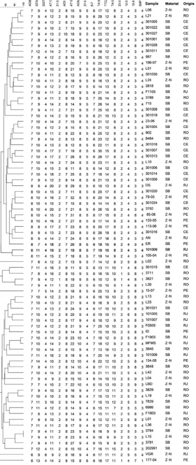
Phylogenetic analysis - For phylogenetic analysis, we selected genotypes obtained from 78 of the 91 sam-ples, excluding 13 that lacked allele definition of one or more VNTR or of the SNP loci. Genotype similarity was determined using the program Bionumerics (ver-sion 6.5, Applied Maths, Belgium) by construction of a similarity matrix using the unweighted pair group method with arithmetic mean (UPGMA) and the Dice similarity coefficient.
RESULTS
Genotyping results using biopsy samples - The 51 bi-opsy samples were subjected to genotyping, using both the MLVA procedure and SNP analysis. All samples yielded an SNP genotype, while 48 of them (94.1%) were characterised by 16 VNTRs, two (3.9%) by 15 VNTRs and a single sample (2%) by 11 VNTRs. The samples genotyped for 15 VNTRs were from CE and RO and the analysis failed to yield the VNTRs 18-8 and AT15, respectively. The sample genotyped for 11 VNTRs was from CE and belonged to a paucibacillary patient; in this instance, the VNTRs 21-3, AC9, AT15, TA10 and 12-5 were not defined (data not shown).
Comparison DNA extraction procedures and geno-typing on Z-N-stained slides - Among the 76 samples processed using the procedure described by van der Zan-den et al. (1998), 23 (30.3%) we could define copy num-bers for 16 (n = 20) or 15 (n = 3) VNTRs and two more samples (2.6%) could be genotyped for eight or nine VNTRs (data not shown). The rest did not generate any VNTR copy numbers (data not shown). Although a lim-ited number of samples generated partial results, we did not observe VNTR-associated genotyping failure. An analysis of the possible relationship between genotyp-ing yield and date of Z-N-stained slide preparations re-vealed that none of the slides from patients diagnosed in 2001, 2005 and 2006 generated PCR products. However, slides that had been prepared from patients diagnosed in 2007, 2008 or 2009 yielded positive PCRs and could be further genotyped for 16 or 15 VNTRs in 10 (33.3%), 11 (44%) and two (50%) of the cases for 2007, 2008 and 2009, respectively, therefore demonstrating a slight in-crease in the genotyping yield efficiency. Among the 25 samples that yielded results for at least eight VNTRs and 24 (96%) yielded SNP-based genotypes. The bacterial index (BI) of samples genotyped by VNTRs and SNPs ranged from 1.75-5.25, but we did not observe a relation-ship between genotyping yield and BI.
Among the 39 Z-N-stained slide samples processed according to Kamble et al. (2010), 11 (28.2%) yielded
genotypes defined by either 16 or 15 VNTRs, while four samples (10.3%) rendered genotypes with 10, 12 or 13 VNTRs. When considering positivity among samples col-lected from patients diagnosed more recently, namely in 2007 and 2008, the genotyping yield was also higher, with genotypes defined by 16 or 15 VNTRs for four (80%) and seven (70%) of the samples, respectively. Among the 15 samples with genotypes defined by at least 10 VNTRs (n = 15), 14 (93.3%) yielded SNP genotypes. The BI of sam-ples genotyped ranged from 0.75-4.5 and again, no rela-tionship between BI and genotyping yield was observed.
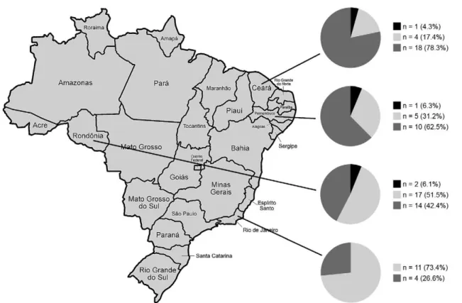
Frequency of genotypes defined by SNP analysis - We obtained conclusive SNP genotypes in 89 (97.8%) of 91 samples, derived from 89 patients from four Brazil-ian states. Two Z-N-stained slides (2.2%), one from RJ and another from PE, generated insufficient PCR prod-ucts. In another two patients with dual samples, the same genotype was obtained, which is suggestive of a single input. Among the 87 patients, SNP genotype 4 was pre-dominant (n = 46, 52.9%), genotype 3 was identified in 37 patients (42.5%) and genotype 1 was detected less frequently in four cases (4.6%). The distribution of each SNP genotype per state is demonstrated in Fig. 1.
Genotypes as defined by analysis of 16 VNTRs and SNPs - All VNTRs used in this analysis were polymor-phic. The smallest discriminatory power was observed for locus 21-3 (0.067), consisting of two alleles (1 and 2 copies), whereas the highest allelic diversity was ob-served at locus AT15 (0.94), consisting of 22 different alleles (between 7-32 copies). The earlier observed cor-relation between the 27-5 and 12-5 allele combination and SNP genotype was also present here: 40 (88.9%) SNP genotype 4 samples possessed the 5:4 copy number allele combination, while 24 cases (72.7%) of SNP geno-type 3 had the 4:5 copy number combination.
The dual samples from the two patients with identi-cal SNP genotypes differed in copy number at three loci each. In both subjects, biopsy samples were collected be-fore Z-N-stained slides. In the first case, where 15 VN-TRs were used for comparison (because the allele defi-nition failed), we observed a decrease of five and four copies for TTC21 and TA18, respectively. In the case of AC9, we observed an increase of one copy; the other 12 loci were identical between the samples. The second case exhibited a decrease of one copy for AT17 and an increase of two and three copies for TTC21 and AT15. The other 13 loci were identical.
three more clusters were formed by isolates derived from the same state: one from RJ (MFMS/F1903) and two from RO (2818/L24; 3788/715). In addition, one cluster was from the Northeast Region (79-05/301024) and two clusters contained isolates from RO and CE (L51/301030; 902/301004). By relating sample type and clusters, we observed that five groups were composed of Z-N-stained slides and skin biopsies, while two were composed of SB samples alone. In addition, all but one of the groups was composed of isolates with the same SNP genotype; the exception included the two samples from RJ that presented SNP genotypes 3 and 4.
Upon construction of a minimum spanning tree (MST) using the same genotypes (Fig. 3), two major branches were observed, showing that isolates from CE had genotypes that were more closely related to those observed in PE, while genotypes from samples from RJ were more similar to those of RO. However, genotypes from the latter state were similarly distributed in both branches of the tree, perhaps due to the larger sample number, which also suggests a higher genetic diversity than that of isolates from other regions. Interestingly, the only cluster was composed of genotypes from Ron-donian isolates and was the central interactive node be-tween both branches of the phylogenetic tree.
An analysis of the relationship between VNTR and SNP-based genotypes revealed a strong correlation be-tween the positioning of the genotypes within the MST (data not shown).
DISCUSSION
The first reliable data on genomic variation between isolates of M. leprae were published just over a decade ago (Matsuoka et al. 2000, Shin et al. 2000) and since then, several polymorphic VNTRs and SNPs have been reported and evaluated in material from leprosy patients residing in different regions around the world (Weng et al. 2007, Young et al. 2008, Cardona-Castro et al. 2009, Mat-suoka et al. 2009, Sakamuri et al. 2009a, b, Srisungnam et al. 2009). Using this genotyping approach in biopsy samples from unrelated leprosy patients from RJ and SP (Fontes et al. 2009), we observed a high genetic variabil-ity of M. leprae; however, because both regions are part of Southeast Brazil, a region that covers only a limited area of the country (10.9% of the Brazilian territory), we investigated genotypes from patients who are residents of the North and Northeast Regions of the country. Addi-tionally, as biopsy sampling is an invasive procedure and because preparing Z-N-stained slides is part of the diag-nostic procedure for leprosy in many health centres of Brazil, we evaluated the usefulness of Z-N-stained slides as a genotyping source of the causative agent.
Two procedures of DNA extraction were used: the first was previously utilised for genotyping of Mycobacterium tuberculosis (Mtb) on Z-N slides prepared from sputum samples of tuberculosis patients (van der Zanden et al. 1998) and the second was reported recently for PCR-me-diated detection of M. leprae DNA on Z-N-stained slides with bacterial counts of 0, but that were PCR-positive in
32.6% (n = 15) of the samples (Kamble et al. 2010). When comparing typing efficiency, both extraction procedures returned similar results, yielding 28-30% complete geno-types. An inverse correlation between the length of time that the slides had been stored and genotype quality was verified, while no direct correlation between the latter and the BI was observed. Complete M. leprae genotypes were sometimes obtained in material with low BI. Our data demonstrate that Z-N-stained slides are an addition-al source for DNA fingerprinting of M. leprae, but more studies are needed to better determine the effect of long-term storage of slides on genotype quality.
We observed 90 unique genotypes from 89 patients, including different genotypes in two samples available from two patients and identical patterns in isolates from two residents of RO. When decreasing stringency for genotype similarity was employed, many of the cases that yielded clusters also had similar origins. Taken together, both results strongly suggest region-associated genotype similarity. Unfortunately, we did not have access to
epi-demiological data of the patients to confirm this associa-tion. The need to better define stringency of definition of genotype similarity was also shown by our observation of the differences in some VNTRs between Z-N-stained slides and skin biopsies from the same patients. Genotype differences in clinical specimens of the same individual have been investigated and some studies demonstrated the existence of an identical pattern in minisatellites and a small variation in microsatellites with the highest allelic diversities, such as AT17, TTC21 (Sakamuri et al. 2009a) and TA10 (Xing et al. 2009). These results suggest that some short tandem repeat loci are most likely prone to stuttering during incubation, treatment and transmission.
Our earlier study also demonstrated that SNP geno-type 3 is the most frequent among M. leprae isolates in Southeast Brazil (Fontes et al. 2009), also occurring pre-dominantly in Europe, North Africa and the Americas (Monot et al. 2005). Here, we confirm the predominance of SNP genotype 3 in RJ and frequently observed this SNP genotype in RO. Conversely, SNP genotype 4 was more predominant in CE and PE, two bordering states in Northeast Brazil. This SNP genotype is characteristic of Africa, the French West Indies, Brazil (Monot et al. 2005), México (Matsuoka et al. 2009) and Japanese Brazilians, who were likely infected in Brazil (Weng et al. 2007). SNP genotype 4 isolates in Latin American countries are theorised to have originated in Africa, in agreement with the notion that leprosy was carried by the slave trade (Monot et al. 2005). The frequencies of SNP genotypes 1, 3 and 4 and the lack of SNP genotype 2 are consistent with the ethnic groups that were present during Brazilian colonisation, as presented by Monot et al. (2005).
In an earlier study, we observed a 4/5 copy number combination of VNTR loci 27-5 and 12-5 and the SNP genotype 3 in 87.5% of isolates and the 5/4 copy number combination in 85.7% of isolates with SNP genotype 4 (Fontes et al. 2009); this result had also been observed in M. leprae samples from Colombia (Cardona-Castro et al. 2009). Associations between different markers, such as SNPs and VNTRs, have also been detected in Mtb (Filliol et al. 2006). Our data demonstrated that this as-sociation was less pronounced for SNP genotype 3 when analysing patients from more regions, now occurring in 72.7% (n = 24) of these genotypes, while the strong asso-ciation of SNP genotype 4 with the 5/4 combined VNTR allele type was maintained (88.9%, n = 40).
In conclusion, our study strongly suggests the exis-tence of geographically related M. leprae genotypes in Brazil and demonstrates that Z-N-stained slides are an al-ternative sample source for the genotyping of M. leprae.
ACKNOWLEDGEMENTS
To the Genomic Platform for DNA Sequencing (PDTIS-Fiocruz).
REFERENCES
Cardona-Castro N, Beltrán-Alzate JC, Romero-Montoya IM, Melén-dez E, Torres F, Sakamuri RM, Li W, Vissa V 2009. Identifica-tion and comparison of Mycobacterium leprae genotypes in two geographical regions of Colombia. Lepr Rev 80: 316-321.
da Silva Rocha A, Cunha dos Santos AA, Pignataro P, Nery JA, de Miranda AB, Soares DF, Brum Fontes AN, Miranda A, Ferreira FIG. 3: minimum spanning tree based on complete variable number
H, Boéchat N, Novisck Gallo ME, Sarno EN, de Oliveira ML, Suffys PN 2011. Genotyping of Mycobacterium leprae from Bra-zilian leprosy patients suggests the occurrence of reinfection or of bacterial population shift during disease relapse. J Med Micro-biol60: 1441-1446.
Filliol I, Motiwala AS, Cavatore M, Qi W, Hazbón MH, Bobadilla del Valle M, Fyfe J, García-García L, Rastogi N, Sola C, Zozio T, Guerrero MI, León CI, Crabtree J, Angiuoli S, Eisenach KD, Durmaz R, Joloba ML, Rendón A, Sifuentes-Osornio J, Ponce de León A, Cave MD, Fleischmann R, Whittam TS, Alland D 2006. Global phylogeny of Mycobacterium tuberculosis based on single nucleotide polymorphism (SNP) analysis: insights into tubercu-losis evolution, phylogenetic accuracy of other DNA fingerprint-ing systems, and recommendations for a minimal standard SNP set. J Bacteriol 188: 759-772.
Fontes AN, Sakamuri RM, Baptista IM, Ura S, Moraes MO, Martínez AN, Sarno EN, Brennan PJ, Vissa VD, Suffys PN 2009. Genetic diversity of Mycobacterium leprae isolates from Brazilian lep-rosy patients. Lepr Rev 80: 302-315.
Groathouse NA, Rivoire B, Kim H, Lee H, Cho SN, Brennan PJ, Vissa VD 2004. Multiple polymorphic loci for molecular typ-ing of strains of Mycobacteriumleprae. J Clin Microbiol 42: 1666-1672.
Kamble RR, Shinde VS, Madhale SP, Kamble AA, Ravikumar BP, Jadhav RS 2010. Extraction and detection of Mycobacterium
leprae DNA from ZNCF-stained skin smear slides for better
identification of negative skin smears. Indian J Med Microbiol 28: 57-59.
Kimura M, Sakamuri RM, Groathouse NA, Rivoire BL, Gingrich D, Krueger-Koplin S, Cho SN, Brennan PJ, Vissa V 2009. Rapid variable-number tandem-repeat genotyping for Mycobacterium
leprae clinical specimens. J Clin Microbiol47: 1757-1766.
Matsuoka M, Gonzalez AV, Estrada I, Carreño-Martinez C, Fafutis-Morris M 2009. Various genotypes of Mycobacterium leprae
from Mexico reveal distinct geographic distribution. Lepr Rev 80: 322-326.
Matsuoka M, Maeda S, Kai M, Nakata N, Chae GT, Gillis TP, Ko-bayashi K, Izumi S, Kahiwabara Y 2000. Mycobacterium leprae
typing by genomic diversity and global distribution of genotypes.
Int J Lepr Other Mycobact Dis 68: 121-128.
Monot M, Honoré N, Garnier T, Araoz R, Coppée JY, Lacroix C, Sow S, Spencer JS, Truman RW, Williams DL, Gelber R, Virmond M, Flageul B, Cho SN, Ji B, Paniz-Mondolfi A, Convit J, Young S, Fine PE, Rasolofo V, Brennan PJ, Cole ST 2005. On the origin of leprosy. Science 308: 1040-1042.
Monot M, Honoré N, Garnier T, Zidane N, Sherafi D, Paniz-Mondolfi A, Matsuoka M, Taylor GM, Donoghue HD, Bouwman A, Mays S, Watson C, Lockwood D, Khamesipour A, Dowlati Y, Jianping S, Rea TH, Vera-Cabrera L, Stefani MM, Banu S, Macdonald M, Sapkota BR, Spencer JS, Thomas J, Harshman K, Singh P, Busso P, Gattiker A, Rougemont J, Brennan PJ, Cole ST 2009. Compara-tive genomic and phylogeographic analysis of Mycobacterium
le-prae. Nat Genet 41: 1282-1289.
MS - Ministério da Saúde 2011. [homepage on the internet]. Han-seníase - Distribuição da hanHan-seníase no Brasil. Tabela de casos novos de hanseníase e detecção geral, registro ativo e coeficiente de prevalência, segundo UF de residência, região – Brasil. [cited 2012 Jan]. Available from: portal.saude.gov.br/portal/arquivos/ pdf/tab_reg_ativo_casosnovos_hans2011.pdf.
Sakamuri RM, Harrison J, Gelber R, Saunderson P, Brennan PJ, Balag-on M, Vissa V 2009a. A cBalag-ontinuatiBalag-on: study and characterisatiBalag-on of
Mycobacterium leprae short tandem repeat genotypes and
trans-mission of leprosy in Cebu, Philippines. Lepr Rev 80: 272-279.
Sakamuri RM, Kimura M, Li W, Kim HC, Lee H, Kiran MD, Black WC 4th, Balagon M, Gelber R, Cho SN, Brennan PJ, Vissa V 2009b. Population-based molecular epidemiology of leprosy in Cebu, Philippines. J Clin Microbiol47: 2844-2854.
Shin YC, Lee H, Lee H, Walsh GP, Kim JD, Cho SN 2000. Variable numbers of TTC repeats in Mycobacterium leprae DNA from leprosy patients and use in strain differentiation. J Clin
Micro-biol 38: 4535-4538.
Srisungnam S, Rudeeaneksin J, Lukebua A, Wattanapokayakit S, Pasadorn S, Mahotarn K, Ajincholapan Sakamuri RM, Kimura M, Brennan PJ, Phetsuksiri B, Vissa V 2009. Molecular epidemi-ology of leprosy based on VNTR typing in Thailand. Lepr Rev 80: 280-289.
Truman R, Fontes AB, de Miranda AB, Suffys P, Gillis T 2004. Ge-notypic variation and stability of four variable-number tandem repeats and their suitability for discriminating strains of
Myco-bacterium leprae. J Clin Microbiol 42: 2558-2565.
van der Zanden AG, Hoentjen AH, Heilmann FG, Weltevreden EF, Schouls LM, van Embden JD 1998. Simultaneous detection and strain differentiation of Mycobacterium tuberculosis complex in paraffin wax embedded tissues and in stained microscopic prepa-rations. Mol Pathol 51: 209-214.
Weng X, Vander Heiden J, Xing Y, Liu J, Vissa V 2011. Transmission of leprosy in Qiubei County, Yunnan, China: insights from an 8-year molecular epidemiology investigation. Infect Genet Evol 11: 363-374.
Weng X, Wang Z, Liu J, Kimura M, Black WC 4th, Brennan PJ, Li H, Vissa VD 2007. Identification and distribution of Mycobacterium
leprae genotypes in a region of high leprosy prevalence in China:
a 3-year molecular epidemiological study J Clin Microbiol 45: 1728-1734.
Xing Y, Liu J, Sakamuri RM, Wang Z, Wen Y, Vissa V, Weng X 2009. VNTR typing studies of Mycobacterium leprae in China: assess-ment of methods and stability of markers during treatassess-ment. Lepr
Rev 80: 261-271.
Young SK, Ponnighaus JM, Jain S, Lucas S, Suneetha S, Lockwood DN, Young DB, Fine PE 2008. Use of short tandem repeat se-quences to study Mycobacterium leprae in leprosy patients in Malawi and India. PLoS Negl Trop Dis 2: e214.