online | memorias.ioc.fiocruz.br
Social and immunological differences among uninfected Brazilians
exposed or unexposed to human immunodeficiency
virus-infected partners
Maria Luiza Silva1/+, Victor Hugo Melo2,Agdemir Waléria Aleixo3, Lúcia Fernandes Aleixo3,
Marcelo Antônio Pascoal-Xavier4, Rafaela Oliveira Silva3, Laís Alves Ferreira3,
Willian Cunha Domingos3, Dirceu Bartolomeu Greco5
1Laboratório Central, Hospital das Clínicas 2Departamento de Ginecologia e Obstetrícia 3Laboratório de Doenças Infecciosas e Parasitárias 4Departamento de Anatomia Patológica e Medicina Legal 5Departamento de Clínica Médica, Faculdade de Medicina,
Universidade Federal de Minas Gerais, Belo Horizonte, MG, Brasil
Understanding the social conditions and immunological characteristics that allow some human immunodefi-ciency virus (HIV)-exposed patients to remain uninfected represents an on-going challenge. In this study, the socio-demographic and sexual behaviour characteristics and immune activation profiles of uninfected individuals exposed to HIV-infected partners were investigated. A confidential and detailed questionnaire was administered and venous blood was tested using HIV-1/enzyme immunoassays, plasma HIV-1 RNA levels/bDNA and immunophenotyping/flow cytometry to determine the frequencies of CD4 and CD8 T cells expressing activation markers. The data analysis showed significant differences (p < 0.05) for immune parameters in individuals who were uninfected, albeit exposed to HIV-infected partners, compared with unexposed individuals. In particular, the exposed, uninfected individuals had a higher frequency (median, minimum-maximum) of CD4+HLA-DR+ (4.2, 1.8-6.1), CD8+HLA-DR+ (4.6, 0.9-13.7), CD4+CD45RO+ (27.5, 14.2-46.6), CD4+CD45RO+CD62L+ (46.7, 33.9-67.1), CD8+CD45RA+HLA-DR+ (12.1, 3.4-35.8) and CD8+CD45RO+HLA-DR+ (9.0, 3.2-14.8) cells, a decreased percentage of CD8+CD28+ cells (11.7, 4.5-24.0) and a
lower cell-surface expression of Fcγ-R/CD16 on monocytes (56.5, 22.0-130.0). The plasma HIV-1 RNA levels demon -strated detectable RNA virus loads in 57% of the HIV-1+ female partners. These findings demonstrate an activation profile in both CD4 and CD8 peripheral T cells from HIV-1 exposed seronegative individuals of serodiscordant couples from a referral centre in Belo Horizonte, state of Minas Gerais.
Key words: HIV-1 discordant couples - flow cytometry - immune activation - immunophenotyping
Heterosexual intercourse is currently the main route of human immunodeficiency virus type 1 (HIV-1) ac-quisition worldwide and the rate of transmission in this manner is increasing throughout Africa, Asia, Latin America and many industrialised countries (Piot et al. 2001, Yang et al. 2005).
The risk of transmission has been associated with a wide variety of behaviours, such as the frequency and type of sexual contact (Quinn et al. 2000, Kaur & Meh-ra 2009) and the use or non-use of condoms, as well as types of interindividual variability, including genetic predisposition, intrinsic cellular defence mechanisms and mucosal and systemic HIV-1-specific cellular and humoral responses (Soriano et al. 2002, Kaslow et al. 2005). These variations in immunity can also impact natural killer (NK) cell activity, HIV-specific immuno-globulin A levels and HIV-specific T cells (Mazzoli et al. 1997, Kaul et al. 2001a, Lo Caputo et al. 2003,
Make-doi: 10.1590/0074-0276140140 Financial support: FAPEMIG
+ Corresponding author: mariasil@hc.ufmg.br Received 24 April 2014
Accepted 7 July 2014
donas et al. 2005). In addition, virus characteristics and levels of plasma viral load (PVL) have been implicated in resistance to infection (Operskalski et al. 1997).
It has been reported that some individuals remain HIV seronegative despite being exposed to HIV, in some cases repeatedly and over long periods of time. These individuals are called HIV-exposed seronegatives (HESN). Although host factors are involved in the pro-duction of this resistance to HIV acquisition, there is no clear explanation for such a low susceptibility to infec-tion (Rowland-Jones & McMichael 1995, Restrepo et al. 2011). Immune activation has been suggested to be criti-cal in the susceptibility to HIV-1 transmission. In vitro, HIV-1 requires activated T cells for a productive infec-tion (Kaur & Mehra 2009) and subjects with an activated immune system show increased in vitro susceptibility to HIV and higher in vivo replication (Lawn et al. 2001).
HIV-1 replicates preferentially in CD45RO+ memory
T lymphocytes rather than immature and immunologi-cally quiescent naïve CD45RA+ lymphocytes (Spina et al.
1997, Woods et al. 1997). Additionally, the upregulation of adhesion molecules during inflammatory processes may further promote virus-induced cell-cell fusion, thereby facilitating the direct spread of the virus between cells (Hildreth & Orentas 1989, Lawn et al. 2001).
the number of discordant couples (in which the male is HIV positive and the female is negative or vice versa) who are interested in pregnancy is increasing (Vernazza et al. 2000).
A better understanding of the genetic and immuno-logic bases of natural resistance to HIV-1 infection could be important for the development of preventive and therapeutic modalities. In this study, we investigated the socio-demographics, sexual behaviour and immune acti-vation and memory status of peripheral blood leukocytes from uninfected individuals exposed to HIV-1-infected partners and compared these results with the results of unexposed individuals using flow cytometry. The aim was to characterise immunophenotyping features that might facilitate an understanding of the immunological features of Brazilian HIV-1 serodiscordant couples.
SUBJECTS, MATERIALS AND METHODS
Study groups - Fourteen individuals who were unin-fected yet exposed to HIV-inunin-fected partners (HESN) as part of a group of HIV-1 discordant heterosexual couples who visited the Clinics Hospital of Federal University of Minas Gerais (UFMG) were invited to participate in the study. The inclusion criteria were an HIV-seronega-tive male partner and an HIV-1 infected female partner, a stable relationship and a history of penetrative sexual intercourse without condom use within the six months prior to the study. Ten volunteer, HIV-1-negative males (10 low-risk laboratory personnel unexposed to HIV-infected partners) who reported no sexual intercourse with an HIV-1-positive partner were recruited to form the negative, HVI-1-unexposed seronegative group (HUSN). All of the subjects were interviewed to ascertain their socio-demo-graphic characteristics {i.e., educational level, family in-come, religion and sexual behaviour [age at 1st intercourse, frequency of intercourse, practice of oral and/or anal sex during the previous 6 months, reported sexually transmit-ted diseases (STDs) and duration of relationship]}.
Blood samples - Peripheral blood samples (5 and 7 mL) were collected by a trained professional at the Central Laboratory of the Clinics Hospital and placed into VacutainerTM tubes containing ethylenediamine
tetraacetic acid (EDTA) as an anticoagulant, a clot acti-vator and gel for serum separation (both purchased from Becton Dickinson, USA). Serologic tests for HIV-1 were used to detect anti-HIV antibodies using microparticle enzyme immunoassays (MEIA) for HIV-1 and HIV-2 (Abbott Laboratories, USA) and were performed at the Central Laboratory. Plasma HIV-1 RNA levels (VLs) were quantified using VERSANT HIV-1 RNA 3.0 As-say (bDNA) (Siemens Healthcare Diagnostics). A flow cytometric analysis of unstimulated whole blood was performed as recommended by Becton Dickinson with the following modifications. First, 50-µL aliquots of EDTA-anticoagulated blood were dispensed into 5-mL polystyrene tubes (Falcon®, BD Pharmingen, USA). The
samples were then individually stained for specific cell-surface markers using three or four-colour immunocyto-metric assays with the following anti-human cell surface marker monoclonal antibodies conjugated to fluorescein isothiocyanate (FITC), phycoerythrin (PE),
peridinin-chlorophyll protein (PerCP), R-PE or cyanine dye Cy5 (PE Cy5.0) and allophycocyanin (APC): anti-CD4 FITC (RPA-T4), anti-CD8 PerCP (RPA-T8), anti-CD3 FITC (UCHT1), anti-CD3 PerCP (SP34-2), anti-CD14 FITC (M5E2), anti-CD16 PE Cy5.0 (3G8), anti-CD18 PE (6.7), CD28 PE (CD28.2), CD38 PE (HIT2), CD45RA PE (HI100), CD45RO PE (UCHL1), CD56 PE (B159), CD62L APC (DREG-56), anti-HLA-DR PE (G46-6), anti-anti-HLA-DR APC (MøP9) and anti-CCR5 APC (2D7/CCR5). These antibodies were purchased from BD Pharmingen.
The samples were gently treated with vortex homog-enisation and incubated in the dark for 30 min at room temperature (RT). Following incubation, the erythrocytes were lysed using 0.45 mL of fluorescence-activated cell sorter (FACS) lysing solution (Becton Dickinson) and re-incubated for an additional 15 min at RT in the dark. After incubation, the cells were washed twice with 2 mL of phosphate-buffered saline (PBS) containing 0.01% so-dium azide (NaN3). After erythrocyte lysis was complete, the samples were centrifuged at 600 g for 7 min at RT. The supernatants were discarded and the cell pellets were washed twice with 2 mL of PBS containing 0.01% NaN3 and stored at 4ºC in the dark prior to flow cytometry anal-ysis. FACS data were acquired within 4 h after staining.
Flow cytometry acquisition and analysis - In total, 3,000 events/tube were acquired within the lymphocyte gate using a FACSCalibur® flow cytometer (Becton
Dickinson) that was properly set up to measure forward (FSC) and side (SSC) light scatters, FITC (FL-1), PE (FL-2), PerCP or PE Cy5.0 (FL-3) and APC (FL4). The CELLQuestTM software (Franklin Lakes, USA), which
was provided by the manufacturer, was used for the data acquisition and analysis and the data were prepared us-ing the FlowJo software (Tree Star, USA). The selective analysis of lymphocytes was carried out by first placing an electronic gate on the FSC vs. SSC dot plot to select small blood lymphocytes based on their morphometric features. The gated lymphocyte population was further characterised based on its relative fluorescent proper-ties to quantify the percentage of fluorescent-positive subpopulations. The selective analysis of NK and NKT cells was initially performed using the lymphocyte scat-ter gate, created according to the FSC vs. SSC dot plots, followed by immunophenotyping using anti-CD3 FITC, anti-CD16 PE Cy5.0 and anti-CD56 PE to determine the subpopulation of interest. The analysis of monocytes was performed by immunophenotyping using SSC vs. FL-1/anti-CD14-FITC dot plots to select SSCLowCD14High
monocytes, followed by immunophenotyping using anti-CD16 PE Cy5 and HLA-DR PE. The results were expressed as the percentage of positive cells within the selected gates. Cell-surface markers presenting a bimo-dal distribution and/or single-colour histograms were used to evaluate the density or expression of cell-surface markers presenting a unimodal distribution, which was expressed as the mean fluorescence intensity, within the gated lymphocytes or monocytes.
HUSN groups using GraphPad Prism software, v.5.0
(USA). Significant differences were considered at p ≤ 0.05.
Ethics - All of the subjects signed an informed con-sent form that was approved by the Ethical Committee of UFMG (UFMG-ETIC 261/2009), Belo Horizonte, state of Minas Gerais, Brazil.
RESULTS
The socio-demographic and sexual behaviour char-acteristics of the individuals enrolled in this study are shown in Table I. The age range of the HESN and HUSN individuals varied from 28-56 and 20-38 years, respec-tively. The median age for first reported sexual inter-course was 10-15 years among the HESN group and 14-18 years among the HUSN group. Among the HESN individuals, the median duration of cohabitation was 8.5 years (range from 5-10 years) and the median number of sexual acts per week was three times; oral and anal sex in the past six months were reported by 57.1% and 28.6% of the subjects, respectively. Among the HESN subjects, 14.3% reported a previous history of any STD and a minority (42.9%) reported regular condom use. For the HUSN group, the median duration of cohabita-tion was seven years and the median number of sexual acts per week was three. Oral sex in the past six months was reported by 40% individuals and all of the subjects denied practicing anal sex. Of the HUSN subjects, 50% reported a previous history of an STD and a majority (55.6%) reported the regular use of condoms. The ma-jority of HESN subjects reported practicing a religion (92.9%). In contrast, half of the HUSN group said they did not practice any religion. With regard to education, at least 64% of the individuals from the HESN group reported seven years of school; the majority (100%) of the individuals in the HUSN group reported completing high school or beyond. Concerning income, 57.1% of the HESN group declared a family monthly income of less
than US$ 800, whereas the majority (60%) of the HUSN group reported a family income above US$ 800.
Regarding laboratory characteristics, all of the HESN and HUNS individuals were HIV-1 seronegative (as test-ed by MEIA). The plasma HIV-1 RNA levels (VL) dem-onstrated detectable RNA virus loads (detection thresh-old 50 copies/mL) in eight out of 14 (57%) of the HIV-1+
female partners. The median of RNA virus loads in cop-ies/mL and minimum-maximum (min-max) was 4,691 and 228-26,302 and in log10 and min-max was 3,269 and 2,358-4,420 (Table II). CD4 counts were determined for all of the subjects and the median CD4 counts observed in the HESN and HUSN groups were 931 (529-2,000) and 760 (573-1,326) cells/µL, respectively (Table II). No differences were observed when the HESN and HUNS data were compared.
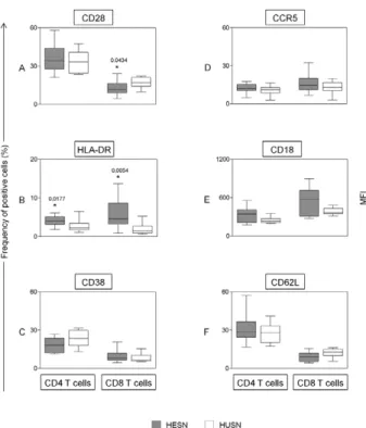
An ex vivo analysis of leukocyte subsets (frequency, activation status and C-C chemokine receptor type 5 ex-pression) present in the peripheral blood was performed using four-colour flow cytometry assays (Figs 1-3). The results of the HESN group were compared to the results of the HUSN group.
No statistically significant difference was observed when comparing the CCR5+CD4+ and CCR5+CD8+
re-sults between the HESN and HUSN individuals (Fig. 1D). The frequency of CCR5 expression in peripheral T cells from the HESN group was 12 (4.9-17.6) for CD4+
cells and 14.7 (6.7-32.5) for CD8+ cells. In the HUSN
group, the frequencies of CD4+CCR5+ and CD8+CCR5+
T cells were 10.9 (2.6-16.3) and 12.9 (2.8-19.9), respec-tively. When the activation status of peripheral T cells was examined, the activation profile of T cells among the HESN group compared with the HUSN group was char-acterised by a lower frequency of CD8+CD28+ cells and
a higher frequency of CD4+HLA-DR+ and CD8+
HLA-DR+ T cells (Fig. 1A, B). An analysis of adhesion
mol-ecule expression among circulating T cells showed no
TABLE I
Socio-demographic and sexual behaviour characteristics of 14 human immunodeficiency virus type 1 (HIV-1) exposed seronegative (HESN) and 10 unexposed seronegative (HUSN) heterosexual men
Characteristics HESN HUSN
Age (years) (mean) 39.1 26.6
Age at first sex (years)(mean) 15.5 18
Average of sexual acts/week 3.0 3.0
Reported oral sex in the past six months (%) 57.1 40
Reported anal intercourse in the past six months (%) 28.6 0
Self reported sexually transmitted disease (%) 14.3 50
Self reported regular condom use (%) 42.9 55.6
Duration of union (years)(mean) 8.5 7
Number of children(mean) 1 < 1
Religiousness (%) 92.8 50
Catholic (%) 35.7 30
Protestant (%) 57.1 10
Education (years)(mean) 6.4 12.6
significant differences in CD18 expression on CD4 and CD8 T cells (Fig. 1E) or in the frequency of CD4+ and
CD8+CD62L+ cells (Fig. 1F).
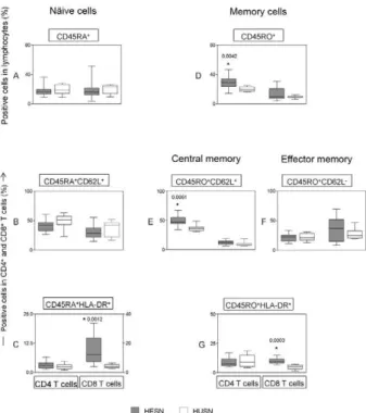
To evaluate the patterns and activation status of naïve and memory T cells in peripheral blood, the fre-quency of CD45RA, CD45RO, CD62L and HLA-DR expression was investigated using multicolour flow cy-tometric assays (Fig. 2). The analysis of the data dem-onstrated an increased frequency of naïve, activated CD8 T cells (CD8+CD45RA+HLA-DR+) in the HESN
group (Fig. 2C), though no difference was found in the frequency of CD62L+CD45RA+ cells among CD4
and CD8 T cells (Fig. 2B). In addition, higher levels of memory cells were demonstrated in the HESN indi-viduals, which were characterised by increased levels of CD4+CD45RO+ cells (Fig. 2D), central memory cells
(CD4+CD45RO+CD62L+) (Fig. 2E) and
memory-activat-ed cells (CD4+ and CD8+CD45RO+HLA-DR+) (Fig. 2G).
No significant difference was observed between the groups in terms of the percentage of effector memory T cells (CD4 and CD8+CD45RO+CD62L-) (Fig. 2F).
The monocyte expression of HLA-DR, CD16 and CCR5 and the frequency of circulating NK (CD3-CD16
+/-CD56+/-) and NKT (CD3+CD16+/-CD56+/) cells was also
analysed (Fig. 3). However, no significant differences were observed in the expression of HLA-DR or CCR5 by monocytes (Fig. 3A, B) or in the frequency of circu-lating NK and NKT cells between the groups (Fig. 3C).
In contrast, decreased cell-surface expression of Fcγ-R
(CD16) was observed on the monocytes obtained from the HESN group (Fig. 3A).
DISCUSSION
Few studies characterising HIV-1-discordant couples have been conducted in our country and only a small
TABLE II
Laboratory characterisation of 14 exposed seronegative heterosexual men and their partners
Exposed seronegative men Human immunodeficiency virus type 1 (HIV-1)-infected women
ID
CD4 (cells/µL)
CD8
(cells/µL) ID
HIV-1 RNA (copies/mL)
CD4 (cells/µL)
CD8 (cells/µL)
M-001 1,160 209 F-001 746 389 468
M-002 600 378 F-002 5,975 550 1,162
M-003 931 537 F-003 < 50 259 433
M-004 685 465 F-004 883 299 1,227
M-005 769 356 F-005 10,111 449 2,617
M-006 969 916 F-006 < 50 877 1,182
M-007 804 2,117 F-007 26,302 374 983
M-008 1,147 626 F-008 12,456 338 309
M-010 1,634 778 F-010 < 50 651 756
M-011 954 813 F-011 3,407 439 854
M-012 2,000 553 F-012 228 1,317 705
M-013 742 672 F-013 < 50 977 590
M-014 529 487 F-014 < 50 499 832
M-015 1,096 555 F-015 < 50 1,228 1,287
data are expressed as cells/µL for CD4 and CD8 T cells and copies/mL for HIV-1 RNA levels. F: females; M: males.
number of them have been performed as an immuno-logical investigation. Aiming to characterise the socio-demographic determinants, sexual behaviours and im-munophenotypic patterns of uninfected individuals exposed to HIV-1-infected partners, this study enrolled 14 heterosexual men from HIV-1-serodiscordant couples who were assisted in a large referral centre in Belo Hori-zonte, as well as 10 individuals not exposed to HIV-1-infected partners.
The demographic and behavioural characteristics observed in this study reflect the current state of the HIV/acquired immune deficiency syndrome epidemic in Brazil, especially with regard to pauperisation (Brito et al. 2001). Individuals who were uninfected, but exposed to HIV-infected partners from HIV-1-discordant hetero-sexual couples demonstrated greater indicators of pov-erty, such as a poor education and insufficient income and, consequently, reported a low adherence to condom use and a large number of children. These characteristics are similar to those reported in other studies performed in Africa and China in HIV-1-serodiscordant couples (Isiugo-Abanihe 2006, Matovu 2010, Duan et al. 2012).
Another intriguing result was the great religious bond of exposed, uninfected partners, mainly those of Protestant religions. Hypothetically, this link to religious practices could influence sexual behaviour and promote safe sex (Muñoz-Laboy et al. 2011); however, our results showed the opposite trend for uninfected partners, as sexual prac-tices with a high risk for HIV transmission remained de-spite the declaration of religious practice. Unfortunately, we did not ask how long the individuals were practicing their religious precepts or whether they ascribed to them after learning the HIV status of their partner.
The transmission and acquisition of HIV-1 have been associated with VL as well as viral and host biological characteristics (Operskalski et al. 1997, Quinn et al. 2000, Kaslow et al. 2005). Gray et al. (2001), in an observation-al study of monogamous, HIV-1-serodiscordant couples, identified plasma HIV-1 RNA as the strongest predictor of HIV-1 transmission, with transmission probabilities that increased with genital ulceration (Gray et al. 2001). Our data demonstrated a detectable RNA virus load in 57% of the infected female partners; however, the VL was low (< 10,000 copies/mL) in these women. Further-more, 87.7% of the HESN group reported no history of any STD. These data together might have contributed to the HIV-1 seronegative status in this study. Although VL
Fig. 3: analysis of monocytes, natural killer (NK) and NKT lym-phocyte subsets in the whole blood of the human immunodeficiency virus type 1 (HIV-1)-exposed seronegatives (HESN) (n = 14) or HVI-1-unexposed seronegative group (HUSN) (n = 10) groups. A triple staining immunophenotyping platform was used to identify the lym-phocyte subsets as CD3-CD16+CD56+ NK-cells or CD3+CD16+CD56+
NKT-cells. The results are expressed in a box plot format. The box stretches from the lower hinge (defined as the 25th percentile) to the upper hinge (the 75th percentile) and therefore, contains the middle half of the scores in the distribution. The median is shown as a line across the box. Therefore, ¼ of the distribution is between this line and the bottom or the top of the box. Significant differences (p < 0.05) are identified with an asterisk.
Fig. 2: status of naïve and memory T cells in the unstimulated peripheral blood of the human immunodeficiency virus type 1 (HIV-1)-exposed seronegatives (HESN) (n = 14) or HVI-1-unexposed seronegative group (HUSN) (n = 10) groups. The results are expressed in a box plot format for the frequency of naïve T cells (CD45RA+ or CD45RA+CD62L+)
(left panels), memory T cells (CD45RO+) (right panels), which are
classified as central memory T cells (CD45RO+CD62L+) and effector
memory T cells (CD45RO+, CD62L-) (left panels) and naïve or
mem-ory-activated (CD45RA+/CD45RO+ HLA-DR+) T cells (left and right
has been associated with an increased rate of seroconver-sion, the same trend was not demonstrated for CD4 cell levels (Pedraza et al. 1999, Fideli et al. 2001, Melo et al. 2008). In our study, there were no significant differences in CD4 counts; however, immune changes characterised by an activation pattern of CD4 and CD8 T cells in the peripheral blood of the HESN individuals were observed. CD8-activated lymphocytes in the peripheral circula-tion play an important role in the host antiviral response (Lawn et al. 2001). It was also reported that cytotoxic T lymphocytes play a role in resistance to HIV infection, though this resistance is believed to be dependent on the persistent exposure to HIV (Kaul et al. 2001b).
Two hypotheses have been suggested to explain the immunophenotypic changes observed in the HESN group. The immunophenotypic changes observed in highly HIV-exposed, uninfected individuals could be a consequence of immunogenic challenge during viral exposure. Mucosal and systemic immune activation was reported in seronegative women who had multiple episodes of unprotected heterosexual intercourse with their HIV-1-infected heterosexual partners, as demon-strated by lower levels of naïve CD4+ T cells and a higher
level of CD4+ memory, CD4+CD28-, CD8+CD28+ and
CD8+CD38+ T cells than in healthy control subjects
(Bia-sin et al. 2000). Suy et al. (2007) observed changes in the memory and activated T cells of highly HIV-exposed, uninfected partners, including lower levels of naïve and CD28+ T cells and higher levels of HLA-DR+ T cells,
CD4+ T cells expressing CCR5 and memory CD4+ T
cells, compared with control subjects. Because immune activation has been suggested to be critical for HIV-1 transmission susceptibility (Pedraza et al. 1999, Camara et al. 2010), it is reasonable to hypothesise that the in-dividuals with lower levels of immune activation would have a decreased susceptibility to HIV infection. Within this context, some investigators have found low levels of CD4+ T cell immune activation and low T cell
respon-siveness in studies involving HESN individuals. In a co-hort of highly exposed, but HIV-seronegative men who had sex with men in Amsterdam, higher naïve (CD45RO
-CD27+) CD4 and CD8 T cell numbers and lower
percent-ages of activated (HLADR+CD38+, CD70+) CD4 and
pro-liferating (Ki67) CD4 and CD8 T cells were observed (Koning et al. 2005). Bégaud et al. (2006) also reported a lower proportion of activation marker-expressing CD4 T lymphocytes (HLA-DR+CD4 and CCR5+CD4 T cells) in
highly exposed, uninfected partners of HIV-1-infected individuals compared to low-risk controls.
Our data revealed an increased activation pattern in naïve and memory CD4+ and CD8+ T cells, as well as an
increased frequency of memory cells, in HESN individu-als compared with HUSN individuindividu-als. We individu-also found a low VL in 57% of the female partners of the HESN group, corroborating the first hypothesis. However, our data did not demonstrate a significant difference in CCR5 expres-sion on CD4+ T cells, as previously described (Biasin et
al. 2000, Koning et al. 2005, Bégaud et al. 2006). De-creased monocyte CD16 expression in the HESN group
was observed in our work; FcγRIII (CD16) is normally
associated with the activation or maturation of monocytes and circulating CD14+CD16+ monocytes are an important
cellular target for HIV-1 entry (Han et al. 2009).
Susceptibility to HIV infection varies among indi-viduals; however, the mechanisms determining HIV transmission are not well understood. Thus, the multiple virological, immunological and host factors that are like-ly involved in protecting against HIV-1 infection should be more extensively investigated.
Our findings demonstrate immunological features (i.e., a higher frequency of activation markers on CD4 and CD8 naïve and memory T cells) in this study of uninfect-ed Brazilians exposuninfect-ed to HIV-infectuninfect-ed partners. The data presented in this paper represent the preliminary results of a pilot study involving a small number of individuals and further investigations are required to obtain a better understanding of these immunological differences.
REFERENCES
Bégaud E, Chartier L, Marechal V, Ipero J, Léal J, Versmisse P, Bret-on G, FBret-ontanet A, Capoulade-Metay C, Fleury H, Barré-Sinoussi F, Scott-Algara D, Pancino G 2006. Reduced CD4 T cell acti-vation and in vitro susceptibility to HIV-1 infection in exposed uninfected central Africans. Retrovirology3: 35.
Biasin M, Caputo SL, Speciale L, Colombo F, Racioppi L, Zagliani A, Blé C, Vichi F, Cianferoni L, Masci AM, Villa ML, Ferrante P, Mazzotta F, Clerici M 2000. Mucosal and systemic immune activation is present in human immunodeficiency virus-exposed seronegative women. J Infect Dis182: 1365-1374.
Brito AM, Castilho EA, Szwarcwald CL 2001. AIDS and HIV infec-tion in Brazil: a multifaceted epidemic. Rev Soc Bras Med Trop 34: 207-217.
Camara M, Dieye TN, Seydi M, Diallo AA, Fall M, Diaw PA, Sow PS, Mboup S, Kestens L, Jennes W 2010. Low-level CD4+ T cell
acti-vation in HIV-exposed seronegative subjects: influence of gender and condom use. J Infect Dis201: 835-842.
Duan S, Ding Y, Yang Y, Lu L, Sun J, Wang N, Wang L, Xiang L, Jia M, Wu Z, He N 2012. Prevalence and correlates of HIV discor-dance and concordiscor-dance among Chinese-Burmese mixed couples in the Dehong prefecture of Yunnan province, China. Sex Health 9: 481-487.
Fideli US, Allen SA, Musonda R, Trask S, Hahn BH, Weiss H, Mu-lenga J, Kasolo F, Vermund SH, Aldrovandi GM 2001. Virologic and immunologic determinants of heterosexual transmission of human immunodeficiency virus type 1 in Africa. AIDS Res Hum Retroviruses17: 901-910.
Gray RH, Wawer MJ, Brookmeyer R, Sewankambo NK, Serwadda D, Wabwire-Mangen F, Lutalo T, Li X, van Cott T, Quinn TC, Rakai Project Team 2001. Probability of HIV-1 transmission per coital act in monogamous, heterosexual, HIV-1-discordant couples in Rakai, Uganda. Lancet357: 1149-1153.
Han J, Wang B, Han N, Zhao Y, Song C, Feng X, Mao Y, Zhang F, Zhao H, Zeng H 2009. CD14highCD16+ rather than CD14lowCD16+
monocytes correlate with disease progression in chronic HIV-infected patients. J Acquir Immune Defic Syndr52: 553-559.
Hildreth JE, Orentas RJ 1989. Involvement of a leukocyte adhesion receptor (LFA-1) in HIV-induced syncytium formation. Science 244: 1075-1078.
Isiugo-Abanihe UC 2006. Sociocultural aspects of HIV/AIDS infec-tion in Nigeria. Afr J Med Med Sci35 (Suppl.): S45-S55.
Kaslow RA, Dorak T, Tang JJ 2005. Influence of host genetic varia-tion on susceptibility to HIV type 1 infecvaria-tion. J Infect Dis191
(Suppl. 1): S68-S77.
Kaul R, Rowland-Jones SL, Kimani J, Dong T, Yang HB, Kiama P, Rostron T, Njagi E, Bwayo JJ, MacDonald KS, McMichael AJ, Plummer FA 2001b. Late seroconversion in HIV-resistant Nairo-bi prostitutes despite pre-existing HIV-specific CD8+ responses.
J Clin Invest107: 341-349.
Kaur G, Mehra N 2009. Genetic determinants of HIV-1 infection and progression to AIDS: susceptibility to HIV infection. Tissue An-tigens73: 289-301.
Koning FA, Otto SA, Hazenberg MD, Dekker L, Prins M, Miedema F, Schuitemaker H 2005. Low-level CD4+ T cell activation is
as-sociated with low susceptibility to HIV-1 infection. J Immunol 175: 6117-6122.
Lawn SD, Butera ST, Folks TM 2001. Contribution of immune activa-tion to the pathogenesis and transmission of human immunodefi-ciency virus type 1 infection. Clin Microbiol Rev14: 753-777.
Lo Caputo S, Trabattoni D, Vichi F, Piconi S, Lopalco L, Villa ML, Mazzotta F, Clerici M 2003. Mucosal and systemic HIV-1-spe-cific immunity in HIV-1-exposed, but uninfected heterosexual men. AIDS17: 531-539.
Makedonas G, Bruneau J, Alary M, Tsoukas CM, Lowndes CM, La-mothe F, Bernard NF 2005. Comparison of HIV-specific CD8 T-cell responses among uninfected individuals exposed to HIV parenterally and mucosally. AIDS19: 251-259.
Matovu JK 2010. Preventing HIV transmission in married and co-habiting HIV-discordant couples in sub-Saharan Africa through combination prevention. Curr HIV Res8: 430-440.
Mazzoli S, Trabattoni D, Lo Caputo S, Piconi S, Blé C, Meacci F, Ruzzante S, Salvi A, Semplici F, Longhi R, Fusi ML, Tofani N, Biasin M, Villa ML, Mazzotta F, Clerici M 1997. HIV-specific mucosal and cellular immunity in HIV-seronegative partners of HIV-seropositive individuals. Nat Med3: 1250-1257.
Melo MG, Santos BR, Lira RC, Varella IS, Turella ML, Rocha TM, Nielsen-Saines K 2008. Sexual transmission of HIV-1 among serodiscordant couples in Porto Alegre, southern Brazil. Sex Transm Dis35: 912-915.
Muñoz-Laboy MA, Murray L, Wittlin N, Garcia J, Terto Jr V, Parker RG 2011. Beyond faith-based organizations: using comparative institutional ethnography to understand religious responses to HIV and AIDS in Brazil. Am J Public Health101: 972-978.
Operskalski EA, Stram DO, Busch MP, Huang W, Harris M, Dietrich SL, Schiff ER, Donegan E, Mosley JW 1997. Role of viral load in heterosexual transmission of human immunodeficiency virus type 1 by blood transfusion recipients. Transfusion Safety Study Group. Am J Epidemiol146: 655-661.
Pedraza MA, del Romero J, Roldán F, García S, Ayerbe MC, Noriega AR, Alcamí J 1999. Heterosexual transmission of HIV-1 is associ-ated with high plasma viral load levels and a positive viral isolation in the infected partner. J Acquir Immune Defic Syndr21: 120-125.
Piot P, Bartos M, Ghys PD, Walker N, Schwartländer B 2001. The global impact of HIV/AIDS. Nature410: 968-973.
Quinn TC, Wawer MJ, Sewankambo N, Serwadda D, Li C, Wabwire-Mangen F, Meehan MO, Lutalo T, Gray RH 2000. Viral load and heterosexual transmission of human immunodeficiency virus type 1. Rakai Project Study Group. N Engl J Med342: 921-929.
Restrepo C, Rallón NI, Carrillo J, Soriano V, Blanco J, Benito JM 2011. Host factors involved in low susceptibility to HIV infec-tion. AIDS Rev13: 30-40.
Rowland-Jones SL, McMichael A 1995. Immune responses in HIV-exposed seronegatives: have they repelled the virus? Curr Opin Immunol 7: 448-455.
Soriano A, Martínez C, García F, Plana M, Palou E, Lejeune M, Aróstegui JI, De Lazzari E, Rodriguez C, Barrasa A, Lorenzo JI, Alcamí J, del Romero J, Miró JM, Gatell JM, Gallart T 2002.
Plasma stromal cell-derived factor (SDF)-1 levels, SDF1-3′A gen
-otype and expression of CXCR4 on T lymphocytes: their impact on resistance to human immunodeficiency virus type 1 infection and its progression. J Infect Dis186: 922-931.
Spina CA, Prince HE, Richman DD 1997. Preferential replication of HIV-1 in the CD45RO memory cell subset of primary CD4 lym-phocytes in vitro. J Clin Invest99: 1774-1785.
Suy A, Castro P, Nomdedeu M, García F, López A, Fumero E, Gallart T, Lopalco L, Coll O, Gatell JM, Plana M 2007. Immunological profile of heterosexual highly HIV-exposed uninfected individu-als: predominant role of CD4 and CD8 T-cell activation. J Infect Dis196: 1191-1201.
Vernazza PL, Troiani L, Flepp MJ, Cone RW, Schock J, Roth F, Bog-gian K, Cohen MS, Fiscus SA, Eron JJ 2000. Potent antiretroviral treatment of HIV-infection results in suppression of the seminal shedding of HIV. The Swiss HIV Cohort Study. AIDS 14: 117-121.
Woods TC, Roberts BD, Butera ST, Folks TM 1997. Loss of inducible virus in CD45RA naïve cells after human immunodeficiency vi-rus-1 entry accounts for preferential viral replication in CD45RO memory cells. Blood89: 1635-1641.