Luísa Côrte
Dissertation presented to obtain the Ph.D degree in Biology
Instituto de Tecnologia Química e Biológica | Universidade Nova de Lisboa
Oeiras,
Insert here an image
with rounded corners
Luísa Côrte
Dissertation presented to obtain the Ph.D degree in Biology
Instituto de Tecnologia Química e Biológica | Universidade Nova de Lisboa
Oeiras, May, 2013
Dissecting the function of the SpoIIIJ and
YqjG membrane protein insertases during
Financial support from Fundação para a Ciência e a Tecnologia through grant SFRH/BD/6489/2001 awarded to Luísa Côrte
The cover contains a treated fluorescence microscopy image of Bacillus subtilis
Supervisor: Prof. Adriano O. Henriques
Exam iners: Prof. Mário A. Santos, Dr. Paulo Tavares, Prof. Isabel de Sá-Nogueira,
ACKNOWLEDGEMENTS
To Instituto de Tecnologia Química e Biológica (ITQB) of the Universidade Nova de Lisboa for receiving me as a PhD student and for providing all the work conditions as well as the scientific environment for the execution of this work.
To Fundação para a Ciência e a Tecnologia (FCT) for the financial support with a PhD fellowship.
To Prof. Adriano O. Henriques, my supervisor, without whom this work would have never been possible. For the opportunity to work in his lab, for his guidance, knowledge and enthusiasm for science.
To Mónica Serrano for her unconditional support, for all the helpful discussions and suggestions, and for her interest, enthusiasm and fantastic humour.
To Prof. Isabel de Sá-Nogueira, with whom we shared the lab, for all the helpful discussions during lab meetings and for her interest in this work. To Anabela Isidro, Cláudia Serra and Irina Franco, for their fantastic disposition, for both our scientific and less scientific discussions, and for their unconditional friendship.
To José Andrade for his unconditional and indefatigable support, for the lively scientific discussions, for his humour, and for his friendship and support, especially during my less healthy times.
To Prof. Rita Zilhão, Prof. Mário Santos, Dr. Tanneke den Blaauwen, Thessa Vinkenvleugel and Marco Roos for making the initiation of my scientific path so appealing.
Joana Lima, Ana Antunes, Rodrigo Saraiva, and Zélia Gouveia, for their friendship, for creating a fantastic work atmosphere and for participating in helpful scientific discussions; to Magda Atilano, Pedro Matos, Helena Veiga, Ana Jorge, Trish Reed, James Yates, Cláudio Alves, Madalena Carido, Luís Ferreira, Assunta Pelosi, Rachele Isticato, Paulo Durão, André Fernandes, Vânia Brissos, Luciana Pereira, Ana Matos, Sandra Viegas, and Vânia Pobre, for creating a great environment both during working hours and out of them.
To Susana Lopes for all the library matters and great mood. To Dr. Cláudio Gomes for his help in this work.
To Sofia Venceslau for helpful suggestions and interest in this work. To Madalena Reis, Joana Gafeira, and Ana Gírio for their friendship and encouragement.
To Ana Taipas, Luísa Fonseca and all the other brilliant dance people for being part of that unique world and for their friendship.
To Andy Lyons for his humour and computer-saving skills.
ABSTRACT
SUMÁRIO
TABLE OF CONTENTS
THESIS OUTLINE xi
CHAPTER I – General Introduction
1
Presenting Bacillus subtilis 3
An overview of sporulation 4
The genetic regulation of sporulation 7
Entry into sporulation 7
Compartmentalisation of gene expression 9
Developmental checkpoints 10
Prespore line of gene expression 10
σF checkpoint 10
σG checkpoint 11
Mother cell line of gene expression 13
σE checkpoint 13
σK checkpoint 14
Protein transport in B. subtilis and other organisms 16
Protein transport systems 16
Targeting signals 17
Targeting factors 22
The Sec pathway 23
The Tat pathway 25
Other secretion systems 26
YidC/Oxa1/Alb3 family 28
Oxa1 and Cox18 28
Alb3 and Alb4 29
Homologues in Archaea 29
YidC 30
Complementation studies 31
GOALS OF THIS WORK 57
CHAPTER II – Genetic plasticity versus species-specific
requirements: the SpoIIIJ paradox 59
Abstract 61
Introduction 62
Materials and Methods 65
Results 72
Discussion 81
Acknowledgements 84
Tables 85
References 89
CHAPTER III – Suppression of the developmental defect
of a spoIIIJ null mutant 93
Abstract 95
Introduction 96
Materials and Methods 98
Results 103
Discussion 116
Acknowledgements 119
Tables 120
Supplemental Data 123
References 124
CHAPTER IV – The two partially redundant membrane protein insertases of Bacillus subtilis, SpoIIIJ and YqjG, have different but dispensable signal peptides 129
Abstract 131
Materials and Methods 135
Results 141
Discussion 154
Acknowledgements 157
Tables 158
References 161
CHAPTER V – The conserved Cys134 residue of Bacillus subtilis
SpoIIIJ is important for its dimerisation and endospore
development 167
Abstract 169
Introduction 170
Materials and Methods 172
Results 177
Discussion 188
Acknowledgements 192
Tables 192
Supplemental Data 194
References 196
CHAPTER VI – General Discussion 201
The malleability of SpoIIIJ 203
On the functionality of YqjG during sporulation 208
Expression of SpoIIIJ and YqjG 210
The signal peptides 215
The oligomerisation of SpoIIIJ 217
A role for Cys134 during sporulation 220
THESIS
OUTLINE
This Thesis is divided into six chapters. Chapter I provides an introduction to Bacillus subtilis, including an overview of the process of sporulation, with emphasis on transcriptional regulation. This chapter also includes an overview of protein transport machinery in Bacillus subtilis and in other organisms, including the Oxa1 family of proteins. Chapter II describes our analysis of the paradox regarding the ability of proteins of the Oxa1 family to accommodate substitutions, which contrasts with an only partial functional overlap of SpoIIIJ and YqjG in B.
subtilis. SpoIIIJ was shown to be resilient to primary sequence alterations apart from changes as severe as truncations; however, Oxa1-like proteins from close members, namely other Bacillus species, exhibited limited complementation capacity.
Chapter III reports studies centred on YqjG, in which full complementation of a spoIIIJ mutant for sporulation is achieved with YqjG variants and/or under certain conditions. The inability of YqjG to support sporulation is suggested to be linked to proteolytic inactivation observed for YqjG at the onset of stationary phase during sporulation.
In the work described in Chapter IV we have pursued the analysis of the role of the signal peptides of YqjG and SpoIIIJ. Based on genetic studies, the proteins were shown to bear signal peptides from distinct classes. In addition, conversion of YqjG’s signal peptide into the same class as SpoIIIJ’s did not enhance its functionality during sporulation. We also show that their signal peptides can be deleted without loss of function regarding viability and sporulation, suggesting that the key determinants for viability and sporulation lie outside the signal peptide regions of these proteins.
This cysteine residue is also shown to have a role during sporulation when SpoIIIJ is present in low amounts.
Chapter
I
Presenting
Bacillus subtilis
Bacillus is a genus of ample significance in human history. The first
antibacterial vaccine consisted of attenuated Bacillus anthracis, produced by Louis Pasteur in 1881. Robert Koch used this organism to demonstrate for the first time that a living organism could cause an infectious disease in 1876 (Barth et al., 2004 and references therein). Bacillus members have been fairly used in a wide range of industrial processes, exploiting both their secretion capacity and the remarkable resistance properties of their spores. Some examples of commercially important products in medical, cosmetics, textile and food industries are alpha-amylase (Palva, 1982), riboflavin (Stahmann et al., 2000), hyaluronic acid (Widner et al., 2005), human interleukin-3 (Westers et al., 2006), insecticides (Smouse and Nishiura, 1997), and peptide antibiotics (Stein, 2005). Spores are used as probiotics for animal and human consumption (Cutting, 2011), in the display of bioactive proteins on the spore surface (Isticato et al., 2001; Potot et al., 2010), being attractive vehicles of vaccine delivery, and also as genetically engineered biosensors of specific compounds (Su et al., 2011). A potential application of bacterial spores from members of the genus
Bacillus has recently been proposed, namely as self-healing agents in
concrete, the currently most used construction material worldwide (Wiktor and Jonkers, 2011).
Members of the genus Bacillus are Gram-positive, rod-shaped, non-pathogenic and endospore-forming aerobic bacteria normally found in the soil (Priest, 1993), although they are ubiquitous in nature, having also been isolated from environments as diverse as freshwater, saline water, plants, animals and air (Maughan and Van der Auwera, 2011). Bacillus
subtilis was initially identified by Ehrenberg in 1835 and later by Cohn in
cell processes: i) the ease of genetic manipulation, superior even to that of
Escherichia coli in respect to the inactivation of chromosomal genes and
gene fusion construction (Cutting and Vander Horn, 1990); ii) non-pathogenicity; iii) sequencing of the whole genome (Kunst et al., 1997), which provided a large body of information that opened major questions and possibilities in fundamental and applied studies; iv) the possibility of using sporulation as bacterial model for cell differentiation.
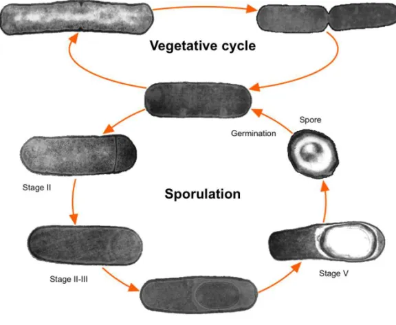
The endospore is a cell type that can survive for extended periods without nutrients, but is able to monitor its environment and readily revive if nutrients become available (Fig. 1; (Moir and Smith, 1990; Piggot and Losick, 2002)). It is thrilling to realise that Bacillus spores have been revived from extinct bees preserved in amber for 25 to 40 million years, and also from another Bacillus species with 250 million years from a salt crystal, although this latter finding is controversial (Cano and Borucki, 1995; Vreeland et al., 2000; Nickle et al., 2002). Such was achieved as spores can bear extreme environmental insults that include wet and dry heat, UV and gamma radiation, extreme desiccation (including vacuum), toxic chemicals, high pressure and oxidising agents. The levels of resistance are such that spores have been suggested as candidates for interplanetary transfer of life (Nicholson et al., 2000; Nicholson et al., 2005; Setlow, 2006, 2007).
An overview of sporulation
Figure 1. The sporulation and vegetative cycles of Bacillus subtilis. A vegetatively growing cell is defined as stage 0. Key morphological changes during sporulation are the asymmetric division (stage II), engulfment of the prespore by the mother cell (stages II-III), assembly of the protective layers (stages IV-V), spore maturation (stage VI), lysis of the mother cell and release of the mature spore (stage VII). In favourable conditions, the spore germinates and the vegetative cycle is restored.
asymmetric positioning of the septum traps two-thirds of the prespore chromosome on the mother cell side, which are then returned to the prespore by a septal DNA translocase (Wu and Errington, 1997; Frandsen
et al., 1999). Next, engulfment of the prespore by the mother cell takes
and outgrowth occur, followed by a resumption of the vegetative growth cycle (Setlow, 2003; Hilbert and Piggot, 2004).
The genetic regulation of sporulation
Entry into sporulation
Grossman, 1995).
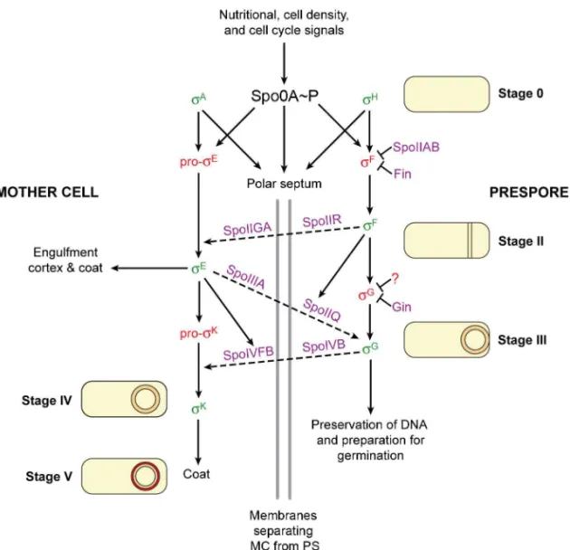
Figure 2. Regulatory network during Bacillus subtilis sporulation. Solid arrows indicate dependence relationships between σ factors (green means active and red means inactive) and target genes whose products bring about morphological change (described in words and simplified representations of the stages of sporulation). The grey vertical lines represent the two membranes that separate the mother cell (MC) from the prespore (PS) after the polar septum forms. Dashed arrows indicate signalling pathways between the two cell types. Key regulatory proteins are shown in purple. Adapted from Kroos, 2007, Camp
et al., 2011, and Serrano et al., 2011.
Both Spo0A production and activity are tightly regulated. Spo0A~P influences its own production and activity: upon entry into stationary phase Spo0A~P stimulates the transcription of its own gene, of the phosphorelay component spo0F, and indirectly stimulates the transcription of spo0H (encoding σH) and of the phosphorelay phosphatase
spo0E (reviewed in Lopez et al., 2009).
physiological outcomes is possible. Since Spo0A~P-dependent genes with a direct role in sporulation possess low affinity for Spo0A~P, only in the presence of high levels of Spo0A-P is sporulation initiated (Fujita et al., 2005).
Compartmentalisation of gene expression
At the beginning of sporulation, Spo0A~P cooperates with σA and σH in directing the transcription of spoIIA, spoIIG and spoIIE which encode key developmental regulators, among other genes (Errington, 2003; Hilbert and Piggot, 2004; Piggot and Hilbert, 2004). Spo0A~P and σH trigger the redirection of septum formation to a polar position, which results in the key event of asymmetric division (Kroos and Yu, 2000; Piggot and Hilbert, 2004). After asymmetric division, distinct but interdependent transcriptional programmes take place in each of the two cells. Their rigorous orchestration depends mainly on the differential usage of several sigma subunits by the RNA polymerase, directing it to specific classes of promoters (Hilbert and Piggot, 2004). Such is achievable due to the transient association between the RNA polymerase and the sigma subunits (Travers and Burgess, 1969).
Each of the four compartment-specific sigma subunits (σF, σE, σG and σK) is inactive at the time of its synthesis and requires subsequent activation. The activation of each sigma factor at the proper location and time is coupled to landmark morphological events by control mechanisms named checkpoints (Losick and Shapiro, 1993; Kroos et al., 1999). Asymmetric division triggers the activation of σF in the prespore and σE in the mother cell, both shaping the initial stages of sporulation. Later, the completion of engulfment of the prespore by the mother cell leads to the activation of σG
Developmental checkpoints
Prespore line of gene expression
σF checkpointσF, encoded by spoIIAC, is the first compartment-specific sigma factor of
sporulation. It is synthesised and kept inactive in the pre-divisional cell until asymmetric division takes place, being selectively activated in the prespore (Margolis et al., 1991; Lewis et al., 1996).
Confining of σF activity to the prespore involves a partner-switching mechanism involving SpoIIAB, SpoIIAA and SpoIIE. SpoIIAB switches between SpoIIAA and σF as binding partners (Alper et al., 1994; Duncan et al., 1995). SpoIIAB is an anti-sigma factor that binds to σF and prevents its association with the RNA polymerase (Duncan and Losick, 1993). SpoIIAA is an anti-anti-sigma factor that is inactive when phosphorylated. The SpoIIE phosphatase produces dephosphorylated SpoIIAA that counteracts the inhibitory effect of SpoIIAB by binding to the SpoIIAB-σF
complex, causing the release of active σF (Duncan and Losick, 1993; Duncan et al., 1995).
SpoIIE is a membrane protein that localises at the polar septum (Barák et al., 1996). SpoIIE is a bifunctional protein, being required for σF activation and for proper asymmetric division (Barák and Youngman, 1996; Feucht
et al., 1996). SpoIIE is considered instrumental in the coupling of gene
expression and morphogenesis, despite the precise mechanisms for preferential desphosphorylation of SpoIIAA in the prespore being still a matter of debate (reviewed in Barák and Wilkinson, 2005).
Another important contribution to the compartmentalisation of σF activity results from the transient exclusion of the gene encoding SpoIIAB from the prespore generated by polar division (Dworkin and Losick, 2001), and from degradation of free SpoIIAB in the prespore (Pan et al., 2001).
σF was found to drive the expression of 48 genes (Wang et al., 2006)
including genes required for σE and σK activation in the mother cell, spoIIR
Londoño-Vallejo and Stragier, 1995; Gomez and Cutting, 1996), genes involved in the regulation of prespore-specific gene expression, spoIIIG, encoding the late prespore sigma factor σG (Karmazyn-Campelli et al., 1989; Sun et al., 1989), spoIIQ, required for both expression of spoIIIG and
σG activity, and involved in engulfment (Londoño-Vallejo et al., 1997; Sun
et al., 2000; Camp and Losick, 2008). Several σF-dependent genes seem to
be transcribed by σG as well (Wang et al., 2006).
The switch from σF to σG was proposed to require a putative anti-sigma factor named Fin (F inhibiting) that is related to the anti-sigma factor for
σG, Gin, in combination with an unknown Fin-independent pathway
(Camp et al., 2011; Serrano et al., 2011).
σG checkpoint
σG is the late sigma factor present in the prespore (Karmazyn-Campelli et
al., 1989; Sun et al., 1989). Co-transcription of spoIIIG (encoding σG) and
spoIIG (encoding σF and some of its regulators) before asymmetric division
occurs with Spo0A and σA but σG is not produced due to a stem-loop structure that blocks the ribosome-binding site (Masuda et al., 1988). Translation occurs later, from transcripts produced by σF and σG from a promoter located immediately upstream of the spoIIIG coding region (Sun
et al., 1989; Sun et al., 1991). Transcription of spoIIIG in the prespore is
delayed relative to other σF-dependent genes as it depends on a poorly understood signal transduction pathway requiring the action of σE in the mother cell (Partridge and Errington, 1993; Evans et al., 2004). Transcription of spoIIIG also requires expression of the σF-controlled gene
spoIIQ (Sun et al., 2000). σG is autoregulatory and thus, once activated, can
recognise its own promoter and maintain its own synthesis (Karmazyn-Campelli et al., 1989; Sun et al., 1991).
σE-controlled spoIIIA operon, and of the prespore-specific, σF-dependent
SpoIIQ (Illing and Errington, 1991; Kellner et al., 1996; Londoño-Vallejo et al., 1997; Camp and Losick, 2008; Meisner et al., 2008; Camp and Losick, 2009), aided by the membrane protein insertase SpoIIIJ (Errington et al., 1992; Murakami et al., 2002; Serrano et al., 2003; Tjalsma et al., 2003; Camp and Losick, 2008; Serrano et al., 2008). This channel, which is formed between the prespore and the mother cell (Camp and Losick, 2008; Meisner et al., 2008), was proposed to function as a “feeding tube” that allows the mother cell to nurture the prespore by providing small molecules needed for biosynthetic activity. The channel would be required for general macromolecular synthesis, rather than specifically activating σG (Camp and Losick, 2009), and to maintain prespore integrity
(Li et al., 2004; Doan et al., 2009).
Three negative regulators of σG are known, the LonA protease and the anti-sigma factors CfsB and SpoIIAB. The latter also inhibits σF prior to asymmetric division (Rather et al., 1990; Duncan and Losick, 1993; Schmidt et al., 1994; Kellner et al., 1996; Karmazyn-Campelli et al., 2008). Both LonA and SpoIIAB are responsible for the inhibition of σG activity under conditions that do not favour sporulation and also in the mother cell during sporulation, contrary to the suggestion that SpoIIAB would have a role in the prespore (Rather et al., 1990; Schmidt et al., 1994; Kellner
et al., 1996; Serrano et al., 2001; Serrano et al., 2004; Chary et al., 2005). CsfB
(controlled by sigma F), also known as Gin (G inhibitor), is an anti-sigma factor that shows specificity to σG, unlike SpoIIAB, and is present in the prespore at early times (Decatur and Losick, 1996; Chary et al., 2007; Karmazyn-Campelli et al., 2008; Rhayat et al., 2009). CsfB is also under σG
transcriptional control of spoIIIG and the nurturing by the SpoIIIA-SpoIIQ channel (Camp and Losick, 2008; Meisner et al., 2008; Camp and Losick, 2009; Doan et al., 2009; Serrano et al., 2011).
The σG regulon includes genes involved in the regulation of prespore-specific gene expression, as its own gene, spoIIIG, and spoVT (Karmazyn-Campelli et al., 1989; Bagyan et al., 1996); genes involved in the activation of the late mother cell sigma factor σK, as spoIVB (Cutting et al., 1991a);
genes involved in spore maturation, as the spoVA operon, and the ssp
genes (Helmann and Moran Jr., 2002; Tovar-Rojo et al., 2002); and in germination, as the gerA and gerB operons, and pdaA (Paidhungat and Setlow, 2001; Fukushima et al., 2002).
Mother cell line of gene expression
σEcheckpointσE is the first mother cell-specific sigma factor during sporulation. σE is
synthesised as pro-σE, an inactive precursor, and is activated by cleavage of the 27 amino acid residue N-terminal “Pro” sequence (LaBell et al., 1987; Stragier et al., 1988; Miyao et al., 1993). This N-terminal sequence is also responsible for tethering Pro-σE to the membrane (Ju et al., 1997; Fujita and Losick, 2002). Proprotein processing is carried out by the membrane-bound SpoIIGA protease (Peters and Haldenwang, 1994; Imamura et al., 2008). σE and SpoIIGA are encoded by the two-gene spoIIG operon which
is expressed in the pre-divisional cell (Kenney and Moran Jr, 1987). However, activation of the SpoIIGA protease occurs only upon receiving a signal from the prespore, the σF-dependent SpoIIR. SpoIIR is predicted to be secreted into the space between the septal membranes and then activate SpoIIGA proteolytic activity towards Pro-σE, thus tying σE
mother cell-specific transcription factor (Fujita and Losick, 2002, 2003); ii) selective degradation of σE in the prespore (Ju et al., 1998; Fujita and Losick, 2002).
The σE regulon has been defined by microchip array in two independent studies that found 171-253 genes under the control of σE (Eichenberger et al., 2003; Feucht et al., 2003). σE-dependent expression is required to prevent a second division at the distal pole which produces cells with two DNA-containing prespore compartments and an anucleate mother cell that subsequently fail to sporulate, the so-called abortively disporic phenotype (Lewis et al., 1994). Three genes under the control of σE are required for the inhibition of this second division, and also for prespore engulfment: spoIID, spoIIM and spoIIP (Lopez-Diaz et al., 1986; Smith et al., 1993; Smith and Youngman, 1993; Frandsen and Stragier, 1995; Pogliano et al., 1999). σE also controls the expression of genes involved in σG
activation, the spoIIIA operon (Illing and Errington, 1991); genes required for initiating cortex synthesis and spore coat assembly, as spoVE, spoIVA,
cotE and spoVID (Zheng et al., 1988; Roels et al., 1992; Beall et al., 1993; Miyao et al., 1993); and to direct synthesis of the late mother cell-specific factor σK, the sigK composite gene, spoIVCA, spoIVF and spoIIID (Kunkel et al., 1989; Kunkel et al., 1990; Cutting et al., 1991b; Sato et al., 1994).
Replacement of σE by σK in the mother cell involves a σK-dependent negative feedback loop that inhibits σA-dependent transcription, including that of the spoIIGB gene encoding σE, and that requires transcriptionally active σK (Zhang et al., 1999).
σK checkpoint
is carried out by the σE-dependent site-specific recombinase SpoIVCA that is encoded in the skin element (Kunkel et al., 1990; Sato et al., 1990; Popham and Stragier, 1992; Sato et al., 1994). Second, the sigK gene is under the control of σE and of σK itself (Kunkel et al., 1988; Kroos et al., 1989). Third, like its mother cell predecessor, the σK protein is translated as an inactive precursor that requires proteolytic removal of an amino-terminal pro-sequence (Kroos et al., 1989). Similarly to the case of σE, the “Pro” sequence prevents interaction with the RNA polymerase and localises the proprotein in the membrane (Zhang et al., 1998). SpoIVFB is the metalloprotease responsible for the proteolytic processing of σK and its activation (Cutting et al., 1991b; Resnekov and Losick, 1998; Rudner et al., 1999). SpoIVFB is negatively regulated by two other membrane proteins, SpoIVFA and BofA (Bypass-of-forespore), all under the control of σE and present in a complex (Cutting et al., 1990; Cutting et al., 1991b; Ricca et al., 1992; Rudner and Losick, 2002). SpoIVFA anchors the complex in the mother cell membrane that surrounds the prespore and acts as a platform bringing BofA and SpoIVFB together, whereby BofA inhibits SpoIVFB processing until a signal has been received from the prespore (Resnekov and Losick, 1998; Rudner and Losick, 2002). Such signal relies on the production of SpoIVB in the prespore under the control of σG (Cutting et al., 1990; Gomez et al., 1995). SpoIVB is a serine protease that is secreted into the space between the prespore membranes (Wakeley et al., 2000). SpoIVB cleaves SpoIVFA, resulting in an alteration in the complex and in the release of SpoIVFB from BofA’s inhibition, being able to process and activate σK (Dong and Cutting, 2003). A second serine protease, CtpB (Pan
et al., 2003), is also able to cleave SpoIVFA and trigger σK activation
(Campo and Rudner, 2006). Activation of σK is coupled to σG activity and engulfment via SpoIVB, which is transcribed by σG and fails to accumulate when engulfment is impaired (Cutting et al., 1990; Gomez and Cutting, 1996; Doan and Rudner, 2007).
and germination, as spoVK, spoVD and gerP (Fan et al., 1992; Daniel et al., 1994; Behravan et al., 2000), and in the regulation of σK-dependent transcription, as gerE (Zheng and Losick, 1990).
Protein transport in
B. subtilis
and other organisms
Protein transport systems
In all domains of life, the lipid membrane is a central feature that preserves the integrity of the cell. The membrane acts not only as a physical barrier, in the maintenance of the composition and concentration of molecules inside the cell, but is also involved in the controlled swap of substances and information between the cell and its surroundings, and with organelles if present. Such was allowed by the emergence of specific protein transport devices that include proteins that either span membranes to create selective pores or that bind the cytosolic face of membranes to create transport vesicles (Pohlschröder et al., 2005b; Odorizzi and Rehling, 2009). In addition, the transport of proteins permitted the appearance of cellular compartments, as the periplasm of Gram-negative bacteria, or the components of the secretory pathway in the cytoplasm of eukaryotes (Pohlschröder et al., 2005b).
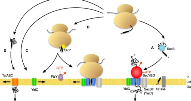
Figure 3. Schematic representation of bacterial protein targeting. The bacterial Sec translocon (blue) is composed of SecY, SecE, and SecG. SecA (red) acts as a peripheral motor protein on the cytoplasmic side. Signal peptides are cleaved by a signal peptidase (SPase). Other ancillary proteins are YidC (green) and SecDF/YajC (light grey). (A) Proteins synthesised at the ribosome (light brown) destined for secretion are mostly post-translationally targeted to the Sec translocase by a targeting sequence which is recognised by SecA. Alternatively, targeting to the translocon can be carried out by the molecular chaperone SecB (light blue). (B) Co-translational targeting of the ribosome with the nascent chain to the translocase complex is attained by the binding of the signal peptide of some preproteins or the signal anchor sequence of membrane proteins by the signal recognition particle (SRP) (yellow), then to the SRP receptor FtsY (purple). Membrane proteins with large hydrophilic periplasmic domains require the presence of SecA. YidC interacts with transmembrane segments as they emerge from the proposed lateral gate of SecYEG. SRP can also deliver proteins directly to YidC. (C) A subset of membrane proteins can insert into the cytoplasmic membrane via YidC after targeting of the ribosome nascent chain to YidC. (D) Translocation of folded precursor proteins occurs via the Tat translocase (orange). For B. subtilis, the majority of the scenario appears to be valid, with the exception of SecB, which does not exist and its function seems to carried out by CsaA. CM, cytoplasmic membrane. Adapted from Natale et al., 2008, and Du Plessis et al., 2011.
Targeting signals
In early cells diffusion may have been sufficient for proteins to reach protein transport devices. However, as cells became increasingly complex, specific piloting factors that enabled more efficient targeting to several protein translocation apparatuses have evolved (Pohlschröder et al., 2005b).
are required for their proper insertion and topology. The cells possess targeting factors that recognise the signals embedded in the proteins and enable targeting to the correct transport apparatus. Several types of targeting/topogenic signals have been identified: (a) signal anchors (type II signal anchor) that initiate translocation of the carboxyl-terminal region of a membrane protein remaining as a membrane anchor with Nin Cout
orientation; (b) reverse signal anchors (type I signal anchor) that initiate the translocation of the amino-terminal region of the protein and remain as a membrane-spanning region of Nout Cin orientation; (c) stop-transfer
sequences which allow translocation arrest and lateral release from the translocation channel, remaining as membrane anchors with Nout Cin
orientation; (d) helical hairpins, two closely spaced hydrophobic regions that insert in a folded manner, having both amino and carboxyl termini in the cytoplasm (Xie et al., 2007; Driessen and Nouwen, 2008; Xie and Dalbey, 2008); (e) signal peptides, which are usually present at the amino-terminus of the precursor protein. Signal peptides are cleaved off from the mature protein and further degraded by signal peptide peptidases (Ichihara et al., 1984; Bolhuis et al., 1999). The existence of an N-terminal region that directed proteins to the correct transport machinery was initially suggested by Blobel and Sabatini in the early 1970s (reviewed in Leslie, 2005). Signal peptides were shown to also perform a role beyond targeting: they are also allosteric activators of the SecYEG translocase by binding to SecA and lowering its activation energy state (Gouridis et al., 2009).
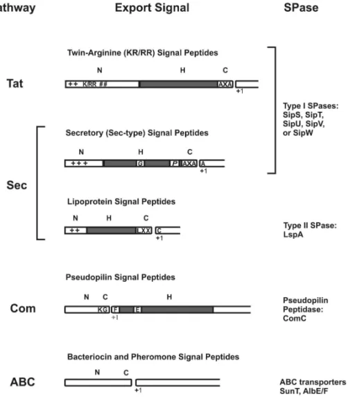
conformation upon insertion in the lipid membrane (Briggs et al., 1986). Helix-breaking residues such as glycine or proline are often found in the middle of this domain, allowing the formation of a hairpin-like structure that facilitates membrane insertion (De Vrije et al., 1990). The C-domain specifies the cleavage site for the signal peptidase (SPase) (von Heijne and Abrahmsén, 1989). Based on the cleavage sites and the export pathways that are expected to be utilised, five major types of signal peptides can be defined for B. subtilis (Figs. 4-5): (a) secretory (Sec-type or type I); (b) twin arginine (RR/KR); (c) lipoprotein (type II); (d) pseudopilin; (e) bacteriocin and pheromone signal peptides.
Figure 5.Protein export pathways in B. subtilis. Proteins can be sorted to various destinations depending on the presence (+SP) or absence (-SP) of an N-terminal signal peptide and specific retention signals. Proteins devoid of a signal peptide are either retained in the cytoplasm or may escape via the flagellar export machinery, the holin systems, or other unidentified systems. Proteins that are retained at the extracytoplasmic side of the membrane can either contain a transmembrane segment (TM) or a lipid modification (+lipobox) and are exported via the Sec or Tat pathways. Proteins that are retained in the cell wall can be exported via either the Sec or Tat pathway. These proteins are retained in the cell wall if their mature parts contain cell wall-binding repeats (+CWB). Proteins can be secreted into the medium via the Sec or Tat pathway or by ABC transporters. Pseudopilins are exported by the Com system. Adapted from Tjalsma et al., 2004.
Signal peptidases are enzymes that catalyse protein maturation by cleaving the signal peptide during or shortly after transport (reviewed in Tjalsma et al., 2004). Signal peptide types (a) and (b) are cleaved by type I SPases, whereas type (c) use LspA (SPase II). Type I SPases recognise a consensus sequence of A-X-A|A at positions -3 to +1 relative to the cleavage site. The vertical line represents the cleavage site. While the E.
coli genome encodes only one type I SPase, in B. subtilis five of these genes
exist, although only sipT or sipS have to be present to ensure viability (Silver and Wickner, 1983; Tjalsma et al., 1998).
arginines (in B. subtilis, R/K-R-X-#-#, # being a hydrophobic amino acid residue and X any residue). On average, the N-domain of Tat signal peptides is longer than Sec-type N-domains (Tjalsma et al., 2004; Natale et al., 2008). The H-domain tends to be less hydrophobic than that of Sec-type signal peptides in E. coli (Cristóbal et al., 1999) but similar in B.
subtilis (Tjalsma et al., 2000). The C-domain may contain a so-called
Sec-avoidance signal, composed of positively charged residues, that prevents interaction with Sec components (Blaudeck et al., 2003).
Lipoprotein signal peptides (c) are lipid-modified by the dyacylglyceryl transferase encoded by lgt, an essential step for cleavage by LspA (type II SPase) both in E. coli and in B. subtilis (Sankaran and Wu, 1994 and references therein; Prágai et al., 1997; Leskelä et al., 1999). SPase II recognises a consensus sequence called “lipobox”, usually L-(A/S)-(A/G)|C at positions -3 to +1 relative to the cleavage site, cleaving before the invariable cysteine residue. In E. coli, but not in B. subtilis, the cysteine residue is further modified by the apolipoprotein N-acyl transferase Lnt (Gupta and Wu, 1991; Kunst et al., 1997).
Prepilin signal peptides were initially identified in bacterial type IV prepilin subunits (reviewed in Strom and Lory, 1993). Pseudopilin signal peptides (d) present a variation on the previously described general theme, as the cleavage site precedes the hydrophobic stretch that becomes thus part of the mature protein (reviewed in Pohlschröder et al., 2005a). In
B. subtilis four proteins bear this class of signal peptides (Tjalsma et al.,
2000), which are required for the binding and uptake of exogenous DNA during genetic competence (reviewed in Dubnau, 1997). They are transported via the specific Com pathway and cleaved by ComC (reviewed in Tjalsma et al., 2000).
B. subtilis bear such signal peptides (reviewed in Tjalsma et al., 2004). The lack of the H-domain is the most distinctive feature of these signal peptides, being thus composed of N- and C-terminal domains (Tjalsma et al., 2000).
Transport of proteins can also occur independently of signal peptides via holins, which form pores in the membrane through which extracellular proteins may reach the cell wall, and by dedicated machinery for the assembly of flagella (Namba et al., 1989; Young and Bläsi, 1995).
Targeting factors
Proper targeting is a critical matter for proteins. When exiting the ribosomal tunnel during synthesis, polypeptides meet several aiding factors, namely chaperones, folding catalysts and targeting factors. Their function is to prevent proteins from aggregating and misfolding and to ensure proper routing to the translocation/membrane insertion machinery (Dalbey et al., 2011).
In E. coli, trigger factor appears to be the default chaperone for nascent
chains, preventing their premature folding, and it also interacts with the ribosome (Ullers et al., 2003 and references therein). Trigger factor is also present in B. subtilis and was show to catalyse in vitro protein folding (Göthel et al., 1998). Another aiding factor is the signal recognition particle (SRP), which mediates the transport of secretory and membrane proteins to the cytoplasmic membrane or to the endoplasmic reticulum, in bacteria, archaea and eukaryotes (reviewed in Yuan et al., 2010). The SRP competes with trigger factor for the ribosome-nascent chain complexes (RNCs), both
in E. coli and in B. subtilis (Ullers et al., 2003; Zanen et al., 2005; Knoops et
al., 2012 and references therein). Nascent polypeptides harbouring more hydrophobic targeting sequences enter the SRP pathway, in E. coli and in
B subtilis (reviewed in Luirink and Sinning, 2004; Zanen et al., 2005). Upon
The SRP is a widely conserved ribonucleoprotein that is composed of a protein part with GTPase activity (the bacterial Ffh, for “fifty-four homologue” of the eukaryotic 54 kDa protein subunit and an RNA part (7S RNA in eukaryotes, 4.5 S RNA in E. coli and the B. subtilis small cytoplasmic RNA) (Walter and Blobel, 1982; Brown and Fournier, 1984; Nakamura et al., 1992). In the latter organism the particle contains still another protein, HBsu (Nakamura et al., 1999). The SRP pathway in archaea appears to be an intermediate between that of eukaryotes and bacteria (reviewed in Yuan et al., 2010). The SRP and its membrane-bound receptor FtsY are essential for cell viability both in E. coli and B. subtilis
(Brown and Fournier, 1984; Nakamura et al., 1992; Phillips and Silhavy, 1992; Luirink et al., 1994; Nakamura et al., 1999; Kobayashi et al., 2003). SecB is a cytosolic chaperone present in many Gram-negative bacteria. When a signal peptide does not display a high level of hydrophobicity it is bound by SecB, which keeps the preprotein in an unfolded state (reviewed in Driessen and Nouwen, 2008). SecB routes preproteins to the Sec pathway by binding to the SecA ATPase. This interaction is strengthened by binding of the signal peptide to SecA (Fekkes et al., 1998). SecB does not exist in B. subtilis, although an analogue may exist (CsaA) (Müller et al., 2000). Thus, both organisms may share a common general mechanism involving co-translational targeting via SRP and post-translational targeting with SecB/CsaA (Tjalsma et al., 2000).
The Sec pathway
The major route for protein transport across and into the cytoplasmic membrane is the Sec pathway. An important step in understanding this pathway was the identification of its major components as well as their structures. Another important contribution came from the functional in
vitro reconstitution of the translocation reaction with purified components
(Brundage et al., 1990; reviewed in Driessen and Nouwen, 2008).
Sec61αβγ and SecYEβ, in bacteria, eukaryotes and archaea, respectively) (Brundage et al., 1990; Görlich and Rapoport, 1993; Pohlschröder et al., 1997). SecY is a highly hydrophobic protein that spans the membrane ten times in M. jannaschii, as well as in E. coli and B. subtilis (Akiyama and Ito, 1987; Nakamura et al., 1990; Van den Berg et al., 2004). SecY forms an hourglass-shaped channel through which the substrate is translocated. The interior of the channel is mostly hydrophilic with the exception of the hydrophobic waist that makes contact with the polypeptide. The pore in SecY has a clamshell-like structure that may open towards the lipid bilayer (Van den Berg et al., 2004; Cannon et al., 2005). SecE has three, one, or two transmembrane (TM) segments in E. coli, B. subtilis and M.
jannaschii, respectively (Schatz et al., 1989; Jeong et al., 1993; Van den Berg
et al., 2004). In E. coliand M.jannaschii, SecE embraces the two SecY halves
in a supportive manner, acting like a molecular clamp (Breyton et al., 2002; Van den Berg et al., 2004). SecG localises at the periphery of the complex and makes little contact with SecY (Breyton et al., 2002). SecG has two TM segments both in E. coli and B. subtilis (Nishiyama et al., 1993; van Wely et al., 1999) and is not strictly required for function in either organism (Nishiyama et al., 1994; van Wely et al., 1999; Breyton et al., 2002).
The oligomeric status of the translocon is a controversial matter. Different techniques suggest that the translocon can be found in a dynamic equilibrium between monomers, dimers or even higher-order oligomers, in eukaryotes, archaea and in bacteria (reviewed in Driessen and Nouwen, 2008; and in Du Plessis et al., 2011).
In terms of subcellular localisation, several Sec components have been seen to form helices both in the rod-shaped E. coli and B. subtilis (Campo et al., 2004; Shiomi et al., 2006), although reports of uniform distribution exist
for E. coli (Brandon et al., 2003; Rubio et al., 2005). Interestingly, in the
coccoid bacterium Streptococcus pyogenes, the Sec system localised to a single microdomain, the ExPortal, adjacent to where a new cell division septum will form (Rosch and Caparon, 2004).
channel, such as SecDFYajC and YidC/SpoIIIJ in bacteria (Bolhuis et al., 1998; Nouwen and Driessen, 2002; Saller et al., 2009), SecDF and possibly YidC in archaea (Eichler, 2003; Pohlschröder et al., 2005a), and Sec62/Sec63/Sec71/Sec72 and TRAM (translocation-associated membrane protein) in eukaryotes (reviewed in Rapoport, 2007).
The energy required for transport is provided by peripheral components, such as the translating ribosome in the case of co-translational membrane insertion in bacteria, eukaryotes and archaea. Post-translational translocation is operated by Sec62/Sec63 and BiP in the endoplasmic reticulum of eukaryotes (reviewed in Du Plessis et al., 2011); Although reports of post-translational translocation in archaea exist, its powering mechanism is not know (Rapoport, 2007 and references therein). In bacteria, the peripherally associated SecA ATPase and proton motive force energise post-translational translocation (Driessen, 1992; Economou and Wickner, 1994). SecA is also required during co-translational insertion of some membrane proteins, both in E. coli and in B. subtilis (e.g. Andersson and von Heijne, 1993; Bunai et al., 2005).
The Tat pathway
The twin-arginine translocation (Tat) pathway catalyses the translocation of proteins in their folded state and/or containing cofactors both into and across membranes (reviewed in Natale et al., 2008). This pathway is present in the three domains of life, being found in bacteria, archaea as well as in chloroplast thylakoid membranes in eukaryotes (reviewed in Natale et al., 2008; and in Yuan et al., 2010). Homologues were also found in mitochondria although not for the complete pathway (Bogsch et al., 1998; Yen et al., 2002).
The composition of the Tat translocase is variable amongst organisms. In
E. coli and in plant thylakoids the minimal Tat translocase is formed by
TatA, TatB and TatC (reviewed in Berks et al., 2005). TatA and TatB are homologues but perform distinct functions (Sargent et al., 1999). The TatBC complex functions in the binding of substrates, with TatC specifically recognising the consensus motif in the signal peptide and TatB interacting with the signal peptide and mediating transfer of the substrate from TatC to the pore (Alami et al., 2003). In one model, TatA complexes constitute the protein-conducting channel of the Tat translocase, where the number of TatA protomers would change to match the size of the substrate (Gohlke et al., 2005). A model with TatA and TatB forming the pore was also proposed (Sargent et al., 2001). In B. subtilis, TatC and TatA constitute the minimal translocase. TatA is bifunctional in this organism as it performs the functions of both TatA and TatB of E. coli and thylakoids. Interestingly, three Tat translocases were found in B. subtilis, with distinct substrate specificities (Jongbloed et al., 2004; Monteferrante et al., 2012). Similarly to most Gram-positive bacteria, TatB is also absent in archaea and multiple copies of Tat components exist (reviewed in Yuan et al., 2010).
Protein translocation by the Tat apparatus is energised exclusively by the proton motive force, both in plant thylakoids and in bacteria (Yahr and Wickner, 2001; Braun et al., 2007).
In E. coli, Tat components seem to localise uniformly throughout the
cytoplasmic membrane, although some punctations at the poles were observed, the significance of which is unclear (Ray et al., 2005). TatCy and the three TatA proteins of B. subtilis showed a dual localisation pattern, both evenly in the membrane as well as in foci, which were more abundant at the cell poles and/or division sites (Meile et al., 2006; Ridder
et al., 2009).
Other secretion systems
secretion systems that export proteins through their multilayered cell envelope and in some cases into host cells, requiring the crossing of three membranes in the latter case. Most of these systems are present in Gram-negative bacteria although some are found in Gram-positive bacteria, which also harbour specific systems (Economou et al., 2006).
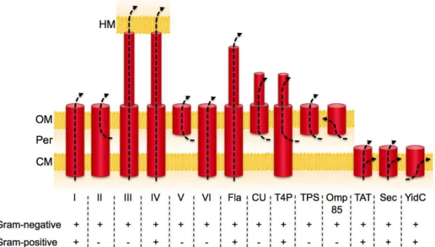
At least six major systems have been discovered so far (T1SS to T6SS, for
Type X Secretion System) (Fig. 6) (reviewed in Economou et al., 2006).
Figure 6. The bacterial protein export and secretion systems zoo. The arrows indicate the path that is taken by the exported protein. Arrows that initiate in the periplasm indicate that Sec (or rarely Tat)-dependent translocation across the cytoplasmic membrane is a necessary first step for these systems. I-VI, secretion systems from types 1 to 6; CM, cytoplasmic membrane; CU, chaperone-usher pathway; Fla, flagellum; HM, host cell membrane; OM, outer membrane; Omp85, also known as YaeT; Per, periplasm; TPS, two-partner secretion; T4P, type IV pili. Adapted from Papanikou et al., 2007, and Economou et al., 2006.
the importance of the additional outer membrane barrier that has to be negotiated in Gram-negative bacteria.
The YidC/Oxa1/Alb3 family
Members of the evolutionarily conserved YidC/Oxa1/Alb3 family of proteins have been shown to perform critical roles in membrane insertion and assembly of energy-transducing complexes in eukaryotic organelles (mitochondria and chloroplast) and in bacteria (for reviews see Kol et al., 2008; Saller et al., 2012). The presence of Oxa1-related proteins in archaea is strongly suggested by genome sequencing (Bonnefoy et al., 2009 and references therein).
The signature feature of this family of proteins is a group of five transmembrane (TM) segments (Yen et al., 2001; Saller et al., 2012) (Fig. 7).
Figure 7. Topology of YidC/Oxa1/Alb3 family of proteins. Topology was mapped with several techniques or predicted for the various proteins herein depicted (for reviews see
Yen et al., 2001; Wang and Dalbey, 2011; Saller et al., 2012). N and C, amino and
carboxyl termini, respectively; IM, inner membrane; TM, thylakoid membrane; CM, cytoplasmic membrane. The structure of the periplasmic domain has been determined for YidC (Oliver and Paetzel, 2008; Ravaud et al., 2008). The N terminus of SpoIIIJ is most likely lipid-modified. Adapted from Oliver and Paetzel, 2008, Tjalsma et al., 2003, and Wang and Dalbey, 2011.
Oxa1 and Cox18
Oxa1 (oxidase assembly 1), present in the inner mitochondrial membrane
of Saccharomyces cerevisiae, is the founding member of this family
the insertion and assembly of energy-transducing respiratory complexes. In particular, Oxa1 directly mediates the co-translational insertion of proteins from the mitochondrial matrix, as the subunit II of cytochrome c
oxidase, Cox2, (Hell et al., 2001) and the assembly of both cytochrome c
oxidase and of the membrane sector of the F1F0 ATPase (He and Fox, 1997;
Hell et al., 2001; Jia et al., 2007).
Mitochondria typically contain proteins from two Oxa1 subfamilies – Oxa1 and Cox18. Cox18 (or Oxa2) was suggested to play a specialised role in the membrane biogenesis of cytochrome c oxidase in several organisms (Wang and Dalbey, 2011; Saller et al., 2012). Cox18 lacks a C-terminal domain present in Oxa1, which was shown to interact with the large ribosomal unit, allowing Oxa1 to act as a ribosome receptor (Jia et al., 2003). Oxa1 was suggested to form dimeric insertion pores on translating ribosomes (Kohler et al., 2009).
Alb3 and Alb4
In chloroplasts of plants and algae Alb3 (albino3) plays an important role in the integration of members of the nuclear-encoded LHCP (light-harvesting chlorophyll-binding protein) family into the thylakoid membrane of chloroplasts (Sundberg et al., 1997; Moore et al., 2000; Bellafiore et al., 2002). alb4 is a homologue of alb3 and it may also be present (Gerdes et al., 2006). The C-terminal domain of Alb3 recruits chloroplast SRP and it is absent in Alb4, which cannot replace Alb3 (Falk
et al., 2010). Whilst both Alb3 and Alb4 are important for chloroplast
membrane protein biogenesis, Alb4 was suggested to be required for assembly and/or stability of the F1F0 ATP synthase complex, being thus
functionally more closely related to YidC and Oxa1 than Alb3 (Benz et al., 2009).
Homologues in Archaea
segments (corresponding to TM4 and 5 of YidC in E. coli), with TMs 2, 3 and 4 being homologous to TMs 2, 3 and 6 of YidC in E. coli, respectively) (Yuan et al., 2010 and references therein).
YidC
The best-studied Oxa1 homologue in bacteria is YidC from E. coli. YidC is essential for viability and constitutes a key component in the biogenesis of membrane proteins (Samuelson et al., 2000). YidC depletion results in a global change in cell physiology (Price et al., 2010; Wang et al., 2010; Wickström et al., 2011). YidC was considered the missing bacterial insertase, as it facilitates the insertion of some Sec-independent proteins that were thought to insert spontaneously (Samuelson et al., 2000). YidC can function independently but also in conjunction with the Sec translocase. In this context, YidC was suggested to transfer polypeptide segments from the SecAYEG complex into the lipid bilayer (Scotti et al., 2000). YidC interacts with the Sec translocase by binding to SecD and SecF (Nouwen and Driessen, 2002).Proteins known to insert via the YidC-only pathway are the F0c subunit of the F1F0 ATPase, MscL and the M13 and
Pf3 phage coat proteins. YidC has been shown to be required for the membrane insertion of several Sec substrates, including the F0a subunit of
the F1F0 ATPase, NuoK (NADH dehydrogenase I subunit K) and subunit
II of cytochrome o oxidase (CyoA) (reviewed in Wang and Dalbey, 2011). YidC has also been implicated in the folding of proteins following their insertion by the Sec translocase, as observed for MalF, belonging to the maltose transport complex, and the LacY lactose permease (Nagamori et al., 2004; Wagner et al., 2008). YidC also has a role in the assembly of multimeric complexes (van der Laan et al., 2004; Kol et al., 2008). YidC and FtsH have been suggested to have a linked role in the quality control of inner membrane proteins (van Bloois et al., 2008).
for insertion (Urbanus et al., 2002) and was shown to integrate TatC and MscL delivered by SRP in vitro (Welte et al., 2012).
YidC revealed a predominantly polar localisation when fused to GFP (Urbanus et al., 2002).
YidC possesses the five TM segments that are conserved in the Oxa1 family plus an additional N-terminal one, linked by a periplasmic loop that is not required for YidC activity. The five conserved C-terminal domains are critical for function, despite being remarkably tolerant to mutations (Sääf et al., 1998; Jiang et al., 2003). Cross-linking studies have shown that TM3 of YidC is in the proximity of the substrate during membrane biogenesis (Klenner et al., 2008; Yu et al., 2008). In addition, TM2 and TM3 were suggested to interact (Yuan et al., 2007). Like Oxa1, YidC was proposed to form dimeric insertion pores on translating ribosomes. TM2 and TM3 of both monomers would form the core of the pore (Kohler et al., 2009). Previously, YidC had already appeared as a monomer and a dimer in Blue Native PAGE (van der Laan et al., 2001). Conflicting reports exist regarding the ability of YidC to contact ribosomes, as it lacks the C-terminal ribosome-binding domain found in Oxa1. Recent work suggests that YidC does bind to ribosomes (Kohler et al., 2009; reviewed in Price and Driessen, 2010; Welte et al., 2012).
Complementation studies
YidC2 and Oxa1 can partially complement each other (Funes et al., 2009). Regarding B. subtilis, both SpoIIIJ and YqjG can functionally complement YidC in E. coli (Saller et al., 2009).
SpoIIIJ and YqjG
Many Gram-positive bacteria, in contrast to Gram-negative bacteria, have two YidC homologues (reviewed in Yen et al., 2001; Funes et al., 2009). In
B. subtilis they are called SpoIIIJ and YqjG (Errington et al., 1992;
Murakami et al., 2002; Tjalsma et al., 2003). Whilst deletion of either spoIIIJ
or yqjG does not result in cell death, the absence of both is lethal. In addition, SpoIIIJ is required for sporulation, a function that YqjG cannot fulfil (Errington et al., 1992; Murakami et al., 2002; Tjalsma et al., 2003). YqjG was shown to be involved in genetic competence development contrary to SpoIIIJ (Saller et al., 2011).spoIIIJ is part of a bicistronic operon with jag (spoIIIJ associated gene). Jag is predicted to be a cytoplasmic protein containing single stranded nucleic acid-binding domains and it is dispensable for sporulation and growth (Errington et al., 1992; Grishin, 1998; Tjalsma et al., 2003). Similarly to spoIIIJ, yqjG is part of a bicistronic operon. mifM (for membrane protein insertion and folding monitor, or
reduced amounts were membrane proteins (Saller et al., 2011). SpoIIIJ and YqjG facilitate insertion of subunit F0c of the ATP synthase and were
found to associate with the whole complex, suggesting a role in its assembly (Saller et al., 2009; Saller et al., 2011). More members of the Oxa1 family are in involved the formation of the F1F0 ATP synthase (reviewed
in Wang and Dalbey, 2011; see above).
In the absence of spoIIIJ, sporulation is blocked after the completion of prespore engulfment. Mutations in spoIIIJ abolish the transcription of prespore-specific genes that use the σG sigma factor of the RNA polymerase but not transcription of the spoIIIG gene encoding σG
(Errington et al., 1992). σG accumulates in a spoIIIJ mutant but is mostly inactive (Serrano et al., 2003). Expression of spoIIIJ in the prespore is sufficient for σG activity and efficient sporulation (Serrano et al., 2003). It
was suggested that the activation of σG after engulfment completion involves the combined action of the spoIIIA-encoded products from the mother cell together with SpoIIIJ from the prespore (Serrano et al., 2003). Indeed, σG activation requires the formation of a channel between both compartments that is composed of two interacting proteins, SpoIIQ from the prespore and SpoIIIAH from the mother cell, as well as the remaining
spoIIIA proteins, including SpoIIIAE (Blaylock et al., 2004; Camp and
Losick, 2008; Meisner et al., 2008; Doan et al., 2009). Genetic evidence points to a functional interaction between SpoIIIAE and SpoIIIJ (Serrano et al., 2008). In addition, SpoIIIAE and SpoIIIJ were shown to directly interact in the membrane, linking the function of the spoIIIJ and spoIIIA
loci in the activation of σG. Such was suggested to take place in the context of the Sec translocon by directing the final stages of insertion and/or folding of SpoIIIAE (Camp and Losick, 2008; Serrano et al., 2008). SpoIIIAE also interacts with YqjG although the interaction appears to be non-functional, blocking YqjG (Serrano et al., 2008). Suppressor mutants that partially bypass the dependence of σG activation on spoIIIJ were
isolated. Interestingly, suppression was more potent regarding σG
SpoIIIJ later in sporulation (Camp and Losick, 2008). The suppressor mutations mapped to pbpG, yqjG, and spoIIIAE. PbpG is a peptidoglycan biosynthetic enzyme involved in cortex formation (McPherson et al., 2001). The PbpG variant that partially bypassed the loss of SpoIIIJ is likely to impair and/or delay cortex synthesis between the membranes surrounding the prespore, leading to partial σG activation and spore formation (Camp and Losick, 2008). YqjG variants might have acquired some of the sporulation-specific functionality of SpoIIIJ, possibly by rendering the interaction with SpoIIIAE more productive (Camp and Losick, 2008; Serrano et al., 2008). SpoIIIAE variants may assemble into the membrane on their own and/or may have acquired the ability to be recognised as a substrate by YqjG (Camp and Losick, 2008).
References
References
Aizawa, S.-I., Zhulin, I.B., Marquez-Magana and Ordal., G.O. 2002. Chemotaxis and motility. In Bacillus subtilis and its closest relatives: from genes to cells, L.A. Sonenshein, J.A. Hoch, and R. Losick, eds. (Washington D.C., USA, American Society for Microbiology), pp. 437-452.
Akita, M., Sasaki, S., Matsuyama, S. and Mizushima, S. 1990. SecA interacts with secretory proteins by recognizing the positive charge at the amino terminus of the signal peptide in Escherichia coli. J Biol Chem 265, 8164-8169.
Akiyama, Y. and Ito, K. 1987. Topology analysis of the SecY protein, an integral membrane protein involved in protein export in Escherichia coli. EMBO J 6, 3465-3470.
Alami, M., Lüke, I., Deitermann, S., Eisner, G., Koch, H.-G., Brunner, J. and Müller, M. 2003. Differential interactions between a twin-arginine signal peptide and its translocase in Escherichia coli. Mol Cell 12, 937-946.
Alper, S., Duncan, L. and Losick, R. 1994. An adenosine nucleotide switch controlling the activity of a cell type-specific transcription factor in B. subtilis. Cell 77, 195-205.
Andersson, H. and von Heijne, G. 1993. Sec dependent and Sec independent assembly of E. coli inner membrane proteins: the topological rules depend on chain length. EMBO J 12, 683-691.
Barák, I., Behari, J., Olmedo, G., Guzmán, P., Brown, D.P., Castro, E., Walker, D., Westpheling, J. and Youngman, P. 1996. Structure and function of the Bacillus SpoIIE protein and its localization to sites of sporulation septum assembly. Mol Microbiol 19, 1047-1060.
Barák, I. and Wilkinson, A.J. 2005. Where asymmetry in gene expression originates. Mol Microbiol 57, 611-620.
Barák, I. and Youngman, P. 1996. SpoIIE mutants of Bacillus subtilis comprise two distinct phenotypic classes consistent with a dual functional role for the SpoIIE protein. J Bacteriol 178, 4984-4989.
Barth, H., Aktories, K., Popoff, M.R. and Stiles, B.G. 2004. Binary bacterial toxins: biochemistry, biology, and applications of common Clostridium and Bacillus proteins. Microbiol Mol Biol Rev 68, 373-402.
Beall, B., Driks, A., Losick, R. and Moran Jr, C.P. 1993. Cloning and characterization of a gene required for assembly of the Bacillus subtilis spore coat. J Bacteriol 175, 1705-1716.
Behravan, J., Chirakkal, H., Masson, A. and Moir, A. 2000. Mutations in the gerP Locus of Bacillus subtilis and Bacillus cereus Affect Access of Germinants to Their Targets in Spores. J Bacteriol 182, 1987-1994.
Bellafiore, S., Ferris, P., Naver, H., Göhre, V. and Rochaix, J.-D. 2002. Loss of Albino3 leads to the specific depletion of the light-harvesting system. Plant Cell 14, 2303-2314.
Benz, M., Bals, T., Gügel, I.L., Piotrowski, M., Kuhn, A., Schünemann, D., Soll, J. and Ankele, E. 2009. Alb4 of Arabidopsis promotes assembly and stabilization of a non chlorophyll-binding photosynthetic complex, the CF1CF0-ATP synthase. Mol Plant 2, 1410-1424.
Berks, B.C., Palmer, T. and Sargent, F. 2005. Protein targeting by the bacterial twin-arginine translocation (Tat) pathway. Curr Opin Microbiol 8, 174-181.
Blaudeck, N., Kreutzenbeck, P., Freudl, R. and Sprenger, G.A. 2003. Genetic analysis of pathway specificity during posttranslational protein translocation across the Escherichiacoli plasma membrane. J Bacteriol 185, 2811-2819.
Blaylock, B., Jiang, X., Rubio, A., Moran, C.P. and Pogliano, K. 2004. Zipper-like interaction between proteins in adjacent daughter cells mediates protein localization. Genes Dev 18, 2916-2928.
Bogsch, E.G., Sargent, F., Stanley, N.R., Berks, B.C., Robinson, C. and Palmer, T. 1998. An Essential Component of a System with Homologues in Plastids and Mitochondria. J Biol Chem 106.