ISSN 1414-431X
BIOMEDICAL SCIENCES
www.bjournal.com.br
www.bjournal.com.br
Volume 45 (10) 875-994 October 2012
Braz J Med Biol Res, October 2012, Volume 45(10) 913-920
doi: 10.1590/S0100-879X2012007500101
Secretory TAT-peptide-mediated protein transduction of LIF
receptor
á
-chain distal cytoplasmic motifs into human myeloid
HL-60 cells
Q. Sun, J. Xiong, J. Lu, S. Xu, Y. Li, X.P. Zhong, G.K. Gao and H.Q. Liu
Institutional Sponsors
The Brazilian Journal of Medical and Biological Research is partially financed by
Faculdade de Medicina de Ribeirão Preto Campus
Ribeirão Preto
Explore High - Performance MS Orbitrap Technology In Proteomics & Metabolomics
Secretory TAT-peptide-mediated protein
transduction of LIF receptor α-chain distal
cytoplasmic motifs into human
myeloid HL-60 cells
Q. Sun
1,2*, J. Xiong
2*, J. Lu
3*, S. Xu
2, Y. Li
4, X.P. Zhong
1,
G.K. Gao
1* and H.Q. Liu
2*
1Department of Hyperbaric Medicine, No. 401 Hospital of PLA, Qingdao, China 2Department of Histology and Embryology, Faculty of Basic Medical Sciences,
Second Military Medical University, Shanghai, China
3Office of Medical Education, Training Department, Second Military Medical University, Shanghai, China 4State Food and Drug Administration of China, Huangdao Branch, Qingdao, China
Abstract
The distal cytoplasmic motifs of leukemia inhibitory factor receptor α-chain (LIFRα-CT3) can independently induce intracellular myeloid differentiation in acute myeloid leukemia (AML) cells by gene transfection; however, there are significant limitations in the potential clinical use of these motifs due to liposome-derived genetic modifications. To produce a potentially therapeutic LIFRα-CT3 with cell-permeable activity, we constructed a eukaryotic expression pcDNA3.0-TAT-CT3-cMyc plasmid with a
signal peptide (ss) inserted into the N-terminal that codes for an ss-TAT-CT3-cMyc fusion protein. The stable transfection of
Chinese hamster ovary (CHO) cells via this vector and subsequent selection by Geneticin resulted in cell lines that express and secrete TAT-CT3-cMyc. The spent medium of pcDNA3.0-TAT-CT3-transfected CHO cells could be purified using a cMyc-epitope-tag agarose affinity chromatography column and could be detected via SDS-PAGE, with antibodies against cMyc-tag.
The direct administration of TAT-CT3-cMyc to HL-60 cell culture media caused the enrichment of CT3-cMyc in the cytoplasm
and nucleus within 30 min and led to a significant reduction of viable cells (P < 0.05) 8 h after exposure. The advantages of using this mammalian expression system include the ease of generating TAT fusion proteins that are adequately transcripted
and the potential for a sustained production of such proteins in vitro for future AML therapy.
Key words: Leukemia inhibitory factor; TAT-HIV1; Protein transduction domain; Acute myeloid leukemia; LIF receptor
Introduction
Correspondence: H.Q. Liu, Department of Histology and Embryology, Second Military Medical University, Shanghai, 200433, China.
Fax: +86-21-8187-0958. E-mail: houqiliu@gmail.com and/or G.K. Gao, Department of Hyperbaric Medicine, No. 401 Hospital of PLA, Qingdao, 266071, China. Fax: +86-532-5187-0818. E-mail: anthonysun@cia.com
*These authors contributed equally to this study.
Received December 12, 2011. Accepted May 29, 2012. Available online June 22, 2012. Published September 3, 2012. Leukemia inhibitory factor (LIF), as a member of the
IL-6 family of cytokines, produces biological effects via binding to its receptor, which consists of a low-affinity LIF receptor α-chain (referred to as LIFRα) and a high-affinity subunit (referred to as gp130), both of which are shared with other IL-6 family cytokines (1). It has been reported that the 136-145-amino acid (aa) region of the distal C-terminal in cytoplasm, which contains 5-tyrosine residues (Y5) and several YXXQ motifs, is genetically conserved among gp130, LIFRα, and G-CSFR in numerous known
914 Q. Sun et al.
we hypothesize that the LIFRα-CT3 polypeptide could also achieve a therapeutic effect intracellularly, thus repesenting a potential future peptide-targeting leukemia therapy.
Advances in protein delivery and the identification of several protein transduction domains have facilitated the delivery of proteins/peptides to cells or organs (7-10). The HIV-trans-activating transduction domain (TAT-PTD) has 11 aa (TAT-PTD49-57: YGRKKRRQRRR) and can perform the intracellular delivery of proteins across the plasma mem-brane (11,12). Although the mechanism of TAT-facilitated cellular uptake remains controversial and uncertain, TAT has been widely applied as a tool for protein transduction
in vivo and in vitro after fusion with various full-length or truncated peptides (10-13). The technology of generating TAT fusion proteins requires the synthesis of a fusion protein in which TAT is linked to the molecule of interest via the use of a bacterial expression vector. In general, the TAT fusion protein is also linked to some sort of tag so as to facilitate its subsequent purification. The purified recombinant fusion protein could be directly added to mammalian cells in culture or injected in vivo into an animal (14). The above technique is generally highly applicable but laborious; in addition, a protein that is derived from a prokaryotic expression system is potentially more limited by its lack of splicing and the associated transcription processing systems or post-translation modifying systems in comparison to eukaryotic expression systems.
In the present study, we have developed an alternative technology that offers advantages in terms of the applica-tion of TAT-mediated transducapplica-tion techniques. We fused TAT-PTD49-57 with LIFRα-CT3 in the recombinant plasmid pcDNA3.0-ss-TAT-CT3-cMyc with a signal peptide (ss) inserted into the N-terminal. Next, the ss-TAT-CT3-cMyc fusion protein was expressed in Chinese hamster ovary (CHO) cells before their culture supernatants were purified through an anti-cMyc agarose affinity column. When we compared the TAT-CT3-cMyc fusion protein to its ss-CT3-cMyc counterpart, the ss-TAT-ss-CT3-cMyc fusion protein was found to be capable of being secreted from CHO cells and subsequently demonstrated a distinct capacity to be delivered into human myeloid leukemia HL-60 cells. Fur-thermore, we foresee that such transformed cells could be a sustained source of protein transduction domain (PTD) fusion peptides and other macromolecules in vivo.
Material and Methods
Reagents
SalI, NheI, XhoI, and BamHI restriction enzymes were purchased from Invitrogen™ (USA). DNA polymerase, PCR purification kit, gel extraction kit, and plasmid mini -prep kit were obtained from Dalian Takara™ (China). The pcDNA3.0 eukaryotic expression vector was purchased from Invitrogen. The rabbit anti-human LIFRα C-terminal antibody was obtained from Sigma™ (USA). All of the
primers that were used in this study were synthesized by Sangon™ (China).
RT-PCR and vector construction
Total RNA was extracted from 1 x 10⁷ HL-60 cells (15). For pcDNA3.0-ss-CT3-cMyc, the ss-CT3-cMyc cDNA was amplified from the pcDNA3.0-gp190CT3 plasmid, which was kindly provided by Yang et al. (5), via a standard polymerase chain reaction with the forward primer F1 (5’-CGCGGATCCGCCGCCACCATGGATTTTCAGGTG CAGATTTTCAGCTTCCTGCTAATCAGTGCCTCAGTC ATAATATCCAGAGGAGCTAGCCGCGTCGACTATC AGCCT-3’) and reverse primer R (5’-CCGCTCGAGCTAC AGATCCTCTTCTG AGATGAGTTTTTGTTCATCGTTTGG TTTGTTC-3’). The forward primer F1 contained BamHI,
NheI, SalI (in bold above), and a 21-amino acid peptide, the sequence of which was originated from a human antibody heavy chain gene at the N-terminals as a signal peptide (16) (as underlined above). The reverse primer R contained XhoI (in bold above) and a 10-aa cMyc epitope tag (17) (EQKLISEEDL) at the C-terminal for further protein purification (as underlined above). An approximately 471-bp amplified fragment was cut with BamHI and XhoI and later ligated into a pcDNA3.0 vector, which had been previously digested with BamHI and XhoI, to create the corresponding pcDNA3.0-ss-CT3-cMyc expression vector.
For pcDNA3.0-ss-TAT-CT3-cMyc, two oligonucleotides were first synthesized and annealed so as to generate a double-stranded oligonucleotide with restriction sites for the NheI and SalI restriction enzymes and to encode 11 aa (YGRKKRRQRRR) from the basic domain of HIV1-TAT. The sequences were: 5’-CTAGCTATGGCAGGAAG AAGCGGAG ACAGCGACGAAGAG-3’ (the TAT sequence is underlined) and 5’-TCGACTCTTCGTCGCTGTCTCCGCTT CTTCGTGCCATAG-3’. We directly ligated the double-stranded nucleotide into the NheI-SalI-digested pcDNA3.0-ss-CT3-cMyc to create pcDNA3.0-ss-TAT-CT3-cMyc.
The proper orientations and sequences of pcDNA3.0-ss-CT3-cMyc and pcDNA3.0-ss-TAT-CT3-cMyc were confirmed by automated sequencing.
Transfection
pre-liminary experiments. The transformed cells were maintained in the same antibiotic concentrations.
Cell culture
CHO cells were maintained in RPMI-1640 medium (Gibco-BRL, Germany) containing 10% fetal bovine serum (FBS, Gibco, Scotland), 100 U/mL penicillin, and 100 µg/ mL streptomycin (Gibco-BRL) at 37°C and 5% CO2 in a humidified incubator (18).
The human myeloid leukemia HL-60 cell line was purchased from Cell Bank, Chinese Academy of Sciences (China), and cultured as described elsewhere (19).
Protein expression and purification
Stably transfected CHO-CT3-cMyc and CHO-TAT-CT3-cMyc cells (1.0 x 10³ cells/mL) were cultured in 20 mL RPMI-1640 medium supplemented with 10% FBS, peni-cillin/streptomycin, and 2600 µg/mL Geneticin in a 75-cm2 culture flask. When the cultures were near confluence, the medium was replaced with 15 mL serum-free RPMI-1640 without Geneticin. The medium was collected after 48-72 h of incubation at 37°C and centrifuged at approximately 2400
g for 15 min. The supernatant was used immediately or kept at -20°C. The process was repeated until 1 L supernatant was collected. After collection, the culture supernatants were combined and filtered through a 0.45-μm filter so as to remove cells and cell debris. Next, the supernatants were loaded onto a gel filtration chromatography column that had been conjugated with anti-cMyc agarose according to manufacturer protocols (A7470, Sigma-Aldrich, Germany). The purified protein was eluted with 0.1 M ammonium hy -droxide at pH 11 to 12 and subsequently neutralized with 1 N acetic acid. The concentration of the fusion protein was determined using a BCA protein assay kit (Pierce, USA) and bovine serum albumin as standard.
Western blots
For the Western blots, we cultured CHO-CT3-cMyc or CHO-TAT-CT3-cMyc cells in serum-free medium. We harvested the medium at near confluence, concentrated it using a 3000 MWCO Microcon Centrifugal Filter Device™ (Millipore, USA) at 4°C (20), separated it by 15% SDS-PAGE, and electroblotted it onto polyvinylidene difluoride (PVDF) membranes (15). For immunoblotting, we used a 9E10 anti-Myc epitope tag monoclonal antibody (SC-40, Santa Cruz Biotechnology, USA) at a dilution of 1:2000 and a secondary peroxidase-labeled anti-mouse IgG antibody at a dilution of 1:5000. For the blocking and dilution of the antibodies, we used 1X TBS/Casein Blocker (Bio-Rad, USA). Protein molecular weight markers were purchased from Beyotime (P0062, China).
Exposure of HL-60 cells to purified fusion proteins
To test the transduction of the CT3-cMyc or TAT-CT3-cMyc fusion protein, we first cultured HL-60 cells as described
in Ref. 19. Before the direct administration of purified fusion proteins, the HL-60 cells were centrifuged and rinsed three times with PBS to eliminate any possible FBS-induced ef-fects. Next, the cells were re-seeded on six-well plates at a concentration of 1 x 10⁵/well with serum-free medium and received fusion proteins at different final concentrations. The same volume of PBS was also added as an internal control. In general, we fixed the corresponding HL-60 cultures after 30 min, 4 h, and 8 h of exposure and examined them under a fluorescence microscope.
Fluorescence microscopy
The pre-staining treatment of non-adherent HL-60 cells (~1 x 10⁵/mL) was carried out as described in Ref. 21. The smears were incubated with a mouse-anti-human cMyc primary antibody that had been diluted in blocking serum (1:50), incubated for 30 min at 37°C, and then rinsed for 5 min in 4X saline-sodium citrate/0.1% Tween 20. This process was repeated two additional times. The blocking step was repeated before the smears were incubated with a fluores -cein isothiocyanate-conjugated secondary antibody that had been diluted in blocking solution (1:200) and subsequently rinsed as above. The smears were eventually mounted in an anti-fade solution for analysis. The nuclei of HL-60 cells were counterstained with DAPI, visualized with a fluorescence microscope (Leica, Germany) at 630X magnification, and photographed using a charge-coupled camera.
Cell viability and statistical analysis
The viability of HL-60 cells was assessed with the cell counting kit-8 (CCK-8; Beyotime) assay as described by Ting et al. (22) and Lang et al. (23). The HL-60 cells were centrifuged and rinsed three times with PBS so as to elimi-nate any possible FBS-induced effect. Next, the cells were re-seeded on a 96-well plate at a concentration of 1 x 10⁴ cells per well and either exposed to the CT3-cMyc protein or to the TAT-CT3-cMyc protein (30 µM) for 8 h with serum-free medium. The same volume of PBS was also added as an internal control. The culture medium was then changed to RMPI 1640 with 10% FBS. The CCK-8 reagents were added at 24, 48, 72, and 96 h, respectively, after adding the CT3-cMyc protein or the TAT-CT3-cMyc protein. Plates were incubated at 37°C for 1 h and absorbance at 450 nm was recorded.
The cell viability tests were performed at least three times, and the data are reported as means ± SEM. The results were considered to be statistically significant if P < 0.05 as determined by the chi-square test. The analysis was conducted using SPSS 17.0.
Results
Transfection and establishment of cell lines that express and secrete TAT-CT3-cMyc and CT3-cMyc
916 Q. Sun et al.
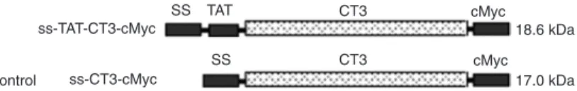
(5). To produce a cell-permeable LIFRα-CT3 with an epitope tag (-cMyc), a eukaryotic expression vector bearing the TAT-CT3-cMyc gene was constructed. A recombinant plasmid without a TAT domain was also constructed as the control (Figure 1). Both pcDNA3.0-ss-TAT-CT3-cMyc and pcDNA3.0-ss-CT3-cMyc plasmids are designed for high-level stable expression in mammalian hosts and for the secretion of proteins that are fused at the N-terminal to the human antibody heavy chain gene.
We established stably transfected cell lines that ex -pressed the two fusion proteins (the ~18.6-kDa ss-TAT-CT3-cMyc and ~17.0-kDa ss-ss-TAT-CT3-cMyc). CHO-ss-TAT-CT3-cMyc and CHO-TAT-CT3-cMyc cells were observed to secrete CT3-cMyc and TAT-CT3-cMyc, respectively, into the culture medium. Western blots of the concentrated, serum-free spent medium of the CHO-CT3-cMyc and CHO-TAT-CT3-cMyc cells, which were prepared using a monoclonal antibody to cMyc, revealed a single band of protein with the same migration as that observed in SDS-PAGE. The serum-free spent medium of wild-type CHO cells was also concentrated and labeled by the same
pro-cedure as an internal control. The molecular weight of CT3-cMyc or TAT-cMyc-CT3-cMyc was approximately 18 kDa, whose specificity was confirmed to one protein band extracted from the spent medium of wild-type CHO cells as the negative control(Figure 2).
TAT-mediated delivery of CT3-cMyc protein to human myeloid HL-60 cells
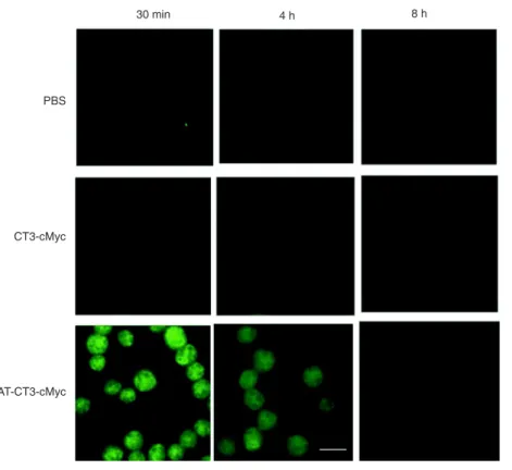
To evaluate the ability of TAT-related CT3 delivery to human myeloid leukemia HL-60 cells, we compared the transmembrane de-livery of CT3-cMyc and TAT-CT3-cMyc after their direct administration to the serum-free medium of the HL-60 cells at various con-centrations or times. Purified CT3-cMyc and TAT-CT3-cMyc fusion proteins were added to a total of 1 x 10⁵ HL-60 cells each so as to achieve final concentrations of 10, 30, and 50 µg/mL at 30 min, 4 h, and 8 h. The identical PBS volumes were added to HL-60 cells to act as controls. The intracellular distributions of these fusion proteins were examined by immunofluorescence.
On a time basis, when the administered doses of both fusion proteins were 30 μg/ mL, which were similar to the PBS controls (Figure 3), no cMyc-positive cells were found in the HL-60 cells that were obtained from the CT3-cMyc-treated group after 4 h (Figure 3); however, cMyc-positive cells were readily detected in the HL-60 cells that were obtained from the TAT-CT3-cMyc-treated group 30 min after their administration (Figure 3).
TAT-CT3-cMyc proteins were primarily found in the nuclei of HL-60 cells. The TAT-CT3-cMyc protein levels, which were calcu-lated based on the number and fluorescence intensity of cMyc-positive cells, peaked at around 30 min to 1 h after their administration and gradually decreased thereafter, although they were still detectable 8 h after administration. These data indicate that TAT-CT3-cMyc but not CT3-cMyc possesses the ability to deliver LIFRα-CT3.
On a dose-dependent basis, because we knew the approximate timing of the transmembrane delivery of TAT-CT3-cMyc, we further explored the dose-response relationships between the fusion protein dosages and fluorescence intensities of cMyc-positive cells after 1 h of TAT-CT3-cMyc fusion protein administration. cMyc-positive cells were readily detected in the HL-60 cells obtained from the TAT-CT3-cMyc-treated group in the 10-μg/mL sample (Figure 4). TAT-CT3-cMyc proteins were primarily found in the nuclei of HL-60 cells. As the protein concentrations were increased, the TAT-CT3-cMyc protein concentrations, which were calculated on the basis of the fluorescence intensity
Figure 1. ss-TAT-CT3-cMyc and control ss-CT3-cMyc fusion proteins.
Figure 2. SDS-PAGE and Western blot of the concentrated spent media of CHO-CT3-cMyc and CHO-TAT-CT3-cMyc cells. The same volume of wild-type (WT) CHO cell spent medium was added as a blank control. The migration of both proteins from the spent medium were similar and indicated an apparent
molecular mass of approximately 18 kDa. Both transformed cells lines were maintained in RPMI-1640 medium supplemented with 10% FBS to near conflu
-ence in 75-cm2 culture flasks. The medium was replaced with 15 mL
serum-free RPMI-1640, which was collected 72 h later, centrifuged, and concentrated.
SDS-PAGE and immunoblots were prepared as described in the Material and Methods section. Lanes 1-4 = Coomassie blue staining; lanes 5-7 =
of cMyc-positive cells, peaked at a concentration of 30 μg/ mL and did not significantly change when compared to the increased concentration up to 50 µg/mL. These data indicate that approximate 30 μg/mL TAT-CT3-cMyc fusion protein was the ideal and saturated dosage close enough
in this experimental design for the transmembrane delivery of 1 x 10⁵ HL-60 cells in vitro.
TAT-CT3-cMyc-induced change in HL-60 cell viability
To assess the effect of delivered LIFRα-CT3 fusion Figure 3. TAT-CT3-cMyc fusion proteins demonstrate transmembrane delivery into HL-60
cells after different times. HL-60 cells were fixed 30 min, 4 h, and 8 h after PBS, CT3-cMyc,
or TAT-CT3-cMyc was added directly to the medium. The intracellular distribution of the
fu-sion protein or control was examined by immunofluorescence using an anti-cMyc antibody.
One hundred cells were analyzed at each time, and the representative images are shown. Scale bar = 15 µm.
Figure 4. Ideal dosage for transmembrane delivery of TAT-CT3-cMyc fusion protein into HL-60 cells. The human myeloid HL-60 cells were cultured with serum-free medium just
before receiving a final concentration of 10, 30, and 50 µg/mL TAT-CT3-cMyc fusion pro
918 Q. Sun et al.
protein in HL-60 cells, we compared the cell viability changes after the direct administration of fusion pro-teins to the serum-free medium of the HL-60 cells at 30 μg/mL for 8 h. No significant difference in cell vi -ability was observed after 24 h of incubation with PBS, CT3-cMyc protein or TAT-CT3-cMyc protein (P > 0.05). A significant decrease in cell viability was observed for HL-60 cells incubated with TAT-CT3-cMyc for 48, 72, and 96 h compared to cells incubated with PBS or CT3-cMyc protein (P <0.05), based on the CCK-8 assay (Figure 5). A significant decrease in cell viability was not observed in the presence of CT3-cMyc or of an equal amount of PBS as the internal control of wild-type HL-60 cells.
Discussion
In the present study, we have shown that an LIFRα-CT3 fusion protein that bears the protein transduction domain of the HIV1-TAT protein, a signal peptide from the human antibody heavy chain gene, and a cMyc-epitope-tag can be derived from stably transformed eukaryotic expression CHO cells and subsequently delivered into human myeloid leukemia HL-60 cells. LIF is so named because it can induce a differ-entiation of the M1 murine myeloid leukemia cell line (24,25). LIFRα-CT3, as reported in our previous research, was observed to have the capacity to activate signal transducer and activator of transcription3 (STAT3) in HL-60 cells by transfection, to initiate LIF-related intracellular signaling, and to facilitate both increasing differentiation and decreasing proliferation (5,26-28); however, transfection or virus-mediated gene delivery may cause an irreversible genetic modification, which makes these deliveries signifi -cantly unacceptable for possible clinical practice (29,30). Peptide-based cell delivery systems are greatly expanded by the recognition of PTDs and synthetic peptides with translocation properties (14,31,32). In the present study, we have shown that the TAT-CT3-cMyc fusion protein can be expressed in eukaryotic system CHO cells, secreted into the medium, and efficiently purified. The purified protein was shown to be able to penetrate not only cells but also the nuclear membranes of HL-60 cells (33).
The technique for the production of TAT-related fusion proteins in general requires the synthesis and purification of such proteins using prokaryotic expression systems (34-36). As an alternative, we have developed a method based on the application of a mammalian expression vector, pcDNA3.0. This vector is designed for high-level expression and can be inserted into other domains so as to regenerate novel properties. In our particular case, the resultant novel property was the ability of the fusion proteins to be secreted out of the CHO cells into serum-free medium supernatants, to be effectively concentrated and directly administered to target leukemia cell. As a prototype, we developed stable
transformed cell lines (37) with easily detected sensitivities in fixed cells to illustrate the advantages of this technique.
The purity of the recombinant TAT-CT3-cMyc was greater than 90%, as determined by SDS-PAGE. Previous studies have shown that secreted TAT fusion proteins are able to transduce target cells but with very low efficiency (37). According to our study, this is not always the case. The original structure of ss-TAT-CT3-cMyc without muta-tions was observed to be qualified enough to ensure the secretion of TAT-CT3-cMyc, which is vital in exploring the structure on which TAT-PTD is based. More specifically, the 30 μg/mL TAT-CT3-cMyc fusion protein concentration used in our study was highly effective in transmembrane location within an hour and was primarily located in the nucleus. We speculate that this phenomenon may have been due first to the location of TAT-PTD, which was located downstream of the signal peptide on the N-terminal, which, in turn, may have somehow transformed itself and may have protected the specific sites from being cleaved by endogenous furins (38,39); second, the signal peptide of our study was never applied to the TAT-based eukaryotic expressing system, which may also contribute to the change in protein confor-mation leading to potential damage.
There are remaining unsolved problems in CHO cell-based TAT-fusion-protein production. The level of protein expression in CHO-transfected cells constantly varies from cell to cell. Some researchers attribute this variation to the inherently stochastic nature of gene expression. The stochastic mechanism in gene expression operates in both prokaryotes and eukaryotes and may explain phenotypic Figure 5. Viability of HL-60 cells measured by the cell counting kit-8 (CCK-8) assay. PBS only, CT3-cMyc or TAT-CT3-cMyc fusion protein was added to serum-free media of HL-60 cells for 8 h before the media were replaced with normal media (RMPI 1640 with 10% FBS). Cell vi-ability was measured using the CCK-8 assay at 24, 48, 72, and 96 h after adding the CT3-cMyc or TAT-CT3-cMyc protein (N = 3 for each
group). A significant decrease in cell viability was observed for HL-60
variations in isogenic cell populations (40). In our observa-tion, TAT-CT3-cMyc and CT3-cMyc were poorly expressed in the transformed CHO cells when determined by immu-nofluorescence using antibodies against cMyc-epitope-tag but highly detectable in a spent medium of corresponding CHO cells (data not shown). Hence, we further speculate that the gene involved in this liposome-based methodology may induce epigenetic alterations of host cells that make it hard to monitor the expression levels in CHO cells but may somehow not affect the efficiency when fusion pro -teins are capable of being secreted outside the host cells. Such evidence and reasoning make CHO cells, which are a eukaryotic expression system, an ideal tool for LIFRα-CT3-based acute myeloid leukemia therapy.
The use of a mammalian secretory system to gener-ate TAT-relgener-ated fusion proteins facilitgener-ates their preparation because of its apparent soluble form and more reliable
transcriptional structure so that they can be directly added to cultured cells. The positive use of LIF in vitro, which is defined as promoting myeloid differentiation and inhibiting cell proliferation, can be achieved by TAT-CT3-based ex -tracellular administration of TAT-CT3-cMyc to HL-60 cells. We have also presumed that the therapeutic application of this technique, whereby parent cells have been transfected with constructs that code for the PTD fusion proteins, could benefit leukemia patients (14).
Acknowledgments
Research supported by grants from the Science and Technology Commission of Shanghai Municipality (No. 09431901100) and Key Program of High and Novel-Tech Building in Clinical Medicine for the People’s Liberation Army (#2010GXJS023).
References
1. Guo SY, Shen X, Yang J, Yuan J, Yang RL, Mao K, et al. TIMP-1 mediates the inhibitory effect of interleukin-6 on the proliferation of a hepatocarcinoma cell line in a STAT3-de-pendent manner. Braz J Med Biol Res 2007; 40: 621-631.
2. Liu H, Dan J, Tang S, Wu S. Involving of the cytoplasmic region of leukemia inhibitory factor receptor alpha subunit, IL-6 related signal transducer-gp130 or fas death domain for
MAPK p42/44 activation in HL-60 cell with LIF or anti-Fas
IgG. Mol Cell Biochem 2001; 217: 113-120.
3. White UA, Stephens JM. Neuropoietin activates STAT3 inde-pendent of LIFR activation in adipocytes. Biochem Biophys Res Commun 2010; 395: 48-50.
4. Cardoso BA, Girio A, Henriques C, Martins LR, Santos C, Silva A, et al. Aberrant signaling in T-cell acute lymphoblastic leukemia: biological and therapeutic implications. Braz J Med Biol Res 2008; 41: 344-350.
5. Yang L, Liu SR, Tang SP, Wang FM, Liu HQ. [Effects of the
box-3 region of the LIFRalpha-chain cytoplasmic domain
(gp190CT3) on the proliferation and differentiation of HL-60 cells]. Zhonghua Xue Ye Xue Za Zhi 2004; 25: 679-682.
6. Tomida M, Heike T, Yokota T. Cytoplasmic domains of the leukemia inhibitory factor receptor required for STAT3 acti-vation, differentiation, and growth arrest of myeloid leukemic cells. Blood 1999; 93: 1934-1941.
7. Abes R, Arzumanov A, Moulton H, Abes S, Ivanova G, Gait
MJ, et al. Arginine-rich cell penetrating peptides: design, structure-activity, and applications to alter pre-mRNA splic-ing by steric-block oligonucleotides. J Pept Sci 2008; 14:
455-460.
8. Torchilin VP. Cell penetrating peptide-modified pharmaceu -tical nanocarriers for intracellular drug and gene delivery.
Biopolymers 2008; 90: 604-610.
9. Johnson LN, Cashman SM, Kumar-Singh R. Cell-penetrat-ing peptide for enhanced delivery of nucleic acids and drugs to ocular tissues including retina and cornea. Mol Ther 2008;
16: 107-114.
10. Pan C, Lu B, Chen H, Bishop CE. Reprogramming human
fibroblasts using HIV-1 TAT recombinant proteins OCT4,
SOX2, KLF4 and c-MYC. Mol Biol Rep 2010; 37:
2117-2124.
11. Jiang L, Ma Y, Wang J, Tao X, Wei D. The transduction of His-TAT-p53 fusion protein into the human osteogenic
sar-coma cell line (Saos-2) and its influence on cell cycle arrest
and apoptosis. Mol Biol Rep 2008; 35: 1-8.
12. Hou Y, Zou J. Delivery of HSF1(+) protein using HIV-1 TAT
protein transduction domain. Mol Biol Rep 2009; 36:
2271-2277.
13. Dietz GP, Bahr M. Delivery of bioactive molecules into the cell: the Trojan horse approach. Mol Cell Neurosci 2004; 27:
85-131.
14. Barka T, Gresik ES, Henderson SC. Production of cell lines secreting TAT fusion proteins. J Histochem Cytochem 2004;
52: 469-477.
15. Fang ZH, Dong CL, Chen Z, Zhou B, Liu N, Lan HF, et al.
Transcriptional regulation of survivin by c-Myc in
BCR/ABL-transformed cells: implications in anti-leukaemic strategy. J Cell Mol Med 2009; 13: 2039-2052.
16. Li B, Wang H, Dai J, Ji J, Qian W, Zhang D, et al. Construc-tion and characterizaConstruc-tion of a humanized anti-human CD3 monoclonal antibody 12F6 with effective immunoregulation functions. Immunology 2005; 116: 487-498.
17. Ren XQ, Furukawa T, Aoki S, Nakajima T, Sumizawa T,
Haraguchi M, et al. Glutathione-dependent binding of a
photoaffinity analog of agosterol A to the C-terminal half of
human multidrug resistance protein. J Biol Chem 2001; 276:
23197-23206.
18. Merjan AJ, Kanashiro CA, Krieger JE, Han SW, Paiva AC.
Ligand-induced endocytosis and nuclear localization of
an-giotensin II receptors expressed in CHO cells. Braz J Med Biol Res 2001; 34: 1175-1183.
19. Pompeia C, Cury-Boaventura MF, Curi R. Arachidonic acid
triggers an oxidative burst in leukocytes. Braz J Med Biol Res 2003; 36: 1549-1560.
20. Dostal L, Chen CY, Wang AH, Welfle H. Partial B-to-A DNA transition upon minor groove binding of protein Sac7d
920 Q. Sun et al.
9600-9609.
21. Bach JP, Borta H, Ackermann W, Faust F, Borchers O, Schrader M. The secretory granule protein syncollin local-izes to HL-60 cells and neutrophils. J Histochem Cytochem
2006; 54: 877-888.
22. Ting L, Bo W, Li R, Chen X, Wang Y, Jun Z, et al. AMP-activated protein kinase supports the NGF-induced viability of human HeLa cells to glucose starvation. Mol Biol Rep
2010; 37: 2593-2598.
23. Lang Q, Zhang H, Li J, Xie F, Zhang Y, Wan B, et al.
3-Hy-droxyflavone inhibits endogenous Aurora B and induces
growth inhibition of cancer cell line. Mol Biol Rep 2010; 37:
1577-1583.
24. Tomida M, Yamamoto-Yamaguchi Y, Hozumi M. Purification
of a factor inducing differentiation of mouse myeloid
leuke-mic M1 cells from conditioned medium of mouse fibroblast
L929 cells. J Biol Chem 1984; 259: 10978-10982.
25. Gearing DP, Gough NM, King JA, Hilton DJ, Nicola NA,
Simpson RJ, et al. Molecular cloning and expression of
cDNA encoding a murine myeloid leukaemia inhibitory factor (LIF). EMBO J 1987; 6: 3995-4002.
26. Liu H, Liu S, Tang S, Ji K, Wang F, Hu S. Molecular analysis of signaling events mediated by the cytoplasmic domain of leukemia inhibitory factor receptor alpha subunit. Mol Cell Biochem 2004; 258: 15-23.
27. Liu H, Tang S, Liu S. [Signal of the cytoplasmic regions of
leukemia inhibitory factor receptor (LIFR) alpha-subunit and
gp130 involves Stat3 activation in leukemic U937 cells].
Zhonghua Xue Ye Xue Za Zhi 1999; 20: 621-623.
28. Sun Q, Wang J, Xiong J, Yang L, Liu H. Free LIF receptor
alpha-chain distal cytoplasmic motifs enhance Jak2-inde-pendent STAT3 phosphorylation and induce differentiation in HL-60 cells. Oncol Rep 2011; 26: 399-404.
29. Schwartz JJ, Zhang S. Peptide-mediated cellular delivery.
Curr Opin Mol Ther 2000; 2: 162-167.
30. Ford KG, Souberbielle BE, Darling D, Farzaneh F. Protein
transduction: an alternative to genetic intervention? Gene Ther 2001; 8: 1-4.
31. Denicourt C, Dowdy SF. Protein transduction technology of-fers novel therapeutic approach for brain ischemia. Trends Pharmacol Sci 2003; 24: 216-218.
32. Zhao J, Gao P, Xiao W, Fan LQ, Wang FJ, Li SX, et al. A novel human derived cell-penetrating peptide in drug deliv-ery. Mol Biol Rep 2011; 38: 2649-2656.
33. Gump JM, June RK, Dowdy SF. Revised role of glycosamino-glycans in TAT protein transduction domain-mediated cellular transduction. J Biol Chem 2010; 285: 1500-1507.
34. Wu Y, Ren C, Gao Y, Hou B, Chen T, Zhang C. A novel
method for promoting heterologous protein expression in
Escherichia coli by fusion with the HIV-1 TAT core domain.
Amino Acids 2010; 39: 811-820.
35. Wang Y, Lin H, Lin S, Qu J, Xiao J, Huang Y, et al. Cell-penetrating peptide TAT-mediated delivery of acidic FGF to retina and protection against ischemia-reperfusion injury in rats. J Cell Mol Med 2010; 14: 1998-2005.
36. Muthumani K, Lambert VM, Shanmugam M, Thieu KP, Choo AY, Chung JC, et al. Anti-tumor activity mediated by protein and peptide transduction of HIV viral protein R (Vpr). Cancer Biol Ther 2009; 8: 180-187.
37. Flinterman M, Farzaneh F, Habib N, Malik F, Gaken J,
Tavassoli M. Delivery of therapeutic proteins as secretable TAT fusion products. Mol Ther 2009; 17: 334-342.
38. Gaken J, Jiang J, Daniel K, van Berkel E, Hughes C, Kuiper M, et al. Fusagene vectors: a novel strategy for the expres -sion of multiple genes from a single cistron. Gene Ther 2000;
7: 1979-1985.
39. Tikhonov I, Ruckwardt TJ, Berg S, Hatfield GS, David PC.
Furin cleavage of the HIV-1 Tat protein. FEBS Lett 2004;
565: 89-92.
40. McAdams HH, Arkin A. Stochastic mechanisms in gene