Analysis of phenotypes altered by temperature stress and
hipermutability in Drosophila willistoni
Marícia F. D’Ávila1, Rosane N. Garcia2, Elgion L. S. Loreto1 & Vera Lúcia da S. Valente1
1. Departamento de Genética, Instituto de Biociências, Universidade Federal do Rio Grande do Sul, Av. Bento Gonçalves 9500, Bloco B, Prédio 43323, Caixa Postal 15053, 91501-970 Porto Alegre, RS, Brazil. ([email protected])
2.Programa de Pós-Graduação em Biologia Animal, Departamento de Zoologia, Universidade Federal do Rio Grande do Sul. Caixa Postal 15053, 91501-970 Porto Alegre, RS, Brazil.
ABSTRACT. Drosophila willistoni (Sturtevant, 1916) is a species of the willistoni group of Drosophila having wide distribution from the South of USA (Florida) and Mexico to the North of Argentina. It has been subject of many evolutionary studies within the group, due to its considerable ability to successfully occupy a wide range of environments and also because of its great genetic variability expressed by different markers. The D. willistoni 17A2 strain was collected in 1991 in the state of Rio Grande do Sul, Brazil (30°05’S, 51°39’W), and
has been maintained since then at the Drosophila laboratory of UFRGS. Different to the other D. willistoni strains maintained in the laboratory, the 17A2 strain spontaneously produced mutant males white-like (white eyes) and sepia-like (brown eyes) in stocks held at
17°C. In order to discover if this strain is potentially hypermutable, we submitted it to temperature stress tests. Eighteen isofemale strains were used in our tests and, after the first generation, all the individuals produced in each strain were maintained at 29°C. Different phenotype alterations were observed in subsequent generations, similar to mutations already well characterized in D. melanogaster
(white, sepia, blistered and curly). In addition, an uncommon phenotype alteration with an apparent fusion of the antennae was observed,
but only in the isofemale line nº 31. This last alteration has not been previously described as a mutation in the D. melanogaster species. Our results indicate that the D. willistoni 17A2 strain is a candidate for hypermutability, which presents considerable cryptic genetic
variability. Different factors may be operating for the formation of this effect, such as the mobilization of transposable elements, effect of inbreeding and alteration of the heat-shock proteins functions.
KEYWORDS. D. willistoni, 17A2 strain, temperature stress, genetic variability, hypermutability.
RESUMO. Análises de fenótipos alterados pelo stress de temperatura e hipermutabilidade em Drosophila willistoni.Drosophila willistoni (Sturtevant, 1916) é uma espécie do grupo willistoni de Drosophila que apresenta ampla distribuição geográfica desde o sul dos Estados Unidos (Flórida) e México até o norte da Argentina. Esta espécie tem sido alvo de muitos estudos evolutivos dentro do grupo devido à sua considerável capacidade de explorar de forma bem sucedida diversos tipos de ambientes e também por sua grande variabilidade genética expressa através de diferentes marcadores. A linhagem 17A2 de D. willistoni foi coletada em 1991 no Estado do Rio Grande do
Sul, Brasil (30°05’S, 51°39’W), e vem sendo mantida desde então no laboratório de Drosophila da Universidade Federal do Rio Grande do Sul. Diferentemente das demais linhagens de D. willistoni mantidas no laboratório, a linhagem 17A2 já produziu espontaneamente
machos do tipo white (olhos brancos)e dotipo sepia (olhos marrons) em estoques mantidos a 170C. A fim de avaliar se esta linhagem é potencialmente hipermutável, nós submetemo-la a experimentos de estresse por temperatura. Dezoito isolinhagens foram utilizadas e, após a primeira geração, todos os indivíduos de cada isolinhagem foram mantidos a 29°C. Diferentes alterações foram observadas nas gerações seguintes, similares com mutações já bem caracterizadas em D. melanogaster (white, sépia, blistered e curly). Além destas, uma
alteração com aparente fusão nas antenas foi encontrada, mas somente em indivíduos da isolinhagem nº 31. Esta última alteração não foi previamente descrita como uma mutação na espécie D. melanogaster. Nossos resultados indicam que a linhagem 17A2 de D. willistoni é
uma candidata a hipermutabilidade, além de apresentar uma variabilidade genética importante. Diferentes fatores podem estar atuando para a formação deste panorama observado, tais como mobilização de elementos transponíveis, seleção por endocruzamento e alteração na função de proteínas de choque térmico (heat-shock proteins).
PALAVRAS-CHAVE.D. willistoni, linhagem 17A2, estresse por temperatura, variabilidade genética, hipermutabilidade.
The 17A2 strain of Drosophila willistoni
Sturtevant, 1916 was collected in 1991 at an agronomic station in Eldorado do Sul, state of Rio Grande do Sul, Brazil (30°05’S, 51°39’W) and since then has been maintained in the Drosophila Laboratory of the
Department of Genetics at UFRGS (Universidade Federal do Rio Grande do Sul, Brazil). This strain has been used in cytogenetic studies that detected a high level of polymorphism for paracentric chromosomic inversions and male recombination (SANTOS-COLARESet al., 2004).
Differing to the findings in the greater part of material collected in natural environments, the 17A2 strain of D. willistoni already presented spontaneous male
mutants white-like (white eyes)and sepia-like (brown
eyes) in stocks, which had been maintained at 17°C in laboratory. These mutations were selected by controlled crossings and stable strains were obtained.
Mutations are very rare events in nature and in
Drosophila the mutation rate has been found to be of
the order of 1 in 100,000, with a variation between 1 in 20,000 and 1 in 200,000 gametes (GARDNER & SNUSTAD,
1986). When a series of spontaneous mutations occur, we can suspect of the hypermutability phenomenon.
Although spontaneous mutations have been reported in previous studies of the D. willistoni species,
stable sibling strains from the altered phenotypes have been difficult to establish. VALIATIet al. (1999), working
Only recently GOÑIet al. (2002) obtained stable
strains of spontaneous mutations in D. willistoni from
individuals collected in Montevideo, Uruguay (34°53’S, 56°16’W). Two mutations yellow-like and dull red-like
were segregated from F2 of the EM1.00 strain. In later generations white and pink mutations appeared. All the
mutant strains demonstrated good viability. By controlled crossings with subsequent cytogenetic analysis, it was shown that the white, pink and yellow lociare located in
theX chromosome.
An alteration in the eye with a phenotype similar to the mutation already described for Drosophila melanogaster Meigen, 1830 known as eyeless (ey) was
also found in D. willistoni (GOÑIet al., 2002). This type of
mutation, inwhich total or partial eye loss occurs, was analyzed at the molecular level by LUDWIGet al. (2003),
but it was not possible to identify the agent responsible for this alteration. These authors suggested that the effect could be related to spot mutations, mutations in genes neighboring the ey region, or even mutations that might
have occurred in another gene having a phenotype similar to the eyeless (review in GEHRING, 2001).
Some environmental factors, such as chemical or physical stress may favor the appearance of morphological alterations due to intrinsic genetic or physiological events, such as: 1) mobilization of the Transposable Elements (TEs) in the host genome, (revision by CAPYet al., 1998); 2)
selection by inbreeding, where a population is generated from only a few individuals (LOESCHCKEet al.,1997); 3)and
by alterations involving Heat -Shock Proteins (HSPs) during development (PETERSEN & YOUNG, 1991). This latter possibility
does not necessarily involve changes in the DNA sequences, but alterations in the expressivity of the cryptic genetic variability (RUTHERFORD & LINDQÜIST, 1998).
As all the organisms are strongly affected by their surrounding environments, the environmental factors play an important part in shaping the ecology and evolution of biological systems. Environmental stresses are especially important at many levels of biological organization (HOFFMANN & PARSONS, 1997; HOFFMANN &
HERCUS, 2000), and one of the ways by which virtually all
organisms combat stress is through the heat-shock response (LINDQUIST & CRAIG, 1988; MORIMOTOet al., 1994;
FEDER & HOFFMANN, 1999).
Heat-shock proteins (HSPs) are the main constituents of this response and typically function as molecular chaperones, which assist in maintaining cellular protein structure. In Drosophila, during thermal stress, occurs the
synthesis of these proteins (SØRENSENet al., 2003) and the
members of the Hsp70, for example family, are capable of mitigate cellular damage (LINDQUIST, 1981; PARSELLet al.,
1993; KREBS & LOESCHCKE, 1994; FEDERet al., 1996; SØRENSEN
et al., 2003; GONG & GOLIC, 2006). However, a cost of
expressing the heat-shock response was identified and the increased survival after acclimation is accompanied by reduced fertility (LOESCHCKEet al., 1997).
The Hsp90 was proposed to act as a general evolutionary capacitor by releasing the effect of cryptic genetic variation under stressful environment (RUTHERFORD & LINDQUIST, 1998). However, more recent
studies have revealed that the influence of Hsp90 on the variation of particular traits was very limited (MILTONet
al., 2005). This suggests that the ability of chaperones,
and Hsp90 in particular, to buffer phenotypic variation is not so general, but might rely on very specific interactions more tightly involved in particular phenotypes. Recent studies in D. melanogaster make a link between
chaperones and chromatin regulators. Cold temperature induces a dark pigmentation, in particular in posterior segments, while higher temperature has the opposite effect. The homeotic gene Abdominal-B (Abd-B) has a major role in the plasticity of pigmentation in the abdomen. Temperature acts on melanin production by modulating a chromatin regulator network, interacting genetically with the transcription factor bric-a-brac (bab), a target of Abd-B and Hsp83, encoding the chaperone Hsp90. Genetic disruption of this chromatin regulator network increases the effect of temperature and the instability of the pigmentation pattern in the posterior abdomen (review in GILBERTet al., 2007). Interesting, evidences show that Drosophila heat-shock genes are especially susceptible
to the insertion of Transposable Elements (TEs) in nature and this can have dramatic fitness consequences, creating variation on which selection can act (WALSERet al., 2006;
CHENet al., 2007).
Transposable Elements (TEs) constitute a substantial component of the Drosophila genome
(LINDSLEY & ZIMM, 1992). According to experimental
estimates (HARADAet al., 1990; NUZHDIN & MACKAY, 1995),
the rates of spontaneous transpositions are approximately 10–5 - 10–6 events per TE copy per sperm
per generation. Direct and indirect evidence has accumulated in favors of the phenomenon of TE transposition being induced by external stress factors, of which temperature treatment and γ-irradiation are the
most important. It was demonstrated that heat shock induces transcription (necessary for retrotransposition) of the copia retrotransposon (STRAND & MCDONALD,
1985). However, the control of TE activity is dependent of the host genome and its interaction with environmental factors. In Drosophila, some retrotransposons were
mobilized after heat-shocks in certain laboratory lines (RATNERet al., 1992; ZABANOVet al. 1994; VASILYEVAet al., 1999), while in other experiments that effect was not
found (ARNAULTet al., 1997; VÁZQUEZet al., 2007).
In view of the appearance of spontaneous mutations in the 17A2 strain and the possibility that this might be a clue to the identification of agents responsible for the variability of this species, we designed this study to identify different alterations in the phenotypes which occur when individuals of this strain are submitted to physical stress (by temperature increase), as well as to describe in detail the type of alteration found in the antenna morphology, herein demonstrated for the first time for the Drosophila genus.
Because the species D. willistoni presents
extensive genetic variability expressed by various markers such as polymorphism chromosomal for paracentric inversions and enzymatic polymorphism (AYALA &
POWELL, 1972), a descriptive investigation such as we
present in this study can provide a base for future research, which could lead to a better understanding of the possible causative agents of this D. willistoni genetic
MATERIAL AND METHODS
The stocks of D. willistoni populations were
maintained by general massal crosses in a standard culture medium (MARQUESet al., 1966) inside a chamber kept at a
constant temperature of 17°C ± 1°C with approximately 60% relative humidity. Two strains were used in this work: WIP-4 (a strain maintained in laboratory since 1966 and that never presented spontaneous mutation) as negative control and 17A2 (a strain colleted in 1991), target of this work. Initially, for obtaining the isostrains of the each strain, forty natural-born virgin females and males were crossed. Only eighteen strains of each population initially selected (the number is indicating in table I), produced offspring for the continuities of the experiment. After the first generation, descendents of each of these strains were submitted to temperature stress being maintained in culture chambers at 29°C. In each new generation, after brief anesthesia in ethylic ether and visualization on stereomicroscope, the individuals were analyzed and counted. All the mutants were removed and submitted to controlled crossings (a mutant phenotype male crossed with a wild type virgin female) in an effort to obtain stable mutant strains. The strains from the population WIP-4 did not show individuals with mutant phenotypes (data
not shown). Thus, the number of controlled crossings is the number of mutant individuals obtained in each generation, for each isofemale line in 17A2 strain, as described in table I.
Individuals of wild phenotype were placed in a new culture medium to breed and deposit their eggs again. The strains were followed-up for eleven generations. The phenotypic alterations detected in each generation were photographed with a stereomicroscope. Ten male individuals of the 17A2 strain and ten of the mutant
blistered strain had been dissected in EPHRUSSI & BEADLE
(1936) serum for the withdrawal of the testes in order to evaluate the structure, appearance and presence/absence of spermatozoa. The results of these comparative analyses were also photographed with a stereomicroscope.
Antennae that showed alterations were analyzed separately on permanent slides mounted with Entellan® (Merck). Slides visualization and image capture was achieved by using a stereomicroscope. Samples of these individuals demonstrating antennae alterations were also dehydrated and fixed for subsequent analyses of the antennae in a Scanning Electron Microscopy (SEM). Normal antennae were processed as controls. The procedures for the preparation for these samples and the image capture were made at the Electronic Microscope
Table I. Total frequency of individuals obtained in each generation in the 17A2 D. willistoni isostrains submitted to temperature stress.
(T, total number of individuals; M, number of individuals with morphological alterations).
Generation
Isostrain F2 F3 F4 F5 F6 F7 F8 F9 F10 F11
1 T:216 T:145 T:158 T:84 T:123 T:88 T:157 T:110 T:77 T:41
M:1 M:2 M:1 M:0 M:2 M:1 M:0 M:13 M:0 M:1
2 T:112 T:238 T:32
M:0 M:1 M:0
4 T:56 T:566 T:40 T:693
M:0 M:2 M:0 M:28
6 T:47 T:472 T:199 T:122 T:114
M:0 M:2 M:0 M:1 M:2
7 T:84 T:664 T:75 T:166
M:0 M:21 M:1 M:3
8 T:124 T:31
M:0 M:0
9 T:72 T:1026 T:462 T:113 T:40
M:0 M:2 M:1 M:2 M:5
1 0 T:67 T:605 T:146 T:133 T:88
M:0 M:3 M:10 M:4 M:0
1 3 T:42 T:1145 T:250 T:154 T:50
M:0 M:12 M:9 M:2 M:8
2 0 T:86
M:0
2 3 T:155 T:23 T:11
M:3 M:0 M:0
2 6 T:8
M:0
2 7 T:256 T:113 T:62
M:2 M:4 M:0
2 8 T:26
M:0
2 9 T:320 T:137 T:73
M:13 M:0 M:2
3 0 T:16
M:0
3 1 T:311 T:92 T:540 T:68 T:363 T:174 T:263
M:4 M:13 M:2 M:10 M:6 M:4 M:37
3 3 T:42
Center (CME) of UFRGS (Porto Alegre, Brazil) in accordance with the standard procedures adapted at that Center. Some individuals of the isostrains presenting reduction in the number of births were dissected to find out if the reproductive structures showed any kind of alteration.
RESULTS
Of the 18 isostrains initially placed in the chamber at 29°C, five were lost in F2, one in F3, four in F4, two in F5, four in F6 and one in F8 - only the 1B isostrain survived to the end of the experiment (Tab. I). In general this loss was mainly due to the gradual reduction in the number of births. The individuals of the isostrains that presented reduction in the number of births had the female gonads and the male testes morphology normal, but no spermatozoa were produced within this last (Fig. 1).
A great number of individuals with different phenotypic alterations appeared in the different isostrains analyzed (indicated by M in table I). The alterations found resembled some D. melanogaster mutants: white (white
eyes), sepia (sepia eyes), blistered (bubbles with
hemolymph in the wings) and Curly (curved wings).
During the experiment two individuals with a double-mutation phenotype were observed: one white/Curly
-like and the other white/blistered-like. Another alteration
with the antennae apparently fused was observed. This latter alteration was found in various individuals but only in those of the isostrain number 31 (Fig. 2).
Although the morphologically altered individuals were removed from the isostrains after the count for the establishment of stable strains, the altered phenotypes continued to show up in subsequent generations (Fig. 3) suggesting that there is some factor intrinsic to the
Fig. 1. Stereo microscope illustrations of the Drosophila willistoni testes. A. normal testes of a wild male of the 17A2 strain. B. Testes of a male of the blistered phenotype obtained from the isostrain 1B. The structure has a transparent aspect because no spermatozoids are
present. Bar, 0.5mm
Fig. 2. Altered phenotypes obtained in individuals of Drosophila willistoni 17A2 isostrain submitted to temperature stress. Picture shows female individuals with altered morphologies: A. white (white eyes); B. sepia (brown eyes); C. blistered (arrow indicates the presence of
genomes of the individuals, that causes the reappearance of thealteration.
Of the controlled crossings carried out with the different altered phenotypes, only one strain with origin in a male blistered-like was established. This individual
had been crossed with a natural-born female. The descendents of this crossing were selected and maintained in stock in the laboratory at 25ºC for approximately eight months. However, after this period,
the number of individuals produced gradually declined until finally the strain was entirely lost.
No previous descriptions of D. melanogaster
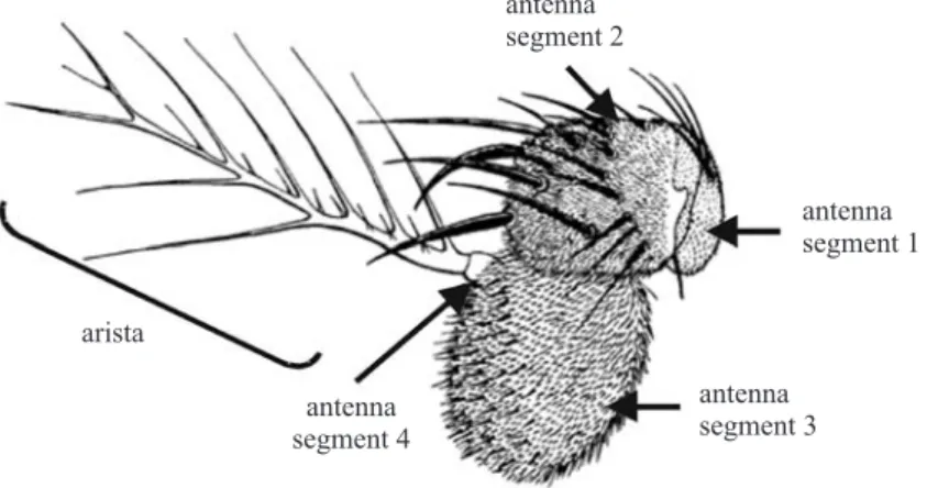
individuals with the antenna alteration phenotype described here were found in the available literature. The pair of antennae is located in the frontal portion of the fly’s head and is divided into four segments. In these, bristles and aristae are found and the entire structure performs hearing (GÖPFERT & ROBERT, 2001; TODIet al.,
2004) and olfactory (FISHILEVICH & VOSSHALL, 2005)
functions. A sketch of the D. melanogaster antenna
structure is provided in figure 4.
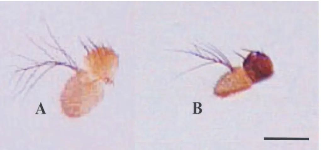
On mutants of D. willistoni isostrain 31it can be
seen that the altered antennae are very close to each other giving the impression when observed at glance that they form a single structure (Fig. 5). We checked that these altered antennae were not different from those observed in wild type flies as regards to the number of segments and bristles of the aristae, but these were always smaller (Fig. 6). Analyses carried out using SEM confirmed the difference in the structures size and further revealed that the antennae became approximated, giving the impression that they constituted only one structure, because they were closely implanted in the head of the flies (Fig. 7).
When crossings were made (at 25°C) between the individuals carrying the antenna alterations and wild type
Fig. 5. Stereo microscope view of Drosophila willistoni antennae. A. An individual with altered antennae (arrow) which appeared in the
isostrain 31. B. View of the antennae of a wild individual (arrows). Note that the altered antennae appear to be a single structure. Bar, 0.5mm
Fig 4. Sketch representing one of Drosophila melanogaster antenna with the respective structures adapted from BRYANT (1978).
0 0,02 0,04 0,06 0,08 0,1 0,12 0,14 0,16 0,18
F2 F3 F4 F5 F6 F7 F8 F9 F10 F11
Generations
isostrain 1 isostrain 6 isostrain 9 isostrain 10 isostrain 13 isostrain 31
Mutant
phenotypes
frequency
flies, the offspring did not show the alterations presented by their parents. Despite of this, generations of the 31º strain maintained at 29°C continued to produce these malformations. In view of this, it was not possible to derive stable strains from these individuals.
DISCUSSION
Our descriptions of the D. willistoni altered
phenotypes produced through temperature stress indicate that an intrinsic factor in the genome of the individuals causes the alterations to appear. This idea is bourn out by the fact that the altered phenotypes continued to show up in subsequent generations, although in each generation the affected individuals were removed from isostrains after the count.
So it was not possible to relate the inviability of strain with the increased number of mutants as observed in the strains 4, 9, 13 and 31 (Tab. I). Besides this, some strains, although presented low number of mutants, were also lost after few generations. However, we cannot completely discard the possibility that the former strains could be lost due to a higher intrinsic genomic instability. Organisms have developed a series of mechanisms to cope with environmental stress, including increased expression of stress proteins and changes in metabolism and hormone concentrations (PLETCHER et al., 2002;
SØRENSENet al., 2005; LANDISet al., 2004). It has been
shown that some of the same mechanisms are also affected by intrinsic stresses, such as inbreeding and aging (KRISTENSEN et al., 2005; PLETCHER et al., 2002;
LANDISet al., 2004; PEDERSENet al., 2005). The deleterious
consequences of inbreeding, environmental stress and the interactions between them are of serious concern in diverse fields of biological sciences, such as evolutionary genetics, conservation biology, and animal breeding. Recently, KRISTENSENet al. (2006) found that the effect of
temperature stress on gene expression is accentuated in inbred compared to out bred lines of D. melanogaster.
On the basis of these results the authors conclude that inbreeding and environmental stress, both independently and synergistically affect gene expression pattern. It also appears to have occurred in the present study. The majority of the alterations observed in the D. willistoni
17A2 strain are mutations well characterized in D. melanogaster.Those which affect the eye pigmentation,
for example, show alterations in gene sequences involved in ommatidia pigmentation metabolic pathways, and may cause the brown (sepia mutant) and white eyes (white
mutant) to appear, contrary to what happens with wild type individuals who have red eyes. The white gene is
involved in the production and distribution of the omochrome pigments (browns) and pteridines (reds) found in the adult flies compounding the eyes and ocelli, as well as in the adult males testes and in the larvae Malpighi tubules. The specific function of the transport
Fig. 7. Frontal view analysis of Drosophila willistoni antennae (SEM). To the left are the badly-formed antennae which occur in the
individuals of the isostrain 31. Note the close implantation of the structures in the head (arrow) which is quite different from that of individuals with the wild 17A2 isostrain phenotype (to the right), where the antennae are implanted farther apart and the structures are well defined (arrows). 400X magnification.
protein, associated to the membrane, is to connect ATP-precursor pigments in the omochrome and pteridines pathways (SULLIVAN & SULLIVAN, 1975; DRESSENet al.,
1988; TEARLEet al., 1989).
The sepia (se) mutant has eyes of a brown color in
the eclosion, which darkens to sepia, and becomes still darker with advancing age. It has normal ocellar pigmentation. Chromographically, the se eyes are
characterized by the absence of red pigments (drosopterin and isodrosopterin, for example), and by an accumulation of yellow pigments. About ten sepia alleles have been
fully described for D. melanogaster (LINDSLEY & ZIMM,
1992).
Other mutations alter the structure of the upper and lower epithelia of the wings causing curvatures(in the curly mutant) or the formation of haemolymph blisters
between the epithelia (in the blistered mutant) (LINDSLEY
& ZIMM, 1992).
The Curly (Cy) is a dominant mutation in D. melanogaster that affects the genes necessary to form
the wing structure in the adult fly. The mutation Curly,
one of the most important tools in genetics analysis in
Drosophila, results in curled wings at 25°C but in normally
straight wings at 19°C (GRIFFITHSet al., 1996). It is used,
for example, as a preferential marker of the second chromosome in genetic laboratories. The curvature of the wings in the Curly fly is caused by an unequal
concentration of the upper and lower epithelia during the dry period after the emergence from the pupa (LINDSLEY & ZIMM, 1992).
The D. melanogaster blistered mutant, on the other
hand, presents formations of haemolymph blisters between the wing surfaces. Some of these mutations are caused by defects in the Drosophila integrin proteins
transmembrane heterodimers, which are part of basal junctions, and maintain the two wing surfaces united (FRISTROMet al., 1993). Mutation in the integrin genes,
such as inflated (if) and myosferoid (mys), cause defects
in the dorsal-ventral adhesion of the wing intervenal cells also resulting in blisters on the wings (BROWER & JAFFE,
1989; WILCOXet al., 1989; ZUSMANet al., 1990).
We cannot definitely state that the phenotypes observed in D. willistoni have their origin in the same
types of mutations previously described for D. melanogaster, because it would be necessary to make a
study at the molecular level of each type of alteration to confirm the similarity unequivocally. Moreover, the alteration resulting in jointed antennae and small aristae, which we found in the isostrain 31 of 17A2 strain, has no similar description in D. melanogaster. This result leads
to the premise that multiple other factors may be involved in the production of the D. willistoni altered phenotypes
which result from exposure to temperature stress. Some hypotheses for the possible agents responsible for this phenomenon are developed below.
The first hypothesis is related to mobilization of the Transposable Elements. High temperatures associate with permissive genomes (VÁZQUEZet al., 2007) may favors
mobilization of these elements by an increase in the production of transposase (ENGELS, 1989) and,
consequently, the phenomenon of gonadal disgenesys (PETERSEN & YOUNG, 1991). In fact, some mutations in D.
melanogaster were characterized as being the result of
the increased mobility of the Transposable Elements. The loco white of D. melanogaster is a hotspot for insertions
presenting, for example, several alleles originated through insertions of Transposable Elements such as Doc, roo
and copia (LINDSLEY & ZIMM, 1992).
LORETOet al. (1998) described a hypermutable strain
of D. simulans Sturtevant, 1919 whose causal agent was
subsequently characterized as being related to the mobilization of a transposable element similar to hobo
(TORRESet al., 2006). Considering that the majority of Drosophila mutations are attributed to transposable
element mobilizations (FINNEGAN, 1990), this would seem
to be an explication pertinent to the phenomenon observed in the D. willistoni 17A2 strain.
Furthermore, some transposable elements are in fact present in the genome of the D. willistoni 17A2 strain.
REGNER et al. (1996) analyzed two populations of D. willistoni (17A2 and WIP-4) for the presence of the P
transposable element, by Southern Blot and in situ
hybridization, and detected the presence of apparently active P elementin the 17A2 strain. LORETOet al. (1998)
and SASSIet al.(2005), using the Southern blot technique,
also detected the presence of sequences similar to hobo
in the genome of the 17A2 strain, although with low homology to those of D. melanogaster.
REGNERet al. (1999) obtained phenotypic alterations
in individuals of the WIP-4 and 17A2 strains in experiments, which associated controlled crossings (between and within strains) and temperature stress. The phenotypes obtained were wing alterations of the Beadex
-like, held-up-like and Notch-like, as well as an eye
alteration similar to extra-eye, all having been previously
described for D. melanogaster. Some individuals with
gonadal disgenesys were also obtained, very similar to what happens with D. melanogaster when there is
mobilization of the P element in its genome. This led the
authors to postulate that P element mobilization might
be the agent causing the mutations.
Our results are similar to those obtained by REGNER
et al. (1999), to the extent that we also obtained alterations
in 17A2 intra-strain crossings under temperature stress. However, in our experiment the alterations are different to those previously described, which suggests that the time period during which the strain was maintained in the laboratory and new putative transpositions may have influenced the obtained results.
The existence of a relationship between environmental conditions and mobilization of TEs, however, still is an open question. In D. simulans natural
populations, the 412 element follows a gradient in copy
number between the South and North hemispheres (VIEIRA
& BIÉMONT, 1996), its copy number being negatively
correlated with minimum temperature (VIEIRAet al., 1998).
Another example of variation in TE number related with natural environmental variation was given for BARE-1 in barley (KALENDAR et al., 2000). Less severe thermal
stresses might have some effect on TE activity, for example high temperature enhances transposition of the P element
in dysgenic crosses (ENGELS & PRESTON, 1980).
the structural mutation rate of 11 TE families (ALONSO
-GONZALEZet al., 2006).
A second hypothesis that could be advanced to explain our results, especially those related to the gradual loss of the isostrains, could be the effect of depression by inbreeding, given that all isostrains were generated from one single couple. The phenomenon of depression by inbreeding is well known, being characterized by a rapid fecundity and viability decline owing to the increase of homozygotes for unfavorable alleles as the population becomes more and more inbred in the laboratory (FUTUYMA, 1992). The evidence that some of the males in
the isostrains produced no spermatozoa supports this hypothesis.
Inbreeding can lead to a homozygozity of development genes mutant alleles which, being sensitive to temperature effects, result in phenotypes altered by stress conditions. This can have been the reason because the strains were lost after the increase of mutant individuals. Inbred populations may hardly suffer the effect of changes in the environment that non-inbred populations perceive as non-stressful, indicating that intrinsic and extrinsic stresses may interact. There is evidence that such interaction can lead to synergism between genetic and environmental stresses causing normally benign environmental conditions to become harmful (review in KRISTENSEN et al., 2006). In D. melanogaster, alleles sensitive to temperature of the
ecdysoneless (ecd) gene lead to third stage mutant larvae
with reduced synthesis or release of the ecdysone hormone in the salivary glands when exposed to temperature stress (HENRICH et al., 1987). A reduction in
ecdysone affects the expression of a proneural gene called atonal (ato), which is responsible for initiating the
process of the development of numberless segment-specific sensorial organs including the eye, the hearing organ (Johnston’s organ located in the antennae) and tension receptors (cordotonal organ located in the paws) (NIWAet al., 2004). The results obtained from the isostrain
31, where the phenotypes with united antennae occurred only at 29°C, may be related to this hypothesis, because when the control crossings were carried out without temperature stress in order to obtain stable strains, altered descendents were not produced.
The final hypothesis, which may explain the appearance of morphological alterations in D. willistoni
17A2 strain under temperature stress, may also be connected to proteins involved in the development processes and to the negative effects that higher temperatures may have on them. The higher temperature may interfere in the regulation pathways of embryonic development genes, in a way that mutations might not necessarily occur. Temperature shock during development induces morphological defects in vertebrates and invertebrates (PETERSEN & YOUNG, 1991).
The Heat Shock Proteins 90 (Hsp90) chaperones the maturation of many regulatory proteins and, in D. melanogaster,buffers genetic variation in morphogenetic
pathways (QUEITSCHet al., 2002). When Hsp90 buffering
activity is compromised, by temperature for example, cryptic variants are expressed and selection can lead to the continued expression of these traits, even when Hsp90
function is restored (RUTHERFORD & LINDQUIST, 1998). The
loss of isostrains in this work may also be alternatively explained by impaired expression of HSPs. In D. melanogaster, alteration in Hsp70 induction is implicated
in reduced fecundity (KREBS & LOESCHCKE, 1994), and
overexpression of Hsp70 is associated with increased larval mortality (KREBS & FEDER, 1997), retarded growth
(FEDER et al., 1992), and reduced egg hatching
(SILBERMANN & TATAR, 2000).
Although the principal objective of the present study is the description of the phenotypes resulting from temperature stress in the 17A2 strain, it is possible to conclude that an intrinsic source of genetic variability exists in this strain, although it still remains to be found. None of the three hypothesis discussed above excludes the others and they may indeed be operating simultaneously. More focused experiments are still necessary in the future to identify with greater accuracy which agents are causing the alterations. However, it can be said that this present work has characterized the D. willistoni 17A2 strain as a hypermutable strain.
Acknowledgments. The authors would particularly like to thank Ardala Breda, Hermes J. Schmitz and Lizandra J. Robe and for English revision and Luis Fernando M. Fonte for helpful suggestions on the pictures. This study was supported by the Brazilian Agencies CNPq and PROPESQ-UFRGS (fellowships and grants).
REFERENCES
ALONSO-GONZALEZ, L.; DOMÍNGUEZ, A. & ALBORNOZ , J. 2006. Direct
determination of the influence of extreme temperature on transposition and structural mutation rates of Drosophila
melanogaster mobile elements. Genetica 128:11-19.
ARNAULT, C.; LOEVENBRUCK, C. & BIEMONT, C. 1997. Transposable
element mobilization is not induced by heat shocks in
Drosophila melanogaster. Naturwissenschaften 84:410-414.
AYALA, F. J. & POWELL, J. R. 1972. Enzyme variability in the
Drosophila willistoni group. Biochemical Genetics
7(3):331-345.
BROWER, D. L. & JAFFE, S. M. 1989. Requirement for integrins
during Drosophila wing development. Nature 342:285-287.
BRYANT, P. J. 1978. Pattern formation in imaginal discs. In:
ASHBURNER, M. & WRIGHT, T. R. F. eds. The Genetics and
Biology of Drosophila, Academic Press. v.2c, p.229-335. CAPY, P.; BAZIN, C.; HIGUET, D. & LAGIN, T. 1998. Dynamics and
evolution of transposable elements. Austin, Texas-Landes Biosciences. 197p.
CHEN, B.; WALSER, J. C.; RODGERS, T. H.; SOBOTA, R. S. ; BURKE, M.
K.; ROSE, M. R. & FEDER M. E. 2007. Abundant, diverse, and
consequential P elements segregate in promoters of small heat-shock genes in Drosophila populations. Journal of Evolutionary Biology 20(5):2056-2066.
DRESSEN, T. D.; JOHNSON, D. H. & HENIKOFF, F. S. 1988. The brown
protein of Drosophila melanogaster is similar to the white
protein and components of active transport complexes.
Molecular Cell Biology 8:5206-5215.
ENGELS, W. 1989. P Elements in Drosophila melanogaster. In:
BERG, D. E. & HOWE, M. M. eds. Mobile DNA. American Society for Microbiology. p.437-484.
ENGELS, W. R. & PRESTON, C. R. 1980. Components of hybrid
dysgenesis in a wild population of Drosophila melanogaster.
Genetics 95:111-128.
EPHRUSSI, B. & BEADLE, G. W. 1936. A technique of transplantation
for Drosophila melanogaster. American Naturalist70:218-225. FEDER, M. E & HOFFMANN, G. E. 1999. Heat-shock proteins,
FEDER, M. E. 1996. Ecological and evolutionary physiology of
stress proteins and the stress response: the Drosophila melanogaster model. In: JOHNSTON, I. A. & BENNET, A. F. eds. Animals and temperature. Cambridge University Press. p.79-102.
FEDER, M. E.; CARTAÑO, N. V.; MILOS, L.; KREBS, R. A. & LINDQUIST,
S. L. 1996. Effect of engineering hsp70 copy number on
Hsp70 expression and tolerance of ecologically relevant heat-shock in larvae and pupae of Drosophila melanogaster.
Journal of Experimental Biology 199:1837-1844. FEDER, M. E.; ROSSI, J. M.; SOLOMON, J.; SOLOMON, N. & LINDQUIST, S.
1992. The consequences of expressing HSP70 in Drosophila
cells at normal temperatures. Genes & Development 6:1402-1413.
FINNEGAN, D. J. 1990. Transposable elements. Drosophila
Information Service 68:371-381.
FISHILEVICH, E. & VOSSHALL, L. B. 2005. Genetic and functional
subdivision of the Drosophila antennal lobe. Current Biology 15(17):1548-1553.
FRISTROM D.; WILCOX, M. & FRISTROM, J. W. 1993. The distribution
of PSintegrins, Laminin A and F-actin during key stages in
Drosophila wing development. Development 117:509-523.
FUTUYMA, D. J. 1992. Biologia Evolutiva. Sociedade Brasileira
de Genética/CNPq, Ribeirão Preto. 631p.
GARDNER, E. J. & SNUSTAD, D. P. 1986. Genética. Guanabara
Koogan, Rio de Janeiro. 756p.
GEHRING, W. J. 2001. The genetic control of eye development
and its implications for the evolution of the various eye types. Zoology 104:171-183.
GIBERT, J. M.; PERONNET, F. & SCHLÖTTERER, C. 2007. Phenotypic
plasticity in Drosophila pigmentation caused by temperature
sensitivity of a chromatin regulator network. PLoS Genetics 3(2):266-280.
GONG, W. J. & GOLIC, K. G.2006. Loss of HSP70 in Drosophila is pleiotropic, with effects on thermotolerance, recovery from heat-shock and neurodegeneration. Genetics 172:275-286. GOÑI, B.; ROHDE, C. & VALENTE, V. L. S. 2002. Genetic
characterization of spontaneous mutations in Drosophila
willistoni. I. Exchange and non-disjunction of the X
chromosome. Drosophila Information Service 85:80-84.
GÖPFERT, M. C. & ROBERT, D. 2001. Turning the key on Drosophila
audition. Nature 411:908.
GRIFFITHS, A. J.; MILLER, J. H.; SUZUKI, D. T.; LEWONTIN, R. C. &
GELBART, W. M. 1996. An Introduction to Genetic
Analysis. W. H. Freeman and Company, New York, 916p.
HARADA, K.; YUKUSHIRO, K. & MUKAI, T. 1990. Transposition rates
of mobile genetic elements in Drosophila melanogaster.
Proceedings of the National Academy of Sciences of USA 87:3248-3252.
HENRICH, V. C.; TUCKER, R. L.; MARONI, G. & GILBERT, L. I. 1987.
The ecdysoneless (ecd 1ts) mutation disrupts ecdysteroid synthesis autonomously in the ring gland of Drosophila
melanogaster. Developmental Biology 120:50-55.
HOFFMANN A. A. & HERCUS M. J. 2000. Environmental stress as an
evolutionary force. Bioscience 50:217-226.
HOFFMANN A. A. & PARSONS P. A. 1991. Evolutionary genetics
and environmental stress. Oxford University Press, New York. 284p.
HOFFMANN, A. A ; DAGHER, H.; HERCUS, M. & BERRIGAN, D. 1997.
Comparing different measures of heat resistance in selected lines of Drosophila melanogaster. Journal of Insect Physiology 43:393-405.
KALENDAR, R.; TANSKANEN, J.; IMMONEN, S.; NEVO, E. & SCHULMAN, A.
H. 2000. Genome evolution of wild barley (Hordeum
spontaneum) by BARE-1 retrotransposon dynamics in
response to sharp microclimatic divergence. Proceedings of the National Academy of Sciences of USA 97:6603-6607. KREBS, R. A. & FEDER, M. E.1997. Deleterious consequences of
HSP70 overexpression in Drosophila melanogaster larvae.
Cell Stress Chaperones 2:60-71.
KREBS, R. A. & LOESCHCKE, V.1994. Costs and benefits of activation
of the heat-shock response in Drosophila melanogaster.
Functional Ecology 8:730-737.
KRISTENSEN, T. N.; SØRENSEN, P.; KRUHØFFER, M.; PEDERSEN, K. S. &
LOESCHCKE, V. 2005. Genome wide analysis of inbreeding effects
on gene expression in Drosophila melanogaster. Genetics 171:157-167.
KRISTENSEN, T. N.; SØRENSEN, P.; PEDERSEN, K. S.; KRUHØFFER, M. &
LOESCHCKE, V. 2006. Inbreeding by environmental interactions
affect gene expression in Drosophila melanogaster. Genetics 173:1329-1336.
LANDIS, G. N.; ABDUEVA, D.; SKVORTSOV, D.; YANG, J.; RABIN, B. E.;
CARRICK, J.; TAVARE, S. & TOWER, J. 2004. Similar gene
expression patterns characterize aging and oxidative stress in
Drosophila melanogaster. Proceedings of The National
Academy of Sciences of USA 101:7663-7668.
LINDQUIST, S. & CRAIG, E. A. 1988. The heat-shock proteins.
Annual Review of Genetics 22:631-677.
LINDQUIST, S.1981. Regulation of protein synthesis during
heat-shock. Nature 293:311-314.
LINDSLEY, D. L. & ZIMM, G. G. 1992. The genome of Drosophila
melanogaster. Academic Press, San Diego. 1133p.
LOESCHCKE, V.; KREBS, R. A.; DAHLGRAARD, J. & MICHALAK, P. 1997.
High-temperature stress and the evolution of termal resistance in Drosophila. In: BIJLSMA, R. & LOESCHCKE, V. eds. Enviromental Stress, Adaptation and Evolution.
Birkhäuser Verlag Basel/Switzerland. p.175-190.
LORETO, E. L. S.; ZAHA, A. & VALENTE, V. L. S. 1998. Characterization
of a hypermutable strain of Drosophila simulans. Cell Molecular Life Science 54:1283-1290.
LUDWIG, A.; PARADA, C.; LORETO, E. L. S. & GOÑI, B. 2003. Genetics
and molecular analysis of a Drosophila willistoni spontaneous
mutation similar to eyeless. Drosophila Information Service 86:138-142.
MARQUES, E. K.; NAPP, M.; WINGE, H. & CORDEIRO, A. R. 1966. A
corn meal, soybean flour, wheat germ medium for Drosophila.
Drosophila Information Service 41:187.
MILTON, C. C.; BATTERHAM P.; MCKENZIE, J. A. & HOFFMANN, A. A.
2005. Effect of E(sev) and Su(Raf) Hsp83 mutants and trans-heterozygotes on bristle trait means and variation in
Drosophila melanogaster. Genetics 171:119-130.
MORIMOTO, R. I.; JURIVICH, D. A.; KROEGER, R. E.; MATHUR, S. K.;
MURPHY, S. P.; NAKAI, A.; SARGE, K., ABRAVAYA, K. & SISTONEN, L.
T. 1994. Regulation of heat shock gene expression by a family of heat shock factors. In: MORIMOTO, R. I.; TISSIERES, A.
& GEORGOPOULOS, C. eds. The biology of heat shock proteins and molecular chaperones. Cold Spring Harbor Laboratory Press, NY. p.417-455.
NIELSEN, M. M.; SØRENSEN, J. G.; KRUHØFFER, M.; JUSTESEN, J. &
LOESCHCKE, V. 2006. Phototransduction genes are upregulated
in a global gene expression study of Drosophila melanogaster
selected for heat resistance. Cell Stress Chaperones 11:325-333. NIWA, N.; HOROMI, Y. & OKABE, M. 2004. A conserved
developmental program for sensory organ formation in
Drosophila melanogaster. Nature Genetics 36(3):293-297.
NUZHDIN, S. V. & MACKAY, T. F. C. 1995. The genomic rate of
transposable element movement in Drosophila melanogaster.
Molecular Biology and Evolution 21:180-181.
PARSELL, D. A.; TAULIEN J. & LINDQUIST S. 1993. The role of
heat-shock proteins in thermotolerance. Philosophical Transactions of the Royal Society of London Series B-Biological Sciences 339:279-285.
PEDERSEN, K. S.; KRISTENSEN, T. N. & LOESCHCKE, V. 2005. Effects
of inbreeding and rate of inbreeding in Drosophila
melanogaster – Hsp70 expression and fitness. Journal of
Evolutionary Biology 18:756-762.
PETERSEN, N. S & YOUNG, P. 1991. Effects of heat shock on
development and Actin mRNA stability in Drosophila. In:
MARESCA, D. & LINDQUIST, S. eds. Heat Shock. Berlin Heidelberg,
Springer-Verlag. p.155-161.
PLETCHER, S. D.; MACDONALD, S. J.; MARGUERIE, R.; CERTA, U.; STEARNS,
S. C.; GOLDSTEIN, D. B. & PARTRIDGE, L. 2002. Genome-wide
transcript profiles in aging and calorically restricted Drosophila
melanogaster. Current Biology 12:712-723.
QUEITSCH, C.; SANGSTER, T. A. & LINDQUIST, S. 2002. Hsp90 as a
capacitor of phenotypic variation. Nature 417:598-599.
RATNER, V. A.; ZABANOV, S. A.; KOLESNIKOVA, O. V. & VASILYEVA, L.
A. 1992. Induction of the mobile genetic element Dm-412
Recebido em junho de 2007. Aceito em janeiro de 2008. ISSN 0073-4721 Artigo disponível em: www.scielo.br/isz
REGNER, L. P.; ABDELHAY, E.; ROHDE, C.; RODRIGUES, J. J. S. &
VALENTE, V. L. S. 1999. Temperature-dependent gonadal hybrid
dysgenesis in Drosophila willistoni. Genetics and Molecular Biology 22(2):205-211.
REGNER, L. P.; PEREIRA, M. S. O.; ALONSO, C. E. V.; ABDELHAY, E. &
VALENTE, V. L. S. 1996. Genomic distribution of P elements in
Drosophila willistoni and search for their relationship with
chromosomal inversions. Journal of Heredity 87(2):190-211.
RUTHERFORD, S. L. & LINDQUIST, S. 1998. Hsp90 as a capacitor for
morphological evolution. Nature 396:336-342.
SANTOS-COLARES, M. C.; DEGRANDI, T. H. & VALENTE, V. L. S. 2004.
Cytological detection of male recombination in Drosophila willistoni. Cytologia 69(4):359-365.
SASSI, A. K; HERÉDIA, F.; LORETO, E. L. S.; VALENTE, V. L. S.; ROHDE,
C. 2005. Transposable elements P and gypsy in natural
populations of Drosophila willistoni. Genetics and Molecular Biology 69(4):734-739.
SHINE, R.; HARLOW, P. S; ELPHICK, M. J.;OLSSON, M. M. & MASON, R.
T. 2000. Conflicts between courtship and thermoregulation: the thermal ecology of amorous male garter snakes (Thamnophilis sirtalis parietalis, Colubridae). Physiological and Biochemical Zoology 73:508-513.
SILBERMANN, R. & TATAR, M.2000. Reproductive costs of heatshock
protein in transgenic Drosophila melanogaster. Evolution 54:2038-2045.
SØRENSEN, J. G.; KRISTENSEN, T. N. & LOESCHCKE, V. 2003. The
evolutionary and ecological role of heat-shock proteins.
Ecology Letters 6:1025-1037.
SØRENSEN, J. G.; DAHLGAARD, J. & LOESCHCKE, V. 2001. Genetic
variation in thermal tolerance among natural populations of
Drosophila buzzatii: down regulation of Hsp70 expression
and variation in heat stress resistance traits. Functional Ecology 15:289-296.
SØRENSEN, J. G.; NIELSEN, M. M. & LOESCHCKE, V. 2007. Gene
expression profile analysis of Drosophila melanogaster
selected for resistance to environmental stressors. Journal of Evolutionary Biology 20(13):1624-1636.
STRAND, D. J. & MCDONALD, J. F. 1985. Copia is transcriptionally
responsive to environmental stress. Nucleic Acids Research 13(12):4401-4410.
SULLIVAN, D. T. & SULLIVAN, M. C. 1975. Transport defects as the
physiological basis for eye color mutants of Drosophila
melanogaster. Biochemical Genetics 13:603-613.
TEARLE, R. G.; BELOTE, J. M.; MCKEOWN, M.; BAKER, B. S. &
HOWELLS, A. J. 1989. Cloning and characterization of scarlet
gene of Drosophila melanogaster. Genetics 122:595-606. TODI, S. V.; SHARMA, Y. & EBERL, D. F. 2004. Anatomical and
molecular design of the Drosophila antenna as a flagellar auditory organ. Microscopy Research and Technique 63:388-399.
TORRES, F. P.; FONTE, L. F.; VALENTE, V. L. S. & LORETO E. L. 2006.
Mobilization of a Hobo-related sequence in the genome of
Drosophila simulans. Genetica 126:101-110.
VALIATI, V. H.; SILVA, N. M. & VALENTE, V. L. S. 1999. Spontaneous
mutant phenotypes found in urban D. paulistorum and D.
willistoni populations. Drosophila Information Service
82:123-125.
VASILYEVA, L. A.; BUBENSHCHIKOVA, E. V. & RATNER, V. A. 1999.
Heavy heat shock induced retrotransposon transposition in
Drosophila. Genetical Research Cambridge 74:111-119.
VÁSQUEZ, J. F.; ALBORNOZ, J. & DOMINGUEZ, A. 2007.Direct
determination of the effects of genotype and extreme temperature on the transposition of roo in long-term mutation
accumulation lines of Drosophila melanogaster. Molecular Genetics and Genomics, 278:653-664.
VIEIRA, C. & BIÉMONT, C. 1996. Geographical variation in insertion
site number of retrotransposon 412 in Drosophila simulans.
Journal of Molecular Evolution 42:443-451.
VIEIRA, C.; AUBRY, P.; LEPETIT, D.; BIÉMONT, C. 1998. A temperature
cline in copy number for 412 but not roo/B104
retrotransposons in populations of Drosophila simulans.
Proceedings of the Royal Society of London Series B-Biological Sciences 265:1161-1165.
WALSER, J. C.; CHEN B. &, FEDER M. E. 2006. Heat-shock promoters:
targets for evolution by P transposable elements in Drosophila.
PLoS Genetics 2(10):e165.
WELTE, M. A.; TETRAULT, J. M.; DELLAVALLE, R. P. & LINDQUIST, S. L.
1993. A new method for manipulating transgenes: engineering heat tolerance in a complex, multicellular organism. Current Biology 3:842-853.
WILCOX, M.; DIANTONIO, A. & LEPTIN, M. 1989. The function of PS
integrins in Drosophila wing morphogenesis. Development 107:891-897.
ZABANOV, S. A.; VASILYEVA, L. A. & RATNER, V. A. 1994. Multiple
inductions of transpositions of mobile genetic element B104
in Drosophila with severe heat shock. Genetika 30:218-224.
ZUSMAN, S.; PATEL-KING, R. S.; FRENCH-CONSTANT, C. & HYNES, R. O.
1990. Requirements for integrins during Drosophila