ORIGINAL
ARTICLE
Comparison of different primes for PCR-based
diagnosis of cutaneous leishmaniasis
Authors
Diego Molina de Oliveira1
Maria Valdrinez Campana Lonardoni2
Ueslei Teodoro2
Thais Gomes Verzignassi Silveira2
1MD; Postgraduate Student, Universidade Estadual de Maringá, PR, Brazil 2MD, PhD; Professor, Departament of Clinical Analysis, Universidade Estadual de Maringá, PR, Brazil
Submitted on: 09/29/2010 Approved on: 02/24/2011
Correspondence to: Thais GV Silveira Departamento de Análises Clínicas e Biomedicina
Universidade Estadual de Maringá
Av. Colombo 5790, Maringá, Paraná, Brazil 87020-900
Financial Support: Fundação Araucária and Conselho Nacional de Desenvolvimento Científico e Tecnológico (CNPq) (Process No. 410550/2006-0).
We declare no conflict of interest.
ABSTRACT
Objective: The objective of this study was to analyze different primers that are commonly used in epidemiological studies for the detection of Leishmania DNA by PCR, and to compare them to the conventional direct parasite search for American cutaneous leishmaniasis (ACL) diagnosis. Mate-rial and methods: Five pairs of primers, four of them derived from Leishmania kDNA sequences (MP3H-MP1L; B1-B2; LBF1-LBR1; 13A-13B), and one derived from the SL RNA (mini-exon) gene repeat (LU5A-LB3C), reported previously, were used. Results: The MP3H-MP1L primers were the best at amplifying the DNA, detecting 2 fg of Leishmania spp. DNA. The 13A-13B primers presented the worst performance, detecting 512 x 103 fg of DNA. Conclusion: The wide variation in the analyti-cal sensitivity of the primers used in the PCR, and the significant differences from the conventional method of ACL diagnosis found in this study, emphasize the importance of standardizing the PCR technique, analyzing sensitivity, and selecting suitable oligonucleotide primers.
Keywords: leishmaniasis; polymerase chain reaction; Leishmania; DNA primers. [Braz J Infect Dis 2011;15(3):204-210]©Elsevier Editora Ltda.
INTRODUCTION
Leishmaniases are infectious-parasitic diseases caused by protozoa of the genus Leishmania.
They are transmitted by the bite of female dip-terans of the family Psychodidae, subfamily Phlebotominae, generally known as sandflies. They have a wide spectrum of clinical forms, in-volving the skin, mucosa, and internal organs.1-3 The leishmaniases are found across four continents, and are endemic in 88 countries, 72 of which are developing countries. It is esti-mated that there is a global human prevalence of 12 million cases, with 600,000 new cases a year for visceral forms and 1-1.5 million new cases for cutaneous forms.4 In Brazil, American Cutaneous Leishmaniasis (ACL) is caused by at least six different species of Leishmania, with the majority of cases involving Leishmania (Viannia) braziliensis,5 especially in the south-ern region of the country.6
The classic techniques of ACL diagnosis have many limitations. Microscopic examina-tions of skin scrapings, although quick and of low cost, have limitations with regard to sen-sitivity, especially for chronic lesions. In vitro
culture techniques are susceptible to
micro-biological contamination, and are difficult to grow due to the particular requirements of the different parasites.7 The Montenegro skin test has good specificity, as it activates the delayed hypersensitivity mechanism, but cannot distin-guish between present and past infections. Se-rological diagnostic techniques using crude an-tigens have the disadvantage of cross-reactivity of Leishmania antigens with antibodies induced by other kinetoplastids, such as Trypanosoma cruzi.8,9 Furthermore, serological techniques have low sensitivity, because of the low antibody titers characteristic of ACL.1 Molecular tech-niques, such as the polymerase chain reaction (PCR), offer an alternative approach to finding the parasites in clinical samples.10 Because of the molecular specificity of PCR, detection and ge-netic characterization of Leishmania can be car-ried out simultaneously.11,12
the detection of Leishmania, and to compare them in the diagnosis of ACL in biological samples taken from dogs and humans in an endemic region.
MATERIALS AND METHODS
Biological samples
Seventy-three DNA samples, all originating from an ACL-endemic region, were used in the study. The samples were stored at -18°C in the Laboratory of Leishmaniases of Universidade Estadual de Maringá. These samples were grouped according to the direct parasite search results. Twenty-six of the samples came from dogs (from blood, lesion scrapings, and lesion biopsies) originating from the municipalities of Mariluz, Abatiá, Rio Bonito do Igua-çu, Santa Cecília do Pavão, and Nova América da Colina (all in the State of Paraná, Brazil). Forty-seven samples (lesion scrapings) came from humans from the northeast region of Paraná, who were treated at the Laboratório de
Ensino e Pesquisa em Análises Clínicas (LEPAC) of the
Universidade Estadual de Maringá, for ACL diagnosis. Blood samples were collected and added to an equal volume of ACD solution (25 mM citric acid; 50 mM sodium citrate; 81 mM glucose). The material was fro-zen at -18°C until DNA extraction. The DNA was ex-tracted from the blood by either the chloroform-phenol method15 or the guanidine-phenol method.16 The DNA sediment was then resuspended in 50 μL of TE buffer (10 mM TRIS; 1 mM EDTA; pH 8.0).
The lesion scraping and biopsy samples were added to 100 μL of STE buffer (10 mM TRIS; 1 mM EDTA; 0.1 M NaCl; pH 8.0). DNA from lesion biopsies was extracted using the Puregene® kit (Gentra, USA). For the extraction of DNA from lesion-scraping samples, the tubes contain-ing the material were incubated at 95°C for 30 min in a Thermocycler (Biometra PC, Germany), centrifuged at 13,000 x g for 1 min, and the supernatant was then trans-ferred to another tube and stored at -18°C until use.
The DNA was quantified by the Quant-iTTM dsDNA HS Assay Kit and the QubitTM fluorometer (Invitrogen, USA).
Direct parasite search (DP)
For the direct parasite search from canine and human sam-ples, slides of lesion material were stained with Giemsa and inspected for the presence of amastigote forms.
Parasites
Promastigote forms of L. (V.) braziliensis (MHOM/BR/1987/ M11272) were cultured in Medium 199 (Invitrogen®, USA) containing 10% bovine fetal serum (Invitrogen®, USA),
Extraction of Leishmania DNA
The parasites (approximately 50 mL of culture) were washed three times by centrifugation (1,600 x g for 10 min) in cold phosphate-buffered saline (PBS). The DNA was extracted by the guanidine-phenol method,16 resuspended in TE buffer, quantified using the Quant-iTTM dsDNA BR Assay Kit and the QubitTM fluorometer (Invitrogen, USA), and serially diluted (1,024 ng/µL to 0.5 fg/µL) for the PCR primer sen-sitivity tests.
Polymerase chain reaction (PCR)
Five different kinetoplast and miniexon sequence pairs of primers were used: MP3H (5’-GAA CGG GGT TTC TGT ATG C-3’) and MP1L (5’-TAC TCC CCG ACA TGC CTC TG-3’),17 B1 (5’-GGG GTT GGT GTA ATA TAG TGG-3’) and B2 (5’-CTA ATT GTG CAC GGG GAG G-3’),18 LU5A (5’-TTT ATT GGT ATG CGA AAC TTC-3’) and LB3C (5’-CGT(C/G)CCGAACCCCGTGTC-3’),19 LBF1 (5’-AAA TTC GCG TTT TTT GGC CTC CCC G-3’) and LBR1
(5’-GCA TAA ACT AGA GAC GGA ACA GAG-3’),20 and
13A (5’-GTG GGG GAG GGG CGT TCT-3’) and 13B (5’-ATT TTA CAC CAA CCC CCA GTT-3’).21 The reac-tion medium (25 µL) contained 1 µM of each of the prim-ers (Invitrogen®, Brazil), 0.2 mM dNTP (Invitrogen®, USA), 1 U Taq DNA polymerase (Invitrogen®, USA), 1.5 mM MgCl2, 1X enzyme buffer, and 2 µL of the extracted DNA. The amplification was carried out in a PC Thermocycler (Biometra, Germany), using an initial heating of 95°C for 5 min. Each cycle was divided into three stages: denatur-ation (95°C – 1.5 min), annealing, and elongdenatur-ation (72°C – 2 min). The PCR conditions were optimized for each of the five primer sets to detect L. (V.) braziliensis DNA template. Table 1 summarizes the specifications for each protocol used in the PCR. After the reaction, the material was kept at 72°C for 10 min, and the amplified product was then stored at 4°C until use. The products of the amplification were submitted to electrophoresis at 10-15 V/cm in aga-rose gel, and stained with 0.1 µg/mL ethyl bromide. For every five samples amplified, one positive control [L. (V.) braziliensis DNA] and one negative control (ultrapure wa-ter) were added. The presence of bands was then observed in a transilluminator (Macro Vue UV-20, Hoefer).
Statistical analysis
Proportions and confidence intervals were calculated by Mid-P exact test using OpenEpi version 2.2.1 software, at a significance level of 5%.
Ethical aspects
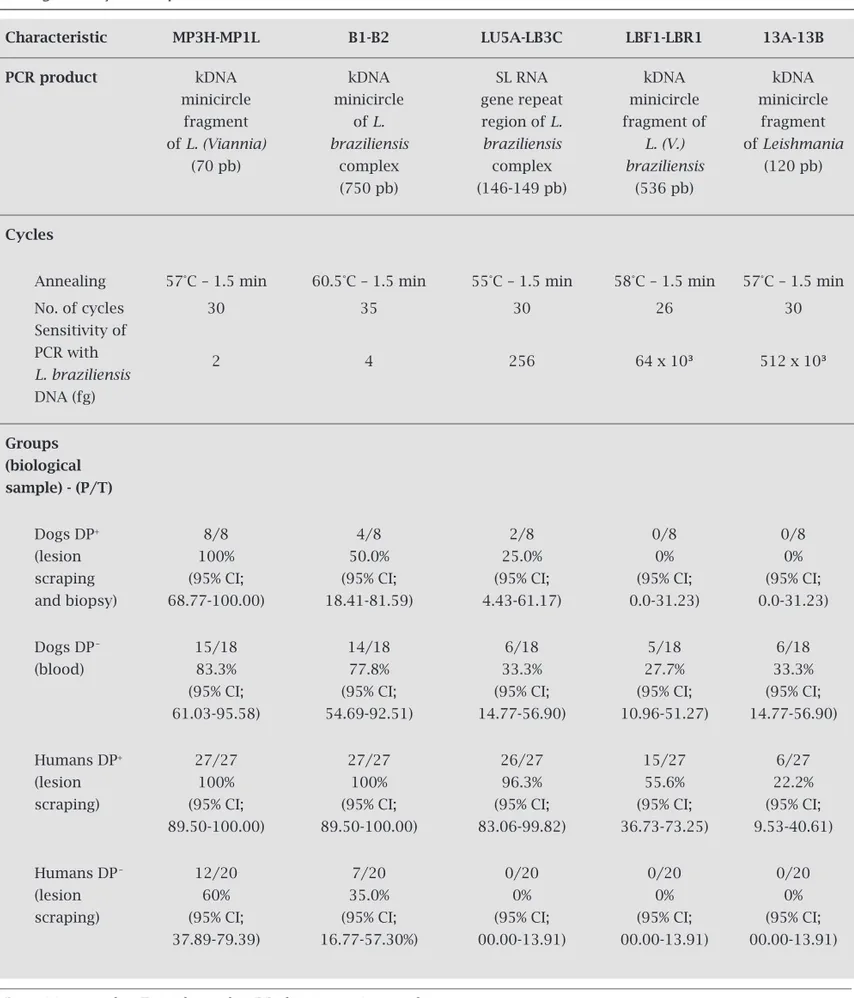
Table 1. Polymerase chain reaction conditions and results with different pairs of oligonucleotide primers on biological samples from dogs and humans with suspected American cutaneous leishmaniasis lesions, according to diagnosis by direct parasite search
Characteristic MP3H-MP1L B1-B2 LU5A-LB3C LBF1-LBR1 13A-13B
PCR product kDNA kDNA SL RNA kDNA kDNA
minicircle minicircle gene repeat minicircle minicircle
fragment of L. region of L. fragment of fragment
of L. (Viannia) braziliensis braziliensis L. (V.) of Leishmania
(70 pb) complex complex braziliensis (120 pb)
(750 pb) (146-149 pb) (536 pb)
Cycles
Annealing 57°C – 1.5 min 60.5°C – 1.5 min 55°C – 1.5 min 58°C – 1.5 min 57°C – 1.5 min
No. of cycles 30 35 30 26 30
Sensitivity of PCR with
2 4 256 64 x 10³ 512 x 10³
L. braziliensis DNA (fg)
Groups (biological
sample) - (P/T)
Dogs DP+ 8/8 4/8 2/8 0/8 0/8
(lesion 100% 50.0% 25.0% 0% 0%
scraping (95% CI; (95% CI; (95% CI; (95% CI; (95% CI;
and biopsy) 68.77-100.00) 18.41-81.59) 4.43-61.17) 0.0-31.23) 0.0-31.23)
Dogs DP_ 15/18 14/18 6/18 5/18 6/18
(blood) 83.3% 77.8% 33.3% 27.7% 33.3%
(95% CI; (95% CI; (95% CI; (95% CI; (95% CI;
61.03-95.58) 54.69-92.51) 14.77-56.90) 10.96-51.27) 14.77-56.90)
Humans DP+ 27/27 27/27 26/27 15/27 6/27
(lesion 100% 100% 96.3% 55.6% 22.2%
scraping) (95% CI; (95% CI; (95% CI; (95% CI; (95% CI;
89.50-100.00) 89.50-100.00) 83.06-99.82) 36.73-73.25) 9.53-40.61)
Humans DP_ 12/20 7/20 0/20 0/20 0/20
(lesion 60% 35.0% 0% 0% 0%
scraping) (95% CI; (95% CI; (95% CI; (95% CI; (95% CI;
37.89-79.39) 16.77-57.30%) 00.00-13.91) 00.00-13.91) 00.00-13.91)
RESULTS
Sensitivity analysis of the five pairs of primers showed differ-ent detection limits for Leishmania DNA. The MP3H-MP1L pair of primers performed best, detecting 2 fg of Leishmania (Viannia) DNA. Conversely, the 13A-13B primers presented the worst performance, detecting only 512 x 103 fg of Leish-mania spp. DNA (Figure 1 and Table 1).
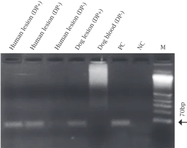
The analyses of eight samples of lesion scrapings and biopsies from dogs with ACL lesions that showed positive DP results had a positivity rate of 100% for PCR using the MP3H-MP1L primers, compared to a positivity rate of 0.0% when using the LBF1-LBR1 or 13A-13B primers. Figure 2 shows amplification in clinical samples using MP3H-MP1L primers.
In 18 samples of DNA extracted from the blood of dogs with suspected ACL lesions, even though DP produced negative results, the positivity rate for PCR when using the MP3H-MP1L primers was 83.3%, and 27.7% when using the least-sensitive primers (LBF1-LBR1).
In the 27 DNA samples from the ACL lesions of hu-mans that turned out positive DP results, the positivity rate for PCR when using the MP3H-MP1L and B1-B2 primers was again 100%. When using the LU5A-LB3C primers, the positivity rate was 96.3%.
In the 20 DNA samples from lesions of humans with suspected ACL, that had negative DP results, the positiv-ity rate for PCR when using the MP3H-MP1L primers was 60.0%, and with the B1-B2 primers was 35.0%. The PCR turned out negative with LU5A-LB3C primers.
The PCR results for canine and human lesion samples using the MP3H-MP1L primers proved be the most sensi-tive, demonstrating superior positivity to those of DP for ACL diagnosis: 100% in the canine and human samples with PD positive and 12/20 (60.0%) in the samples from humans with lesion suspected of the ACL (Table 1).
High rates of positivity were found for PCR, using the different primers to amplify DNA from blood samples of
2048 x 10
3 fg
2048 x 10
3 fg
1024 x 10
3 fg
1024 x 10
3 fg
512 x 10
3 fg
512 x 10
3 fg
128 x 10
3 fg
128 x 10
3 fg
256 x 10
3 fg
256 x 10
3 fg
64 x 10
3 fg
64 x 10
3 fg
32 x 10
3 fg
2048 fg
128 fg
64 fg 1024 fg
64 fg
32 fg 512 fg
32 fg
16 fg 128 fg
8 fg
4 fg 256 fg
16 fg
8 fg 64 fg
4 fg
2 fg 32 fg
2 fg
1 fg
32 x 10
3 fg
120bp
146-149bp
536bp
70bp
70bp
750bp
E
D B
C
M M
M
M M
A
Human lesion (DP+)Human lesion (DP-)Human lesion (DP-)Dog lesion (DP+)Dog blood (DP-)PC NC M
Figure 1: Representative gel showing analytical sensitivity of PCR using as the template a twofold serial dilution of DNA from promastigote forms of L. (V.) braziliensis (MHOM/ BR/1987/M11272). Panel A: 120 bp fragment of the kDNA minicircle region of the genus Leishmania, using 13A-13B primers. Panel B: 536 bp fragment of the kDNA minicircle region of L. (V.) braziliensis, using LBR1-LBF1 primers. Panel C: 146-149 bp fragment of the SL RNA gene repeat region of the L. braziliensis complex, using LU5A-LB3C primers. Panel D: 70 bp fragment of the k-DNA minicircle
DISCUSSION
We selected five pairs of primers, four of them derived from kDNA sequences and one from SL RNA (mini-exon). The kDNA minicircles are present in a very high copy number (10,000 per cell), and the SL RNA gene is present in the nuclear genome as a tandem repeat sequence of ap-proximately 200 copies.19 These selected pairs of primers are intended to detect L. braziliensis, the main species causing ACL in southern Brazil.
PCR using the MP3H-MP1L primers, which only ampli-fy fragments of the kDNA of species of the subgenus Leish-mania (Viannia), proved be very sensitive, being capable of detecting 2 fg of DNA, which is similar to the results re-ported by Lopes17 and Velasquez,15 who detected 0.14 fg and 0.9 fg of DNA, respectively. The other pair of primers that showed good sensitivity was B1-B2, which amplifies frag-ments of the kDNA of species of the L. braziliensis complex, detecting 4 fg of DNA, a value very close to that found by De Bruijn and Baker.18
The 13A-13B primers that amplify fragments of the kDNA of species of Leishmania showed low efficiency, detecting only concentrations higher than 512 pg of DNA. Manna,22 using these same primers, but with DNA from Leishma-nia infantum, demonstrated a sensitivity of 1 pg of DNA. A more significant value of 10 fg of L. (V.) braziliensis DNA was found by Rodger.21 However, these latter authors used the DNA hybridization method, which results in high-er sensitivity and thhigh-erefore efficiency of the primhigh-er. Recently, Marcussi20 developed a pair of primers (LBF1-LBR1) capa-ble of amplifying a specific sequence of the kDNA minicircle of L. (V.) braziliensis. Data obtained in the present study us-ing this pair showed that it was capable of detectus-ing 64 pg of DNA, showing greater sensitivity in DNA detection com-pared to the limit of 4 ng reported by Marcussi.20
The mini-exon gene is of great value to molecular biol-ogy techniques applied to studies of leishmaniasis; in addi-tion to the large number of copies in the genome, it contains conserved and variable regions that differ among the Leish-mania complexes.23 Derived from this gene, the LU5A-LB3C pair of primers has the ability to amplify fragments of DNA from various species of Leishmania of the L. braziliensis
complex, including L. (V.) braziliensis, L. (V.) guyanensis,
L. (V.) panamensis, and L (V.) peruviana.21 For this pair of primers, the present study showed low sensitivities (256 fg) compared to previous studies carried out by Harris,19 study-ing samples from lesion scrapstudy-ings from patients in South and Central America, and Gomes,24 in dogs in Brazil, with a detection level above 1 fg of DNA.
The higher sensitivity obtained with the MP3H-MP1L primers can be attributed in part to the smaller size of the amplicon, which is amplified more efficiently than larger fragments.19 Interestingly, the B1-B2 primers showed the same sensitivity (4 fg) as the MP3H-MP1L primers, despite
the size of the amplicon (750 bp), which is 10-fold larger. It is not surprising that LU5A-LB3C primers were less sensi-tive compared to the primers for kDNA, because there are 50 times fewer copies of the SL RNA gene than of the kDNA minicircle,19 which explains the 100-fold lower sensitivity. The differences in sensitivity observed among the several reports that have used the same pair of primers, can be ex-plained by different DNA extraction methods that affect the quality of DNA.25
The conclusive laboratory diagnosis of ACL is made by demonstrating amastigote forms of the parasite in mate-rial from scrapings or biopsies from lesions, and samples of bone marrow.5 PCR has shown several advantages in the diagnosis, clinical characterization, and epidemiology of leishmaniases. Several studies of ACL caused by parasites of the L. braziliensis complex in Central and South America have compared diagnoses by PCR with the conventional technique. Except in a few cases, the tests using the PCR technique were significantly more sensitive than those using parasitological diagnostic methods.13,26 However, there is great variation in the sensitivity of PCR, especially in re-lation to the method of DNA extraction, the choice of the oligonucleotide primers,25,27,28 the clinical samples used, and the length of infection.16
In the present study, 26 samples from dogs (lesion scrapings, biopsy, and blood) and 47 samples from humans (lesion scrapings), from an endemic area of ACL caused by
L. (V.) braziliensis, were tested by PCR using these five pairs of primers. PCR using the MP3H-MP1L primers, which amplify a fragment of the kDNA minicircle region of the subgenus Leishmania (Viannia), showed high positivity in human and canine lesions, detecting all samples that had positive parasite search. Demonstration of amastigote forms of the parasite in material from ACL lesions shows low sensitivity. Silveira6 demonstrated that only 59.4% of the patients with positive laboratorial diagnostic for ACL showed positive direct parasite search in lesions. In this way, PCR was significantly more efficient than the PD, de-tecting DNA in 12/20 (60.0%) human lesion samples with negative DP. Although the B1-B2 primers detected higher number of infected samples from human than PD, its per-formance in canine samples was not good. On the other hand, PCR carried out with the LU5A-LB3C, in spite of a good performance in samples from human lesions with positive DP, detected only 25.0% of dog samples with posi-tive DP and none from human samples with negaposi-tive DP. It is important to emphasize that all canine and human samples analyzed had DNA quantities higher than the sen-sitivity limits to these primers. The presence of sufficient quantities of DNA in samples only shows that it was ade-quate to carry out the assay and the dilution of Leishmania
clini-cal samples from different sources, but as all samples from DP positive lesions could be detected by MP3H-MP1L, it is supposed there were no PCR inhibitors in these samples.
LBF1-LBR1 and 13A-13B primers were less effec-tive than DP to detect Leishmania DNA in human le-sion scrapings probably due the small DNA quantities in these samples. It shows the importance of the choice of the primers pairs mainly to analyze samples that, by the characteristics from the samples collection, presents small quantities of DNA like lesion scrapings. All lesion samples detected by PCR using the less-sensitive prim-ers were also detected by PCR using the more-sensitive primers.
Detection of DNA from Leishmania spp. in the blood samples of dogs from an endemic area has been reported by several investigators.15,25,29,30 In the present study, all PCR primers were capable of detecting Leishmania spp. DNA, with different positivity rates, in blood samples from dogs with suspected ACL lesions, whereas the DP technique produced negative results. All the canine blood samples detected by PCR using the less-sensitive primers were also detected by PCR using the more-sen-sitive primers, with the exception of one, which was not detected by the LBF1-LBR1 primers but was detected by the 13A-13B primers.
The diagnostic positivity rate obtained with PCR primers was corroborated by the analytical sensitivity. The MP3H-MP1L and B1-B2 primers, both targeting the kDNA minicircle, detected smaller quantities of L. (V.)
braziliensis DNA and they showed higher positivity in
the diagnosis. The MP3H-MP1L amplify a conserved se-quence17 and the B1-B2 amplify the entire kDNA minicir-cles of L. (V.) braziliensis,18 showing that the size of the amplicon did not influence the PCR. The LU5A-LB3C also showed good positivity, but only in lesion scrapings from humans. The other two pairs of primers targeting the k-DNA minicircle, 13A-13B for the genus Leishmania
and LBF1-LBR1 for the species L. (V.) braziliensis, may be directed to an unconserved region in the genus and spe-cies, respectively. More studies are necessary with a larger number of Leishmania isolates, to establish the real am-plitude of DNA detection for the latter pairs of primers.
CONCLUSION
In light of these results, it can be concluded that the PCR primers used in the diagnosis and epidemiology of cuta-neous leishmaniasis showed a wide variation in sensitivity. Of the pairs of primers analyzed, MP3H-MP1L and B1-B2, targeting the kDNA minicircles, performed best. Compared to the direct parasite search, however, they performed
sig-ACKNOWLEDGEMENTS
The authors thank the Fundação Araucária and the Conse-lho Nacional de Desenvolvimento Científico e Tecnológico
(CNPq) for financial support to carry out this study (Process No. 410550/2006-0).
REFERENCES
1. Grimaldi Jr G, Tesh RB. Leishmaniasis of the New World: cur-rent concepts and implications for future research. Clin Micro-biol Rev 1993; 6:230-50.
2. Lainson R, Shaw JJ. New World Leishmaniasis. The neotropi-cal Leishmania species. Collier L, Balows A, Sussman M, eds. Topley & Wilson’s Microbiology and Microbial Infectious Dis-eases. Ninth edition. Arnold, London, 1998.
3. Murray HW, Berman JD, Davies CR, Saravia NG. Advances in Leishmaniasis. Lancet 2005; 366:1561-77.
4. World Health Organization - WHO. Available at: http://www. who.int/Leishmaniasis/en/ (Accessed in 15/01/2009). 5. Gontijo B, Carvalho MLR. Leishmaniose Tegumentar
Ameri-cana. Rev Soc Bras Med Trop 2003; 36(Suppl.1):71-80. 6. Silveira TGV, Arraes SMAA, Bertolini DA et al. Observações
sobre o diagnóstico laboratorial e a epidemiologia da leish-maniose tegumentar no Estado do Paraná, sul do Brasil. Rev Bras Med Trop 1999; 32:413-23.
7. Armijos RX, Chico ME, Cruz ME et al. Human cutaneous Leishmaniasis in Ecuador: identification of parasites by en-zyme electrophoresis. Am J Trop Med Hyg 1990; 42:424-8. 8. Camargo ME, Rebonato C. Cross-reactivity in
immunofluo-rescence test for Trypanosoma and Leishmania antibodies. A simple inhibition procedure to ensure specific results. Am J Trop Med Hyg 1969; 18:500-5.
9. Badaro R, Reed SG, Barral A, Orge G, Jones TC. Evaluation of the micro enzyme-linked immunosorbent assay (ELISA) for antibodies in American visceral Leishmaniasis: antigen selec-tion for detecselec-tion of infecselec-tion-specific responses. Am J Trop Med Hyg 1986; 35:72-8.
10. White T, Madej R, Persing D. The polymerase chain reaction: clinical applications. Adv Clin Chem 1992; 29:161-96. 11. Harris E, Lopez M, Arevalo J et al. Short courses on DNA
de-tection and amplification in Central and South America: the democratization of molecular biology. Biochem Educ 1993; 21:16-22.
12. Harris E, Belli A, Agabian N. Appropriate transfer of molecu-lar technology to Latin America for public health and biomed-ical sciences. Biochem Educ 1996; 24:3-12.
13. Medeiros ACR, Rodrigues SS, Roselino AMF. Comparison of the specificity of PCR and the histopathological detection of Leishmania for the diagnosis of American cutaneous leish-maniasis. Braz J Med Biol Res 2002; 35:421-4.
14. Garcia FCB, Santos SSR, Chociay MF, Medeiros ACR, Roselino AMF. Métodos subsidiários para o diagnóstico da leishmaniose tegumentar americana (LTA): compa-ração dos resultados do seqüenciamento de DNA e da
PCR-RFLP para determinação da espécie de Leishmania
16. Venazzi EA, Roberto AC, Barbosa-Tessmann IP, Zanzarini PD, Lonardoni MV, Silveira TG. Polymerase chain reaction with lesion scrapping for the diagnosis of human American tegumentary leishmaniasis. Mem Inst Oswaldo Cruz 2006; 101:427-30.
17. Lopez M, Ingá R, Cangalaya M et al. Diagnosis of Leishmania using the polymerase chain reaction: a simplified procedure for field work. Am J Trop Med Hyg 1993; 49:348-56.
18. De Bruijn MHL, Barker DC. Diagnosis of new world Leish-maniasis: specific detection of species of the Leishmania brazil-iensis complex by amplification of kinetoplast DNA. Acta Trop 1992; 52:45-58.
19. Harris E, Kropp G, Belli A, Rodriguez B, Agabian N. Single-step multiplex PCR assay for characterization of New World Leishmania complexes. J Clin Microbiol 1998; 36:1989-95. 20. Marcussi VM, Marcussi LM, Barbosa-Tessmann IP, Lonardoni
MV, Silveira TG. Leishmania (Viannia) braziliensis: New prim-ers for identification using polymerase chain reaction. Exp Parasitol 2008; 120:300-5.
21. Rodgers MR, Popper SJ, Wirth DF. Amplification of kineto-plast DNA as a tool in the detection and diagnosis of Leishma-nia. Exp Parasitol 1990; 71:267-75.
22. Manna L, Vitale F, Reale S et al. Comparison of different sam-pling for PCR-based diagnosis and follow-up of canine viscer-al leishmaniosis. Vet Parasitol 2004; 125:251-62.
23. Fernandes O, Bozza M, Pascale JM, de Miranda AB, Lopes UG, Degrave WM. An oligonucleotide probe derived from kDNA minirepeats is specific for Leishmania (Viannia). Mem Inst Oswaldo Cruz 1996; 91:279-84.
24. Gomes AH, Ferreira IM, Lima ML et al. PCR identification of Leishmania in diagnosis and control of canine Leishmaniasis. Vet Parasitol 2007; 144:234-41.
25. Reithinger R, Lambson BE, Barker DC, Davies CR. Use of PCR to detect Leishmania (Viannia) sp. in dog blood and bone mar-row. J Clin Microbiol 2000; 38:748-51.
26. Rodriguez N, Guzman B, Rodas A, Takiff H, Bloom BR, Con-vit J. Diagnosis of cutaneous Leishmaniasis and species dis-crimination of parasites by PCR and hybridization. J Clin Mi-crobiol 1994; 32:2246-52.
27. Ikonomopoulos J, Kokotas S, Gazouli M, Zavras A, Stoitsiou M, Gorgoulis VG. Molecular diagnosis of Leishmaniasis in dogs: comparative application of traditional diagnostic meth-ods and the proposed assay on clinical samples. Vet Parasitol 2003; 113:99-113.
28. Bensoussan E, Nasereddin A, Jonas F, Schnur LF, Jaffe CL. Comparison of PCR assays for diagnosis of cutaneous Leish-maniasis. J Clin Microbiol 2006; 44:1435-9.
29. Llanos-Cuentas EA, Roncal N, Villaseca P et al. Natural infec-tions of Leishmania peruviana in animals in the Peruvian An-des. Trans R Soc Trop Med Hyg 1999; 93:15-20.