Full paper published online: August 31, 2009 ISSN 1678-9199.
GENOTYPING OF Clostridium perfringens ASSOCIATED WITH SUDDEN
DEATH IN CATTLE
Miyashiro S (1), Baldassi L (1), Nassar AFC (1)
(1) Animal Health Research and Development Center, Biological Institute, São Paulo,
São Paulo State, Brazil.
ABSTRACT: Toxigenic types of Clostridium perfringens are significant causative
agents of enteric disease in domestic animals, although type E is presumably rare,
appearing as an uncommon cause of enterotoxemia of lambs, calves and rabbits.
We report herein the typing of 23 C. perfringens strains, by the polymerase chain
reaction (PCR) technique, isolated from small intestine samples of bovines that have
died suddenly, after manifesting or not enteric or neurological disorders. Two strains
(8.7%) were identified as type E, two (8.7%) as type D and the remainder as type A
(82.6%). Commercial toxoids available in Brazil have no label claims for efficacy
against type E-associated enteritis; however, the present study shows the
occurrence of this infection. Furthermore, there are no recent reports on Clostridium
perfringens typing in the country.
KEY WORDS: Clostridium perfringens, iota toxin, sudden death, PCR, cattle.
CONFLICTS OF INTEREST: There is no conflict.
CORRESPONDENCE TO:
SIMONE MIYASHIRO, Instituto Biológico, Av. Conselheiro Rodrigues Alves, 1252,
Vila Mariana, São Paulo, SP, 04014-002, Brasil. Phone: +55 11 5087 1721. Fax: +55
INTRODUCTION
The aim of this work is to describe the typing of Clostridium perfringens strains
isolated from cattle, a widely occurring pathogenic bacterium and certainly the most
important cause of clostridial enteric disease (enterotoxaemia) in domestic animals
(1).
The pathogenicity of Clostridium perfringens is associated with several toxins. The
alpha, beta, epsilon and iota toxins are the major lethal poisonous substances
produced by the organism and are closely related to its virulence, even though they
produce several minor extracellular toxins. Usually, C. perfringens has been
classified into five toxigenic types (A through E) on the basis of their ability to
produce the major lethal toxins (2, 3). Two other major toxins (i.e. enterotoxin and
beta-2) can also be produced by all types of C. perfringens, although they are not
used for its typing (4).
C. perfringens type A is consistently recovered both from the intestinal tracts of
animals and from the environment, while others (types B, C, D and E) are less
common in animal intestinal tracts (5).
In sheep, type D is widely regarded as the causative agent of fatal enterotoxemia or
“overeating disease” (3), but is also important in calves, goats and adult cattle (1). In
Brazil, Lobato et al. (6) reported the detection of C. perfringens type D in a bovine
enterotoxaemia case, but C. perfringens typing has been rarely described in the
country.
Type E is a putatively uncommon cause of enterotoxemia of lambs, calves and
rabbits (7). Little is known about the pathogenesis of type E infections, although it is
assumed that, in keeping with the pattern set by isolates of other toxin types, iota
toxin plays an important role (8). Recently, Songer and Miskimmins (8) reported two
cases of bovine enterotoxemia caused by C. perfringens, determined as genotype E
by PCR analysis.
Bovine enterotoxaemia is characterized by a high case fatality rate, sudden deaths,
lesions of hemorrhagic enteritis of the small intestine and, quite often, an absence of
other clinical signs (1). In the present trial, among the C. perfringens strains isolated
from intestinal samples obtained in post-mortem examination of 23 bovines – 60.86%
presented only sudden death (14/23), 21.74% of which were preceded by enteric
disorders (5/23), 13.05% by neurological symptons (3/23) and 4.35% by both enteric
polymerase chain reaction (PCR). Macroscopically, hemorrhagic small intestines
were reported in 39.13% of cases (9/23), hemorrhage in other tissues such as heart,
kidney or liver in 21.74% (5/23), while icteric liver was observed in 17.4% (4/23) and
pethechiae in 21.74% (5/23). Macroscopic pathologies were absent in six animals
(26%). The samples were collected in five Brazilian states (São Paulo, Minas Gerais,
Mato Grosso do Sul, Goiás and Paraíba) from 2005 to 2007, and the age of the
animals varied from 2 to 10 years.
MATERIAL AND METHODS
Isolation and Biochemical Identification of C. perfringens
The samples were cultured in Cooked Meat Medium (CMM) at 37°C for 18 to 24
hours and 10 µL of this culture was streaked in a plate containing Mueller-Hinton
agar with 5% defibrinated sheep blood and incubated under anaerobic conditions in
McIntosh and Fields jars submitted to vacuum conditions before hydrogen inoculation
at 37°C for 18 to 24 hours.
After incubation, colonies were analyzed according to the shape, color, production
and type of hemolysis. Bacterial morphology was microscopically assessed in
gram-stained smears. Colonies presenting C. perfringens characteristics were isolated,
cultured in CMM and incubated at 37°C for 18 to 24 hours. These cultures were
submitted to the following biochemical tests for species identification: production of
catalase, lecithinase and gelatinase, fermentation of glucose and lactose, and skim
milk coagulation. Interpretation was performed according to Cowan (9).
All strains were incubated in CMM, and after 18 to 24 hours of incubation at 37°C,
cultures were stored at room temperature
.
Polymerase Chain Reaction (PCR)
The strains biochemically identified as C. perfringens isolates were typed by PCR
with toxin-specific primers. DNA extraction was performed for each strain kept in
CMM by the guanidine isothyocianate methodology adapted from Boom et al. (10).
The extracted DNA was used for multiplex PCR detection of alpha, beta, epsilon and
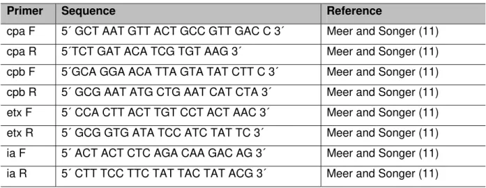
iota toxin genes with specific primers described by Meer and Songer (11) (Table 1)
which amplifies 324 bp, 196 bp, 655 bp and 446 bp fragments, respectively, in a
hybridization at 53°C for 30 seconds and extension at 72°C for 40 seconds. As
positive control, three other C. perfringens strains from the Biological Institute (types
A, C and E) were used.
Amplification reactions were carried out in a Peltier Thermal Cycler-200 (MJ
Research) and the analysis of the amplified products was performed by means of
electrophoresis in 1.3% agarose gel with TBE 0.5 X running buffer (0.045 M
TRIS-Borate and 1 mM of EDTA ph 8.0). Gel was stained with ethidium bromide, visualized
with a UV transiluminator (300-320 nm) and photographed by a photodocumentation
system (Kodak Digital Camera DC/120® Zoom, Brazil) and analyzed with the
software 1D Image Analysis® (Kodak Digital Science, Brazil).
Table 1. Primer sequences
Primer Sequence Reference
cpa F 5´ GCT AAT GTT ACT GCC GTT GAC C 3´ Meer and Songer (11)
cpa R 5´TCT GAT ACA TCG TGT AAG 3´ Meer and Songer (11)
cpb F 5´GCA GGA ACA TTA GTA TAT CTT C 3´ Meer and Songer (11)
cpb R 5´ GCG AAT ATG CTG AAT CAT CTA 3´ Meer and Songer (11)
etx F 5´ CCA CTT ACT TGT CCT ACT AAC 3´ Meer and Songer (11)
etx R 5´ GCG GTG ATA TCC ATC TAT TC 3´ Meer and Songer (11)
ia F 5´ ACT ACT CTC AGA CAA GAC AG 3´ Meer and Songer (11)
ia R 5´ CTT TCC TTC TAT TAC TAT ACG 3´ Meer and Songer (11)
RESULTS
Biochemical Characterization of the Strains
Twenty-three strains were biochemically identified as C. perfringens, due to the
production of catalase, lecithinase and gelatinase, and to fermentation of glucose,
lactose and tumultuous fermentation of skim milk.
Polymerase Chain Reaction (PCR) for C. perfringens Strain Typing
Among the 23 C. perfringens strains isolated, nineteen (82.6%) were differentiated as
type A with presence of only alpha toxin gene, two strains (8.7%) as type E that
besides alpha toxin presented an iota toxin gene, and two strains (8.7%) as type D
Table 2. Results of C. perfringens strain typing by PCR
Sample number Locality (state) Type of C. perfringens
1 Paraíba E
2 Minas Gerais E
3 São Paulo D
4 Minas Gerais D
5 São Paulo A
6 São Paulo A
7 São Paulo A
8 São Paulo A
9 São Paulo A
10 São Paulo A
11 São Paulo A
12 São Paulo A
13 São Paulo A
14 São Paulo A
15 São Paulo A
16 São Paulo A
17 Goiás A
18 Minas Gerais A
19 Minas Gerais A
20 Minas Gerais A
21 Minas Gerais A
22 Mato Grosso do Sul A
23 Mato Grosso do Sul A
DISCUSSION
The samples submitted to the Biological Institute (São Paulo, Brazil) for diagnostic
screening are not necessarily representative; nevertheless, there have been no
reports regarding C. perfringens typing in Brazil.
Most strains were identified as type A, which is reported to be easily isolated from
tissues, effusions and intestinal tract of cadavers within a few hours after death;
furthermore, it grows rapidly in culture and may mask other organism growth. Also,
prevention of infections or intoxications caused by type A is complicated for at least
two reasons: firstly, the organism is so ubiquitous that it is impractical to resort to
mass immunization and, secondly, the enzymatic nature of the majority of the
to other proteins (3). However, recently a C. perfringens type A toxoid for cattle in the
US was developed since standard clostridial vaccines do not offer protection against
the alpha toxin of C. perfringens type A, nor there is any cross-protection.
The amplification of toxin genes from different types of C. perfringens is specific
when compared with the in vivo methodology by toxin seroneutralization tests as
described by Kadra et al. (12), and is also shown to be more rapid and reliable.
It has been reported that C. perfringens type E is an apparently uncommon cause of
enterotoxemia in calves (1), and most reports describe other types of the
microrganism involved in these cases (13,14). In the present trial, most strains
(82.6%) were found to be C. perfringens type A, similarly to a recent report by
Manteca et al. (15). In Brazil, Baldassi et al. (16) reported the typing of 89 C.
perfringens strains from bovines by ELISA test and, besides cross-reactivity among
samples, the presence of C. perfringens type E was not evaluated.
The fact that all these strains had originated from a single host type and sudden
death condition is notable, and this report provides some information about which
types of C. perfringens are causing enterotoxaemia in Brazilian herds, thus alerting
investigators to the occurrence of type E. More rigorous epidemiological and
diagnostic pursuit of similar cases may be warranted.
It is also important to note that currently available commercial toxoids in Brazil will
likely offer little or no protection against type E infections.
The inclusion of genotyping as part of the diagnostic approach will provide more
information on the true importance of the occurrence herein reported.
REFERENCES
1. Songer JG. Clostridial enteric diseases of domestic animals. Clin Microbiol Rev.
1996;9(2):216-34.
2. Hatheway CL. Toxigenic clostridia. Clin Microbiol Rev. 1990;3(1):66-98.
3. Niilo L. Clostridium perfringens in animal disease: a review of current knowledge.
Can Vet J. 1980;21(5):141-8.
4. Garmory HS, Chanter N, French NP, Bueschel D, Songer JG, Titball RW.
Occurrence of Clostridium perfringens beta 2-toxin amongst animals, determined
using genotyping and subtyping PCR assays. Epidemiol Infect. 2000;124(1):61-7.
5. Carter GR, Wise DJ. Essentials of veterinary bacteriology and mycology.
6. Lobato FCF, Assis RA, Abreu VLV, Souza Jr MF, Lima CGRD, Salvarani FM.
Enterotoxemia em bovino. Arq Bras Med Vet Zootec. 2006;58(5):952-4.
7. Hart B, Hooper PT. Enterotoxaemia of calves due to Clostridium welchii type E.
Aust Vet J. 1967;43(9):360-3.
8. Songer JG, Miskimmins DW. Clostridium perfringens type E enteritis in calves: two
cases and a brief review of the literature. Anaerobe. 2004;10(4):239-42.
9. Cowan ST. Cown and Steel’s manual for the identification of medical bacteria. 2nd
ed. Great Britain: Cambridge University Press; 1974. p. 238.
10. Boom R, Sol CJ, Salimans MM, Jansen CL, Werthein-van Dillen PM, van der
Noordaa J. Rapid and simple method for purification of nucleic acids. J Clin Microbiol.
1990;28(3):495-503.
11. Meer RR, Songer JG. Multiplex polymerase chain reaction assay for genotyping
Clostridium perfringens. Aust J Vet Res. 1997;58(7):702-5.
12. Kadra B, Guillou JP, Popoff M, Bourlioux P. Typing of sheep clinical isolates and
identification of enterotoxigenic Clostridium perfringens strains by classical methods
and by polymerase chain reaction (PCR). FEMS Immunol Med Microbiol.
1999;24(3):259-66.
13. Kalender H, Kiliç A, Atil E. Enterotoxemia in a cow due to Clostridium perfringens
type A. Turk J Vet Anim Sci. 2007;31(1):83-4.
14. Yoo HS, Lee SU, Park KY, Park YH. Molecular typing and epidemiological survey
of prevalence of Clostridium perfringens types by multiplex PCR. J Clin Microbiol.
1997;35(1):228-32.
15. Manteca C, Daube G, Pirson V, Limbourg B, Kaeckenbeeck A, Mainil JG.
Bacterial intestinal flora associated with enterotoxaemia in Belgian Blue calves. Vet
Microbiol. 2001;81(1):21-32.
16. Baldassi L, Barbosa ML, Bach EE, Iaria ST. Toxigenicity characterization of
Clostridium perfringens from bovine isolates. J Venom Anim Toxins.