!
!
!
CHARACTERIZATION OF THE CLINICAL,
HISTOLOGICAL AND GENETIC PROFILE OF
ARTICULAR DAMAGE IN HEREDITARY
HEMOCHROMATOSIS
ANTÓNIO MANUEL MENDONÇA ROMÃO DE BRITO CAMACHO
Tese para obtenção do grau de Doutor em Medicina
na Especialidade de Biomedicina
na Faculdade de Ciências Médicas
! iii!
CHARACTERIZATION OF THE CLINICAL,
HISTOLOGICAL AND GENETIC PROFILE OF
ARTICULAR DAMAGE IN HEREDITARY
HEMOCHROMATOSIS
Nome do autor: António Manuel Mendonça Romão de Brito Camacho
Orientadora: Maria Leonor Cancela, Professora Catedrática da
Universidade do Algarve
Co-orientadores: Jaime da Cunha Branco, Professor Catedrático da
Universidade Nova de Lisboa
Pascal Richette, Professor Associado da
Universidade de Paris 7
Tese para obtenção do grau de Doutor em Medicina na Especialidade de
Biomedicina
Declaração de Compromisso de Anti-Plágio
Declaro por minha honra que os trabalhos que apresento são todos da minha autoria e originais e que todas as minhas citações estão corretamente identificadas. Tenho consciência de que a utilização de elementos alheios não identificados constitui uma grave falta ética e disciplinar.
Lisboa, _________________________________
Nome:
____________________________________________________________________
________________________________________ (Assinatura)
26 de Setembro de 2015
Contents
Acknowledgments xvii
Summary xix
Resumo xxi
List of Papers xxiii
1 Introduction 1
1.1 Prevalence and incidence of osteoarthritis . . . 1
1.2 Causes of osteoarthritis . . . 3
1.3 Hemochromatosis . . . 4
1.3.1 Characteristics and molecular causes . . . 4
1.3.2 Clinical presentation . . . 6
1.3.3 Diagnosis . . . 6
1.3.4 Treatment . . . 7
1.3.5 Hemochromatosis and arthropathy . . . 7
1.4 Pathogenesis of osteoarthritis and tissues affected . . . 8
1.5 Risk factors for osteoarthritis . . . 12
1.6 Diagnosis of osteoarthritis . . . 16
1.7 Treatment of osteoarthritis . . . 17
1.8 Animal models in osteoarthritis research . . . 18
2 Aims of the study 21 3 Methods 23 3.1 Paper I . . . 23
3.1.1 Animal model, experimental procedure, feeding and housing . . . . 23
3.1.2 Assessment of iron accumulation . . . 26
3.1.3 Grading of osteoarthritic changes . . . 26
3.1.4 Morphological characterization of the knee joint by micro-CT . . . 27
3.1.5 Histological evaluation of undecalcified samples . . . 28
3.1.6 Evaluation of gene expression . . . 29
3.1.7 Immunohistochemistry . . . 32
3.1.8 Statistical analysis . . . 32
3.2 Paper II . . . 33
3.2.1 Patient recruitment and evaluation . . . 33
3.2.2 Statistical analysis . . . 33
4 Results 35 4.1 Paper I . . . 35
4.2 Paper II . . . 45
5 Discussion 49 5.0.1 Limitations of the study . . . 49
5.0.2 Changes in cartilage and bone in iron overloaded joints . . . 50
5.0.3 Genetic expression in iron overloaded joints . . . 51
5.0.4 Role of genotype in musculoskeletal complications . . . 52
5.0.5 Future directions . . . 53
6 Conclusions 55
References 57
A Paper I 87
B Paper II 99
List of Figures
1.1 Iron metabolism . . . 5
1.2 Structural changes and signalling in osteoarthritis . . . 9
3.1 Paper I flowchart . . . 24
3.2 Surgical procedure . . . 25
3.3 Process of micro-CT acquisition and analysis . . . 28
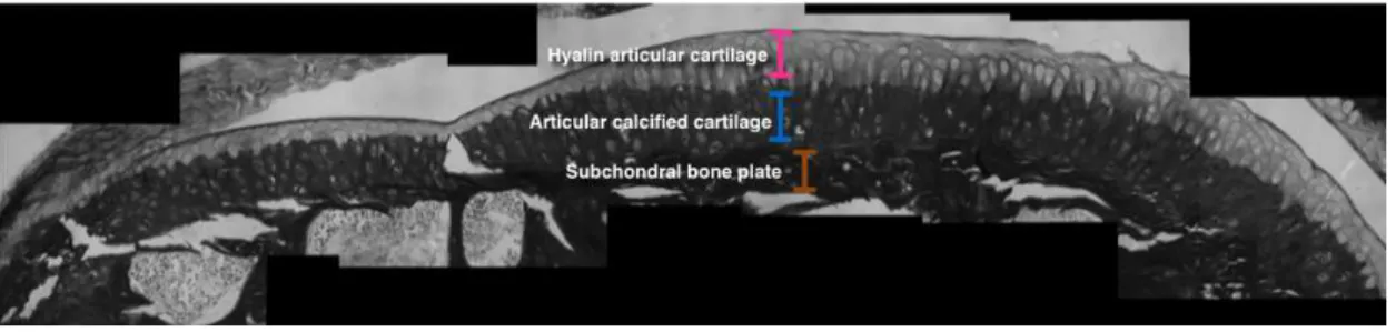
3.4 Measurement of calcified cartilage . . . 29
3.5 Samples for molecular biology . . . 29
4.1 Iron accumulation in the knee of WT and Hfe-KO mice . . . 36
4.2 Iron accumulation in the knee of non-operated Hfe-KO mice . . . 36
4.3 Iron parameters in WT and Hfe-KO mice . . . 37
4.4 Cartilage changes following meniscectomy . . . 39
4.5 Articular cartilage calcification in the knee joints of WT and Hfe-KO mice 40 4.6 Subchondral bone changes following meniscectomy in WT and Hfe-KO mice 41 4.7 Changes in gene expression of pro-inflammatory cytokines . . . 42
4.8 Changes in gene expression following meniscectomy . . . 43
4.9 Immunohistochemistry . . . 44
List of Tables
1.1 Prevalence of osteoarthritis . . . 2 1.2 Causes of secondary osteoarthritis . . . 3 1.3 Types of hereditary hemochromatosis . . . 6
3.1 Sequence of primers used in the real-time polymerase chain reaction . . . 31
4.1 Characteristics of patients with hereditary hemochromatosis by genotype . 45 4.2 Prevalence of self-reported symptoms at time of survey by genotype . . . . 46 4.3 Prevalence of osteoarthritis, joint replacement, back pain and sciatica by
genotype . . . 46 4.4 Comparison of the prevalence of osteoporosis and associated fractures by
genotype . . . 47
Acronyms
3D three dimensional.
Acan aggrecan.
ACC articular calcified cartilage.
ADAMTS5 a disintegrin and metallopeptidase with thrombospondin motifs 5.
Adamts5 a disintegrin-like and metallopeptidase with thrombospondin type 1 motif, 5.
BMD bone mineral density.
Bmp6 bone morphogenetic protein 6.
C282Y cysteine-to-tyrosine substitution at amino acid 282.
cDNA complementary deoxyribonucleic acid.
CI Confidence Interval.
Col10a1 collagen, type X, alpha 1.
Col2a1 collagen, type II, alpha 1.
DALYs disability-adjusted life years.
DEXA dual-energy X-ray absorptiometry.
dGEMRIC delayed gadolinium-enhanced MRI of the cartilage.
ECM extracellular matrix.
EDTA ethylenedinitrilotetraacetic acid.
FPN ferroportin.
GAG glycosaminoglycan.
H63D histidine-to-aspartic acid substitution at position 63.
HA hyaluronic acid.
HAC hyaline articular cartilage.
HAMP hepcidin antimicrobial peptide.
HFE hemochromatosis.
Hfe hemochromatosis.
HH Hereditary Hemochromatosis.
HIC hepatic iron concentration.
HJV hemojuvelin.
HKA hip-knee-ankle.
HLA human leukocyte antigen.
IL1 interleukin 1.
Il1b interleukin 1 beta.
Il6 interleukin 6.
IQR interquartile range.
KBD Kashin-Beck disease.
KL Kellgren-Lawrence.
LBA load-bearing axis.
M-MLV Moloney Murine Leukemia Virus.
MHC major histocompatibility complex.
MMP13 matrix metallopeptidase 13.
Mmp13 matrix metallopeptidase 13.
MMP3 matrix metallopeptidase 3.
Mmp3 matrix metallopeptidase 3.
MRI magnetic resonance imaging.
Acronyms xv
NHANES National Health and Nutrition Examination Survey.
NSAID non-steroidal anti-inflammatory drugs.
NTBI non-transferrin bound iron.
OA osteoarthritis.
OARSI Osteoarthritis Research Society International.
OP osteoporosis.
PBS phosphate buffer saline.
RNA ribonucleic acid.
ROI region of interest.
ROS reactive oxygen species.
Rpl13a ribosomal protein L13A.
RQI RNA quality indicator.
RT reverse transcription.
RT-PCR real-time polymerase chain reaction.
Runx2 runt related transcription factor 2.
TBS Tris buffered saline.
TFR2 transferrin receptor 2.
Tfrc transferrin receptor.
TNF-α tumour necrosis factor a. VCAM-1 vascular adhesion molecule 1.
Acknowledgments
I would like to thank the following people:
Leonor Cancela, my supervisor, for her generosity in welcoming me into her labo-ratory and for her guidance in the design and execution of the project.
Jaime Branco, my co-supervisor, for his ready availability, and for the counselling provided during the entire PhD programme.
Pascal Richette, my other co-supervisor, for his valuable orientation in the field of clinical research.
Márcio Simão for his friendship, for his help with laboratory techniques, and for all the valuable discussions we had about the direction of this project.
Hang Korng-Ea for his help with the surgical interventions in the mice.
Martine Cohen-Solal for receiving me in her department at INSERM 1132, Paris, and allowing the use of its resources.
Paulo Gavaia, Agnés Ostertag and Caroline Marty for their technical expertise.
Eduarda Vidal, my clinical supervisor and Luís Branco Amaral, the head of the Orthopaedics Department at Hospital Curry Cabral, Lisbon, for supporting my decision to suspend the residency in order to complete the PhD programme.
All my colleagues at the BioSkel lab at CCMAR - Universidade do Algarve, and those at the Orthopaedic Department of Hospital Curry Cabral for their friendship and valuable discussions.
My parents, for all their help and encouragement through the years.
My wife, Maria João, for her love, guidance and support.
Summary
Osteoarthritis (OA) is the most common joint disease in humans. It affects the joint as a
whole and is characterized by progressive articular cartilage destruction, abnormal sub-chondral bone remodelling, formation of osteophytes, ligament and periarticular muscle weakening and in some cases synovial inflammation, which ultimately lead to a painful and impaired joint. There are several known risk factors for the development of OA but the exact sequence of events that lead to the destruction of the articular cartilage is not yet fully understood.
Hereditary hemochromatosis (HH), a disease caused by mutations in the HFE gene, is characterised by systemic iron overload, toxic accumulation of iron in parenchymal cells of liver, heart, and endocrine glands. It is also associated with musculoskeletal complications, namely an increased prevalence of OA. The role of iron overload in the development of OA is still undefined.
To further understand the molecular mechanisms involved in the pathology of HH-related OA, we surgically induced OA in the knee of a murine model of hereditary hemochromatosis and studied the changes to cartilage and bone. Also, in order to un-derstand how the different mutations in the HFE gene affect systemic iron overload
and related musculoskeletal complications, we studied the prevalence of musculoskeletal complications in a cohort of patients with different HH genotypes.
Hfe-KO mice showed a systemic iron overload and an increased iron accumulation in the knee synovial membrane following surgery. The histological OA score was signifi-cantly higher in the Hfe-KO mice at 8 weeks after surgery. Micro-CT study of the proxi-mal tibia revealed increased subchondral bone volume and increased trabecular thickness. Gene expression and immunohistochemical analysis showed a significant increase in the expression of matrix metallopeptidase 3 in the joints of Hfe-KO mice compared with control mice at 8 weeks after surgery.
Among our cohort of HH patients the majority were homozygous for the C282Y mu-tation. The serum ferritin concentration and serum transferrin saturation at diagnosis were significantly higher in C282Y homozygous patients compared with those who were compound heterozygous (C282Y/H63D). Also the overall prevalence of self-reported mus-culoskeletal complications was significantly higher in patients with C282Y homozygosity. The findings of this study suggest that systemic iron overload does not cause OA directly but acts as a susceptibility factor. The systemic and synovial iron overload both contribute to increase the catabolic response of the articular cartilage to mechanical
Resumo
A osteoartrose (OA) é a patologia articular mais frequente nos humanos. Afecta a arti-culação como um todo e as suas características principais são a destruição progressiva da superfície articular, a remodelação anormal do osso subcondral, a formação de osteofitos, o enfraquecimento das estruturas ligamentares, a atrofia muscular e, em alguns casos, a inflamação da membrana sinovial. Todas estas alterações levam a uma articulação dolorosa e incapaz de cumprir a sua função. Existem vários factores que aumentam o risco de vir a desenvolver OA mas a sequência exacta de acontecimentos que levam à destruição de uma articulação ainda não está totalmente esclarecida.
A Hemocromatose Hereditária (HH) é uma doença causada por mutações no gene HFE e caracteriza-se por causar uma sobrecarga sistémica de ferro e acumulação tóxica de ferro nas células parenquimatosas do fígado, do coração e das glândulas endócrinas. Está também associada a uma maior prevalência de patologias do aparelho osteoarticular, nomeadamente OA. Ainda não está definido qual o papel que a sobrecarga de ferro desempenha na génese da OA secundária à HH.
Para melhor entender os mecanismos moleculares responsáveis pelo aparecimento da OA secundária à HH, no decorrer do presente trabalho, foi induzida cirurgicamente OA no joelho de um modelo murino de hemocromatose e foram estudadas as alterações verificadas ao nível da cartilagem articular e do osso. Para além disso, para perceber se as diferentes mutações no gene HFE influenciavam a sobrecarga sistémica de ferro e as complicações osteoarticulares desta doença, foi estudada a prevalência de patologia osteoarticular em coortes de doentes com diferentes genótipos de HH.
Os ratinhos Hfe-KO apresentaram uma sobrecarga sistémica de ferro e uma deposi-ção aumentada de ferro na membrana sinovial do joelho após a cirurgia. Às 8 semanas após cirurgia os ratinhos Hfe-KO apresentavam uma pontuação histológica da OA signi-ficativamente superior aos seus controlos saudáveis, traduzindo-se numa maior degenera-ção da cartilagem articular. Utilizando um aparelho de microtomografia computorizada foi possível estudar as alterações do osso subcondral ao nível da tíbia proximal. Esta apresentava-se com um volume ósseo aumentado e as trabéculas que a constituíam eram mais espessas do que as do grupo de controlo. Ao nível da expressão genética e imu-nohistoquímica observámos nos joelhos dos ratinhos Hfe-KO, um aumento significativo da expressão da metaloproteinase da matriz 3.
No estudo das coortes de doentes com HH, a maioria dos doentes eram homozigóti-cos para a mutação C282Y. A concentração de ferritina sérica e a saturação da
ferrina sérica na altura do diagnóstico eram significativamente mais altas no grupo dos doentes homozigóticos quando comparadas com a dos doentes heterozigóticos compos-tos (C282Y/H63D). Para além disso os doentes homozigóticos para a mutação C282Y referiam uma maior prevalência de complicações osteoarticulares.
List of Papers
The thesis is based on the following papers:
1. Camacho A, Simão M, Ea H-K, Cohen-Solal M, Richette P, Branco J, Cancela ML, Iron overload in a murine model of hereditary hemochromatosis is associated with accelerated progression of osteoarthritis under mechanical stress. Osteoarthritis and Cartilage (2015), doi: 10.1016/ j.joca.2015.09.007.
2. Camacho A, Funck-Brentano T, Simão M, Cancela L, Ottaviani S, Cohen-Solal M, Richette P (2015) Effect of C282Y Genotype on Self-Reported
Musculoskele-tal Complications in Hereditary Hemochromatosis. PLoS ONE 10(3): e0122817.
doi:10.1371/journal.pone.0122817
Chapter 1
Introduction
Osteoarthritis (OA) is the most common joint disease in humans (Glyn-Jones et al.,
2015). It affects the joint as a whole and is characterized by progressive articular cartilage
destruction, abnormal subchondral bone remodelling, formation of osteophytes, ligament and periarticular muscles weakening (Arden & Nevitt, 2006) and in some cases synovial inflammation (Sellam & Berenbaum, 2010) that ultimately lead to a painful and impaired joint.
1.1
Prevalence and incidence of osteoarthritis
Prevalence
Systematic autopsy studies report that cartilage lesions, subchondral sclerosis and osteo-phytes are present in the knees of 60% of men and 70% of women aged over 70 (Arden & Nevitt, 2006).
Large-scale population health surveys, measuring either radiographic or self-reported OA (Table 1.1) provide current information about the prevalence of this disease. There is considerable variation among studies, which may be due to different disease definitions,
anatomical locations and characteristics of the population sample such as age, genetic background (Lawrenceet al., 2008), occupation (O’Reilly et al., 2000) or environmental exposures to toxins (Sunet al., 2012).
OA prevalence increases indefinitely with age, and it is estimated that up to 8% of adults aged 25 and older in the United States of America have clinical OA of some joint (Lawrence et al., 2008). Worldwide OA affects 9.6% of men and 18% of women aged
>60 years (Woolf & Pfleger, 2003). It is the sixth leading cause of disability-adjusted life years (DALYs) accounting for 3% of the total global DALYs (Woolf & Pfleger, 2003) and it is projected that in high-income countries this figure will remain similar until 2030 (Mathers & Loncar, 2006).
Table 1.1: Prevalence of osteoarthritis in large-scale surveys. National Health and Nutrition Examination Survey (NHANES);
Source Age Population Site Criteria Overall %
(Dillonet al., 2006) >60 NHANES-III,
USA
Knee Radiographic 37.4%
Symptomatic 12.1% (Felsonet al., 1987) >63 Framingham,
USA
Knee Radiographic 33 %
Symptomatic 9.5%
(Kimet al., 2014) >50 Framingham,
USA
Hip Radiographic 18.5%
Symptomatic 4.0%
(Jordanet al., 2007) >45 Johnston
County, USA
Knee Radiographic 28%
Symptomatic 16%
(Jordanet al., 2009) >45 Johnston
County, USA
Hip Symptomatic 10%
Radiographic 28%
(van Saaseet al., 1989) >45 Zoetermeer,
Netherlands
Knee Radiographic 19.2%
Hip Radiographic 8.1%
(Dahaghinet al., 2005) >55 Rotterdam,
Netherlands
Hand Radiographic 28.3%
Hand pain 16.8%
(Vavken & Dorotka, 2011) >15 Austria Entire body Self-reported 18.8%
Incidence
The incidence of OA depends on the disease process that initiated the cartilage degen-eration. Since it is impossible to know for certain the onset of the disease process, most of the studies base their results on radiographic criteria to assess the incidence and pro-gression of OA, giving a skewed estimate of the true incidence of this disease. Also most of the incidence rates reported reflect a mix of primary and secondary OA.
Estimates from the Australian population suggest that osteoarthritis affects women
more frequently than men across all age groups (2.9 per 1000 vs. 1.7 per 1000) (Mathers
et al., 1999), findings corroborated by other authors in a meta-analysis (Srikanth et al., 2005). The incidence of radiographic disease is higher than of symptomatic OA (Felson
et al., 1995) but both increase with age, approaching 1% per year for symptomatic knee
1.2. CAUSES OF OSTEOARTHRITIS 3
1.2
Causes of osteoarthritis
Usually, OA is classified as primary (idiopathic) or secondary, according to the mechanism that initiates the cartilage degradation (Ferri, 2015).
Primary OA is considered a process that occurs with ageing and normal usage of the joint. However, some authors defend that this classification is not valid, and that all OA is secondary (Brandtet al., 2009a), since the degenerative process derives from either an
abnormal joint structure or an abnormal distribution of force across the different joint
tissues (Murray, 1965; Solomon, 1976; Mitchell & Cruess, 1977). In this context, primary OA could be classified as a case of normal forces acting on an abnormal joint structure. Indeed, the ageing process leads to chondrocyte senescence undermining their ability to maintain the cartilage matrix (Martin et al., 2004) and causing a decrease in matrix
proteoglycan content. This is accompanied by a decrease in water content, altering the force-absorbing characteristics of the cartilage, making it more susceptible to degenera-tion from normal joint use (Buckwalter et al., 2005).
Several disorders cause direct or indirect damage to articular cartilage leading to joint degeneration (Buckwalter & Mankin, 1998). A brief summary of these conditions is presented in Table 1.2. The presumed mechanisms leading to OA development are either damage to articular cartilage or alterations to joint alignment, congruity or stability that eventually lead to joint degeneration.
Since the focus of this dissertation is the role of iron overload in the development of OA, the relationship between Hereditary Hemochromatosis (HH) and OA will be described in more detail.
Table 1.2: Causes of secondary OA and their presumed initiating mechanism
Cause Presumed Mechanism
Acute trauma (Intra-articular fracture, joint surgery)
Damage to articular cartilage or incongruity of joint or both
Chronic joint overload (sports, occupa-tional)
Damage to articular cartilage or subchondral bone or joint incongruity
Hemochromatosis Mechanism unknown
Inflammatory arthritis Synovial membrane inflammation induces bone, ligament and cartilage destruction
Ochronosis Deposition of homogentisic acid polymers in
ar-ticular cartilage
Gaucher’s disease Bone necrosis or pathological fracture leads to incongruity of joint
Dysplasia of joint and cartilage (develop-mental or congenital)
Abnormal shape of the joint or abnormal articu-lar cartilage or both
Acromegaly Overgrowth of cartilage produces joint
incon-gruity
Calcium pyrophosphate deposition disease Accumulation of crystals in articular cartilage
Table 1.2: Causes of secondary OA, cont.
Cause Presumed Mechanism
Neuropathic arthropathy (Charcot joints due to diabetes mellitus, syphilis, sy-ringomyelia, myelomeningocele, amyloido-sis)
Loss of proprioception and joint sensation
Avascular necrosis Bone necrosis leads to collapse of the articular surface and incongruity of the joint
Hemophilia Recurrent hemarthroses leads to proliferative
synovitis and cartilage degradation
Ligament injuries Instability of the joint
1.3
Hemochromatosis
1.3.1 Characteristics and molecular causes
There are at least five recognised subtypes of hemochromatosis (Table 1.3), each with distinct genetic and molecular profiles (Kanwar & Kowdley, 2013). These can be grouped into those associated to hemochromatosis (HFE) gene mutations and those independent of HFE gene mutations, with the latter being more relevant in the Asia-Pacific popu-lations. We will focus on the Type I, or classical hemochromatosis, resulting from the mutation of the HFE gene, since it is the most common type on the European population. Hereditary hemochromatosis (HH) (Figure 1.1) is an autosomal recessive disorder characterized by increased absorption of dietary iron, and rapid iron release from intra-cellular storage sites, which leads to abnormal accumulation of iron in several organs, particularly in the liver, heart, endocrine organs and joints (Allen et al., 2008;
Guggen-buhlet al., 2011b). Large-scale screening studies in populations of Northern European descent have estimated that the prevalence of the disease is 0.5 to 11.5 per 1,000 persons and that it affects predominantly males (Tanner et al., 1985; Karlsson et al., 1988;
Ed-wardset al., 1988; Leggettet al., 1990; Phataket al., 1998; Asberget al., 2001; Steinberg et al., 2001), making it one of the most common genetic disorders among caucasians.
Trousseau (Trousseau, 1865) originally reported the triad of cirrhosis, diabetes and skin tanning, and Von Recklinghausen completed the description, reporting the presence of strong iron deposits within the liver of those patients, suggesting that iron played a central role in the development of the disease (Von Recklinghausen, 1889).
The research around this disease continued, but it was only in 1977 that Simon et al. were able to link it to a region in chromosome 6, close to the human leukocyte antigen (HLA) genes (Simonet al., 1977).
More recently the role of genetic factors was elucidated when Feder (Feder et al.,
1996) identified two missense alterations: cysteine-to-tyrosine substitution at amino acid 282 (C282Y) and histidine-to-aspartic acid substitution at position 63 (H63D), in an major histocompatibility complex (MHC) class I-like gene in 83% of their HH patients.
1.3. HEMOCHROMATOSIS 5
Figure 1.1: Iron metabolism in healthy patients and in hemochromatosis. Adapted from P. C. Adams and J. C. Barton. Haemochromatosis. Lancet, 370(9602):1855–60, Dec 2007.
affected the ability of the HFE protein to interact with the transferrin receptor (Waheed
et al., 1997; Federet al., 1998), thus failing to form the membrane-associated iron-sensing
complex composed of HFE, transferrin receptor 2 (TFR2) and hemojuvelin (HJV). That, in turn, leads to low levels of hepcidin antimicrobial peptide (HAMP) transcription (Bri-dleet al., 2003; Giannetti & Björkman, 2004; Waheed et al., 2008; Schmidtet al., 2008;
D’Alessio et al., 2012).
HAMP is the main regulator of iron homeostasis (Viatte & Vaulont, 2009). It is a peptide produced by the liver in response to anaemia, hypoxia and inflammation (Nicolas
et al., 2002) and acts by inducing the degradation of the iron-exporter protein ferroportin
(FPN) (Nemeth et al., 2004). The HAMP mediated internalization and degradation of FPN causes intracellular iron retention in enterocytes, macrophages and hepatocytes, lowering blood serum iron levels (Pantopoulos et al., 2012). Inversely the lack of HAMP
Table 1.3: Types of hereditary hemochromatosis, adapted from D. Ekanayake et al. , Recent advances in hemochromatosis: a 2015 update,Hepatol Int, Apr 2015
Type Gene Function Prevalence Associated features Type I HFE Upregulates
hep-cidin
Most common form worldwide; accounts for 90% of cases
Classical hemochro-matosis
Type IIA HJV Upregulates hep-cidin
Rare; more com-mon than type IIB
Severe early onset
Type IIB HAMP Inhibits iron up-take by enterocyte
Rare
Type III TFR2 Hepatic transferrin receptor, possible role in hepcidin regulation
Rare in Europe. Most common form in Japan. Present in Italy and Brazil.
Can have juvenile or adult onset. Most cases are adult and have a more severe course than type I. Type IV FPN Iron export Rare Reduced end-organ
damage
1.3.2 Clinical presentation
Classical hemochromatosis usually manifests itself in middle-aged patients and the signs can range from simple biochemical abnormalities such as elevated serum ferritin to severe organ damage and disease (Pietrangelo, 2010). The variations in signs and symptoms occur because the HFE mutations only predispose the individual to hemochromatosis; additional factors such as simultaneous mutations in other genes, gender, alcohol intake, obesity and concomitant liver disease are required (Olynyk et al., 1999). Some organs
such as heart, liver, pancreas, pituitary gland and joints, are more readily affected by the
iron overload (Bacon & Sadiq, 1997). The classic presentation of liver cirrhosis, bronze-colored skin, diabetes, heart disease and joint inflammation is rare nowadays, due to the increased awareness for the disease and better screening programs (Ekanayakeet al.,
2015). Most of the patients nowadays present nonspecific symptoms such as weakness, lethargy, arthralgia, and also unspecific signs such as hepatomegaly. The transferrin saturation is frequently elevated and, in later stages of the disease, the serum ferritin also increases, indicating iron deposition in soft tissues (Pietrangelo, 2010). Patients with serum ferritin levels >1000µg l−1 at diagnosis have an increased risk of cirrhosis and death (Bartonet al., 2012).
1.3.3 Diagnosis
1.3. HEMOCHROMATOSIS 7
symptomatic, has hyperferritinemia or a first degree relative with hemochromatosis, de-termination of transferrin saturation and of serum ferritin levels is indicated (Crownover & Covey, 2013).
After ruling out other causes that affect body iron levels and other causes of liver
damage such as viral and alcoholic hepatitis, a transferrin saturation >45% and ferritin levels >300µg l−1 in men or >200µg l−1 in women, is a strong indication for HFE genetic screening (Crownover & Covey, 2013). Homozygotes for C282Y mutation with elevated iron parameters do not need a confirmatory biopsy and can start treatment. Patients with other HFE genotypes should undergo a liver biopsy to assess the need for treatment. Liver biopsy is the best determinant of fibrosis and cirrhosis and it has a great value in determining patient prognosis. An hepatic iron concentration (HIC) >4000µgis diag-nostic of HH phenotype and is a formal indication to begin treatment with phlebotomy (Kanwar & Kowdley, 2014).
With the advent of less invasive methods for measuring HIC such as T2* magnetic resonance imaging (MRI) (St Pierre et al., 2005) and for staging liver disease such as transient elastography (Adhoute et al., 2008), the necessity to perform a liver biopsy is
diminishing.
1.3.4 Treatment
Once the disease is diagnosed a treatment or surveillance protocol is initiated. For patients with serum ferritin in the normal range a yearly follow-up with transferrin saturation and serum ferritin measurement suffices (European Association For The Study
Of The Liver, 2010).
If the ferritin levels are elevated, treatment with phlebotomy, in order to bring serum ferritin to a level between 50 µg l−1 and 100 µg l−1, is indicated (European Association For The Study Of The Liver, 2010).
Phlebotomy is the only widely accepted treatment. It works by directly reducing the haemoglobin stores of iron and by inducing erythropoiesis, which mobilises stored iron (Ekanayake et al., 2015). Even though no randomised trial documented the efficacy of
this treatment, it is known that it has beneficial effects in some symptoms of the disease.
Treatment with iron chelators or with erythrocytapheresis have been described in the literature (Kanwar & Kowdley, 2014).
Liver transplantation is a curative treatment option, and post-transplant outcomes are comparable with other diagnoses. However, when compared to phlebotomy, it has a much higher cost and entails chronic immunosuppression (Bardou-Jacquet et al., 2014).
1.3.5 Hemochromatosis and arthropathy
At the time Schumacher speculated that iron could act as a toxin causing physical or metabolic alterations in the cartilage.
Since the original description, several studies have focused on the prevalence of mus-culoskeletal complications in hemochromatosis, reporting (i) an increased prevalence of
joint pain and arthritis (McDonnell et al., 1999; Richette et al., 2010; Sahinbegovic et al., 2010b) and (ii)increased rates of joint replacement surgery (Sahinbegovic et al., 2010a; Wang et al., 2012; Elmberg et al., 2013) in patients with mutations of the HFE
gene. However, the mechanism by which iron overload damages the joints in HH is still undefined.
Histological studies of synovial tissue in HH-related arthritis show superficial de-posits of hemosiderin, minor proliferation of synovial cells, and less inflammatory cell infiltrate when compared to rheumatoid arthritis (Muirden & Senator, 1968), but more macrophages and neutrophils when compared to primary OA (Heilandet al., 2010). The
latter study suggests that the increased accumulation of neutrophils may lead to produc-tion of matrix-degrading enzymes causing cartilage degradaproduc-tion. Another study (Carroll
et al., 2010) found a two to threefold increased ferritin concentration in the synovial fluid
of OA patients who were heterozygous for HFE gene mutations, compared to patients without any HFE mutation, but they found no differences in the concentration of selected
inflammatory cytokines or matrix metallopeptidase 3 (MMP3) between the two groups. Patients with HH were found to have increased non-transferrin bound iron (NTBI) concentrations in serum (de Valket al., 2000). This potentially toxic iron form could also
be present in excess in the joints of HH patients and cause cellular damage to synovial membrane and chondrocytes due to its propensity for generating reactive oxygen species (ROS) (Brissotet al., 2012).
In conclusion, while significant progress has been made in understanding the molecu-lar mechanisms of iron overload in HH the pathogenesis of HH-related OA is still poorly understood (Husar-Memmeret al., 2014).
1.4
Pathogenesis of osteoarthritis and tissues a
ff
ected
Osteoarthritis is the pathophysiological response of a synovial joint to insult, either me-chanical, metabolic or inflammatory (Brandtet al., 2009b). It is a disease of the joint as
a whole, meaning that lesions in either cartilage, bone, ligaments, synovial membrane or periarticular muscles can ultimately lead to the destruction of the extracellular matrix of articular cartilage, subchondral bone sclerosis, osteophyte formation, joint effusion and
loss of function (Figure 1.2).
The chondrocytes are responsible for the synthesis and maintenance of the articular extracellular matrix (ECM) (Muir, 1995). Their metabolism, and subsequent articular cartilage growth and function, is influenced by mechanical stimuli (Lee et al., 2000;
Bougault et al., 2012) and by mediators from the synovial membrane (Pelletier et al.,
1.4. PATHOGENESIS OF OSTEOARTHRITIS AND TISSUES AFFECTED 9
Figure 1.2: Structural changes and selected signalling and effector molecules in the
development of osteoarthritis. ADAMTS: a disintegrin and metalloproteinase with thrombospondin-like motifs; IL: interleukin; MMP: matrix mettaloproteinase; TGF: transforming growth factor; TNF: tumor necrosis factor.Adapted from S. Glyn-Jones et al.Osteoarthritis. Lancet, Mar 2015
Changes to cartilage
Articular hyaline cartilage is mainly constituted by a mesh of type II collagen fibres, stabilised by other collagen types and non-collagenous proteins, which provide shear and tensile strength to the cartilage. Hyaluronic acid and proteoglycans are embedded in this structure and they retain water in the cartilage providing compressive resistance (Glyn-Jones et al., 2015). Water is the most abundant component of articular cartilage and most of it is contained within the intrafibrillar space of the matrix, held in place by the negative charge of the proteoglycans (Pearle et al., 2005).
Chondrocytes, which are highly differentiated cells, have a limited capacity for
pro-liferation or migration since they are encased in this dense extracellular matrix. In the event of a lesion, they are unable to reach the damaged areas and the matrix components that they would produce cannot fill the extracellular matrix (ECM) defects (Buckwalter
et al., 2005). Although chondrocytes synthesise sufficient macromolecules to maintain
the structure of the ECM, their capacity to further increase the synthesis of proteoglycans or collagen is limited and insufficient to repair a significant tissue defect (Muir, 1995).
dense and avascular tissue. Therefore disruption of the tissue does not cause fibrin clot formation or migration of undifferentiated cells to the site of tissue damage, where they
could then proliferate, differentiate and synthesise a new matrix.
The progression of the cartilage damage occurs in three overlapping phases: damage to the chondrocytes or to the extracellular matrix, chondrocyte response to the insult, and the decline of chondrocyte anabolic processes and eventual loss of cartilage tissue. The initial damage can result from several causes (Table 1.2), but most frequently results from a mechanical insult.
The damage to the collagen network, which usually begins in the superficial layer of the cartilage, is accompanied by loss of the articular cartilage proteoglycans (Radin
et al., 1973; Squires et al., 2003). This disrupts the extracellular matrix framework,
increasing the permeability and decreasing the tensile strength of the tissue (Banket al., 2000), allowing the proteoglycans to expand and increasing the water concentration of the extracellular matrix (Inerotet al., 1978). These alterations of the cartilage are clinically
referred to as chondromalacia, and are accompanied by fibrillation of the surface layer, predisposing the remaining cartilage to further mechanical insult (Setton et al., 1993).
These high-strain mechanical stimuli that act on the chondrocytes are transduced by mechanosensitive ion channels leading to biochemical-metabolic responses (Lee et al., 2014).
One of the responses to mechanical stress is increased production of nitric oxide (NO), an inflammatory mediator, (Fermoret al., 2002) which increases apoptosis (Blanco et al., 1995), and leads to a decrease in chondrocytes proteoglycan production, replicative capacity and telomere length (Yudoh et al., 2005). Mechanical insult also induces the
expression of genes encoding for several collagenases (metallopeptidase 1, 3 and 13), and aggrecan-degrading enzymes(ADAMTS 4 and 5) (Lee et al., 2005; Burleigh et al., 2012), resulting in degradation of the collagen network and of the proteoglycan structure, further weakening the mechanical properties of articular cartilage. There is a small repair component associated with this stage of OA, with an increase in proteoglycan (Venn
et al., 1995) and type II collagen (Hermansson et al., 2004) synthesis, and chondrocyte
proliferation (De Ceunincket al., 2001). If the insult to the cartilage is maintained the
cartilage damage will progress since newly synthesised proteoglycans will fail to aggregate (Moskowitzet al., 1979) and the type II collagen will not be incorporated in the existing
collagen network (Buckwalter et al., 2005).
The final stage of OA occurs when cartilage is unable to recover from the mechanical and chemical insults, resulting in chondrocyte hypertrophy, calcification of the ECM, and complete destruction of the articular cartilage (van der Kraan & van den Berg, 2012). With ageing, the chondrocytes are less responsive to anabolic growth factors, and synthesise smaller and less functional proteoglycans (Martin & Buckwalter, 2003). In addition there is an accumulation of advanced glycation end-products that affect the
1.4. PATHOGENESIS OF OSTEOARTHRITIS AND TISSUES AFFECTED 11
Changes to subchondral bone
Articular cartilage is seated on a subchondral plate, a thin layer of cortical bone supported by the trabecular bone of the metaphysis. Articular cartilage is too thin to effectively
absorb the shock of impulsive loads. The bone and soft tissues attenuate these forces far better (Radin & Paul, 1970).
The subchondral bone acts as a cushion to dampen the forces acting upon the articular cartilage, and the probable mechanism for shock absorption is limited trabecular fracture (Simonet al., 1972). This would result in the formation of a fracture callus and increased
remodelling of the subchondral bone (Radin & Rose, 1986; Hayami et al., 2004).
The remodelling process caused by repetitive and unattenuated impulsive loads leads to increased trabecular thickness and reduced size and number of the intertrabecular spaces, resulting in sclerosis and stiffening of the subchondral plate (Radinet al., 1991).
With MRI imaging it is possible to detect bone-marrow lesions related to trabecular microfractures at different stages of healing that are localized in the areas with most
cartilage damage (Taljanovic et al., 2008). Some authors (Imai et al., 1989; Brandt et al., 2009b) propose that the rigid subchondral bone increases the shear stress at the
cartilage/bone interface, causing splitting between the layers of bone and cartilage which results in cartilage degeneration. Conversely, loss of cartilage integrity could lead to increased loading of the subchondral bone and subsequent remodelling (Brandt et al.,
2009b). The exact sequence of events is still undefined. Features of endochondral os-sification such as chondrocyte hypertrophy, cartilage degradation and vascular invasion are replayed in osteoarthritis, resulting in tidemark advances (Oegemaet al., 1997) and
osteophyte formation (Moskowitz & Goldberg, 1987; Hashimotoet al., 2002). These os-teophytes can restrict joint motion and be a source of pain at the limits of joint motion.
Changes to periarticular tissues
The cells in the synovial membrane produce the synovial fluid, which contributes to cartilage nourishment, and lubricates and protects the articular surfaces. Synovial fluid includes lubricants such as hyaluronic acid (HA) (Smith & Ghosh, 1987) and lubricin (Jayet al., 2007), but in patients with OA the lubrication capability of the synovial fluid
is diminished (Ludwig et al., 2012), possibly owing to lower concentration and lower
molecular mass of the HA (Moreland, 2003).
The cartilage and subchondral bone degradation products found on OA act on the synovial membrane, leading to a persistent low grade synovitis (Brandt et al., 2009a),
and resulting in the production of proinflammatory cytokines, such as interleukin 1 (IL1) and tumour necrosis factor a(TNF-a) (Smithet al., 1997). These cytokines are released
into the synovial fluid and act on the chondrocytes by inducing the biosynthesis of NO (Stadler et al., 1991). In turn this results in reduced proteoglycan synthesis (Taskiran
et al., 1994), reduced type II collagen synthesis (Goldring et al., 1988), and activation
of metalloprotease activity (Murrell et al., 1995), with the end result being damage
muscle atrophy can also be seen in late-stage OA, since joint pain leads to decreased mobilization causing secondary muscular atrophy.
1.5
Risk factors for osteoarthritis
Mechanical overload of the joint not only literally crushes the articular cartilage and subchondral bone (Brandtet al., 2009b),but also leads to the expression of proteases that cause ECM degradation (Burleigh et al., 2012). Not all joints subjected to mechanical
overload develop OA. Follow-up studies of patients with anterior cruciate ligament or meniscus tears, report that 20 years after the diagnosis, the prevalence of OA was on average 50% (Lohmander et al., 2007). This was also observed among subjects with
paediatric orthopaedic hip conditions in whom the prevalence of hip OA 30 years after the diagnosis is 60-70% (Weinstein, 2000).
Although this does not mean that the patients which did not develop OA will not develop it in the future, it reinforces the hypothesis that there are other factors that are likely to contribute to the development of OA, namely environment (Hunteret al., 2002), gender (Yoshimuraet al., 2009), diet (La Grangeet al., 2001) and genetics (Tsezou, 2014).
From the biomechanical point of view several factors can cause mechanical overload of the joint, with the most common being joint incongruity, joint instability, loss of limb alignment, diminished muscle strength or excess body weight. Each of these will now be briefly described.
Joint incongruity
Joint incongruity may arise from intra-articular fractures (Marsh et al., 2002; Murray et al., 2004); from congenital abnormalities such as hip dysplasia (Jacobsen & Sonne-Holm, 2005); from osteonecrosis of the femoral head (Ohzonoet al., 1991), humeral head
(Hasan & Romeo, 2002) or femoral condyles (Lotke & Ecker, 1988); from meniscectomy (Rooset al., 1998) or from altered geometry of the metaphysis such as femoroacetabular impingement (Ganzet al., 2003), among other causes. It results in abnormal load
distri-bution within the joint and is associated with a very high risk of developing osteoarthritis of the affected joint.
Joint instability
Experimental data has shown that joint instability causes an abnormal distribution of forces across the articular cartilage (Papageorgiouet al., 2001), leading to extracellular
matrix lesions and subsequent cartilage degeneration (Kamekura et al., 2005).
1.5. RISK FACTORS FOR OSTEOARTHRITIS 13
massive rotator cuff tears (Neeret al., 1983) or traumatic anterior instability (Buscayret et al., 2004) and of ankle OA in patients with traumatic ligament injury (Valderrabano et al., 2006).
Limb alignment
In the lower limb, the ground reaction force is transmitted across a linear axis extend-ing from the centre of the hip to the centre of the talus, which is often referred to as the load-bearing axis (LBA). The hip-knee-ankle (HKA) angle describes how closely the mechanical axes of the femur and tibia are lined up with each other. Perfect alignment would have the femoral and tibial long axes in line with each other. Surveys show that in healthy adults the HKA is 1º varus (Cooke et al., 1997) positioning the knee centre
marginally lateral to the LBA, causing 60-70% of the ground reaction force to be trans-mitted through the medial compartment (Andriacchi, 1994). The increased compressive stress is associated with increased incidence and progression of knee OA (Sharma et al.,
2010), and this could explain the greater prevalence of medial compartment involvement in OA(McAlindon et al., 1992).
Muscle strength
The quadriceps is the main muscle of the anterior compartment of the thigh and acts as a brake, decelerating the descent of the leg during the swing phase of gait. Quadriceps weakness (Slemenda et al., 1997) and atrophy (Fink et al., 2007) are common clinical findings in patients with knee OA, but little is known about why it develops.
One hypothesis is that diseased joints, due to altered proprioception or pain, transmit afferent inputs that inhibit the motor neuron stimulation (Rutherfordet al., 1986) causing a diminished quadriceps muscle activation (Lewek et al., 2004). This leads to altered
movements when walking and to different patterns of muscle activation in the lower
limb, particulary an imbalance between the quadriceps and the hamstring (Hortobágyi
et al., 2005), which may interfere with the joint’s ability to dissipate the forces arising
from contact with the ground, ultimately contributing to OA progression (Childset al.,
2004).
Individuals with hip OA also exhibit a decrease in the cross-sectional areas and strength of pelvic and thigh muscles, when compared to healthy age-matched controls (Arokoskiet al., 2002) making it clear that alterations in muscle function are not limited to the knee. Muscle strength is also associated with the initiation of hand OA, but it seems to have an inversed effect, in that, men with high grip strength are at increased
risk for developing hand OA (Chaissonet al., 1999).
Age and gender
Age is the biggest risk factor for the development of OA (van Saase et al., 1989; Felson
et al., 2000; Lawrence et al., 2008). While the exact mechanism is not clear, it could
with corresponding decline in function (Martin et al., 2004), or a reduced capacity to
respond to biomechanical stresses owing to age-related decrease in neuromuscular joint protective mechanisms (Mau-Moeller et al., 2013; Maden-Wilkinson et al., 2014), or a combination of all these factors. Osteoarthritis is more common in women than in men (Lawrenceet al., 2008). The role of oestrogen in the development and symptoms of OA
in humans is still undefined (Nevittet al., 2001), but a recent study found evidence that in mice, testosterone accelerated the progression of surgically induced OA (Ma et al.,
2007) suggesting that chondrocyte metabolism may be influenced by hormones.
Genetic background
Osteoarthritis seems to have a genetic basis, as pointed out by genome-wide associa-tion studies which have found several genes to be differentially expressed between cases
and controls, namely up-regulation of the apoptosis pathway (Ramos et al., 2014),
up-regulation of the Wnt pathway (Velasco et al., 2010) and down-regulation of oxidative
defence genes (Aigneret al., 2006). These large scale genome-wide studies also identified several loci associated with increased OA susceptibility (Kerkhof et al., 2010; Demirkan et al., 2012; Evangelou et al., 2014), but the molecular mechanisms by which each of
these genetic variations increase OA risk are still not clear (Tsezou, 2014).
Bone mineral density
Increased bone mineral density (BMD) has been identified as a potential risk factor for hip and knee OA in several epidemiological studies (Hannan et al., 1993; Burger et al.,
1996; Hochberg et al., 2004; Hardcastle et al., 2015). While the association between
radiographically diagnosed OA and increased BMD is widely accepted, it is possible that confounding factors such as activity levels or bone size may explain this association (Javaid & Arden, 2013). In addition, increased BMD is more strongly related to the presence of osteophytes than to joint space narrowing (Nevitt et al., 1995), suggesting that increase BMD may predispose to the bony features of OA (appearance of osteophytes and subchondral sclerosis) rather than directly to cartilage loss (Hardcastleet al., 2014).
Since most epidemiological studies use the Kellgren-Lawrence (KL) criteria, in which osteophytes are considered as a radiographic sign of OA (Schiphofet al., 2008), the bony
features of OA seen in individuals with increased BMD would lead to a diagnosis of radiographic OA, yet cartilage damage could in fact be minimal.
Obesity
Weight gain is strongly associated with increased progression of cartilage damage (Buc-knor et al., 2015), and obesity is a well-established risk factor for knee OA (Hochberg et al., 1995; Oliveriaet al., 1999), although the effect could also depend on knee alignment
carti-1.5. RISK FACTORS FOR OSTEOARTHRITIS 15
lage and ligaments. Quadriceps muscle strength seems to be maintained in the obese individuals at risk for knee OA (Segal et al., 2011). Metabolic alterations associated
with obesity, namely hyperglycaemia, raised triglycerides, hypertension or reduced high-density lipoproteins are also associated with an increased risk of OA (Monira Hussain
et al., 2014). Cartilage metabolism could also be altered in obese individuals, explaining
the increased prevalence and incidence of OA in non-weight bearing joints, such as hand and wrist, in obese individuals (Carman et al., 1994).
Diet
Oxidative damage to cartilage and periarticular tissues caused by reactive oxygen species could increase the susceptibility to OA (Afonso et al., 2007), so an increased intake of
antioxidants might be associated with a lower rate of OA (McAlindon et al., 1996a). Longitudinal population studies found a reduced progression rate of knee OA in indi-viduals with a high reported intake of vitamin C (McAlindon et al., 1996a; Peregoy &
Wilder, 2011), but a more recent study found that individuals who were in the highest tertile of circulating vitamin C levels had an increased incidence of radiographic knee osteoarthritis, and similar results were found for circulating vitamin E levels (Chaganti
et al., 2014).
Vitamin D metabolites influence the development and maturation of calcifying car-tilage (Schwartz et al., 1989), and it has been postulated that low levels of vitamin D
could increase the risk of incident hip and knee OA (McAlindonet al., 1996b; Laneet al., 1999), however a large longitudinal study (Felsonet al., 2007) and a randomized clinical
trial(McAlindonet al., 2013) both found that vitamin D supplementation failed to reduce
knee pain or OA progression.
Kashin-Beck disease (KBD) is a condition that affects the chondrocytes of the
articu-lar cartilage and growth plate, leading to secondary deformities of the long bones and to cartilage degeneration (Kolsteren, 1992). While the aetiology is unknown, several studies show a relationship with selenium (Sun et al., 2012; Zhanget al., 2011) and iodine (Yao et al., 2011) deficiencies, mycotoxins on grain (Liu et al., 2014), and the presence of
organic material in drinking water (La Grange et al., 2001) reinforcing the importance of nutrients in the pathogenesis of OA.
Occupation and physical activity
Mechanical overload of joints caused by repetitive motions could lead to OA. Occupations such as construction work and mining, which require repetitive knee-bending and heavy lifting, have been associated with knee OA in a middle-aged working population (Ander-son & Fel(Ander-son, 1988; O’Reillyet al., 2000). Hand OA was also associated with occupations
demanding increased manual dexterity, such as cleaning work, clothing industry works and masonry (Rossignolet al., 2005).
of hip and knee OA among former elite athletes when compared to untrained controls (Martiet al., 1989; Kujala et al., 1994) and to non-elite athletes (Lindberget al., 1993;
Roos et al., 1994), but for most individuals practising recreational sports at a normal level there is no increased risk of developing OA (Lequesneet al., 1997). It is still unclear
if the increased prevalence of OA in elite athletes is related with high-intensity loading of the joints or if it is secondary to an increased prevalence of joint injuries (Hunter & Eckstein, 2009).
1.6
Diagnosis of osteoarthritis
Osteoarthritis is classically diagnosed by clinical examination and plain film radiographs. Patients in the early phases of OA usually have minimal signs and symptoms, but there may be discrete loss of motion at the extremes or soreness after excessive exercise that resolves with a few days of rest or anti-inflammatory medication. With the progression of the disease the symptoms can aggravate. There will be pain with ordinary daily activities, and physical examination will reveal joint pain, loss of motion, crepitus, deformity and joint effusion. By this stage the disease is probably irreversible. Symptoms fluctuate
with time and are influenced by comorbid pathologies. Patients usually only seek medical attention when symptoms affect their daily lives, and the fluctuating course hinders the
diagnosis of the disease in its early stages.
The features of osteoarthritis seen in plain radiographs include joint space narrowing, osteophyte formation and the development of subchondral sclerosis and cysts (Keyes
et al., 1992). Scoring systems for OA features in plain radiographs include those proposed
by Kellgren and Lawrence (Kellgren & Lawrence, 1957), by Ahlbäck (Ahlbäck, 1968) and by the Osteoarthritis Research Society International (OARSI) (Altman & Gold, 2007). When compared to newer imaging modalities such as MRI, plain radiographs lack sensitivity and cannot detect localized cartilage damage (Kimet al., 2003). Also the
radiographic findings in plain radiographs may not correlate adequately with the severity of the symptoms (Creameret al., 2000). Nevertheless, plain radiographs are inexpensive
and readily available, making them very useful in clinical and research settings.
Newer MRI techniques such as delayed gadolinium-enhanced MRI of the cartilage (dGEMRIC), T1rho, sodium MRI, T2 mapping and (Jazrawi et al., 2011) are currently
being used to study the early stages of OA. Physiological MRI methods such as dGEM-RIC allow us to study the glycosaminoglycan (GAG) concentration of cartilage (Kim
et al., 2003) and have a good correlation with the histological grade of osteoarthritis (Zilkenset al., 2013) and with the development of future knee OA (Owmanet al., 2008).
The main downsides of this technique are the long scan times and the use of high doses of intravenous nephrotoxic contrast agent (Marckmann et al., 2006). Advantages of T2 mapping are that it does not require contrast, has acceptable scanning times, and its values are correlated with histological changes (Nishioka et al., 2012). Moreover, the
addition of a T2 mapping sequence to a routine MRI improves the detection of knee cartilage lesions (Kijowskiet al., 2013).
1.7. TREATMENT OF OSTEOARTHRITIS 17
used being the Mankin (Mankinet al., 1971) score system and the OARSI (Pritzkeret al.,
2006). These systems focus on cellular changes, proteoglycan content and architectural changes of the articular cartilage (fissures, erosion, osteophyte formation). Since they rely on a tissue biopsy of the affected joint, and require a specialized and time-consuming
tissue processing, their usefulness in the clinical setting is limited, being more often used in the research setting.
1.7
Treatment of osteoarthritis
The treatment of OA is diverse, ranging from patient education to surgical intervention, the main purpose being to prevent further cartilage damage and progression to end-stage disease that usually is accompanied by debilitating symptoms.
Lifestyle modifications
Many risk factors for OA are amenable to lifestyle changes (Messier et al., 2004). For
instance, weight loss in obese patients reduces the risk of developing OA and improves symptoms once the disease is established. Also, specific physical therapy, low-impact aerobic exercises and muscle strengthening improve symptoms in the early stages of OA (Richmondet al., 2010). The use of a cane or a walker can also be beneficial by reducing the joint load and providing stability.
Pharmaceutical drugs
Medications currently used to treat patients are unable to reverse the cartilage damage and are used for treating the symptoms. Paracetamol and non-steroidal anti-inflammatory drugs (NSAID) are frequently used for symptom control as they provide good pain con-trol (Richmond et al., 2010; Hochberg et al., 2012). However, there are some concerns
over the long term use of NSAIDs due to the risk of gastrointestinal bleeding and of cardiovascular events. Glucosamine and chondroitin appear to have anti-inflammatory and anabolic properties in-vitro (Chanet al., 2005), but the oral administration of these
molecules does not yield clinically relevant effects (McAlindon et al., 2000). Therefore,
its use is not recommended in guidelines published by international bodies (Richmond
et al., 2010; Hochberget al., 2012; McAlindonet al., 2014).
HA is a GAG found in the synovial fluid and acts as a lubricant. Intra-articular injection of this molecule has been used as viscosupplementation but no clinically relevant benefit has been found in terms of pain or function (Rutjeset al., 2012). Intra-articular
steroid injections are useful for short-term relief of pain and inflammation, but do not prevent cartilage degradation (Richmond et al., 2010).
Surgery
However, the newly formed cartilage does not have the same mechanical properties as hyaline cartilage (Mollonet al., 2013). Therefore, the relief provided by these treatments
is temporary (Minaset al., 2014).
An arthroscopic debridement of the joint is not recommended as a standard treatment in OA, except if there are associated intra-articular loose bodies or symptomatic meniscal lesions (Richmondet al., 2010).
In selected cases, osteotomies are useful in restoring limb alignment and in unloading areas of damaged cartilage, effectively delaying the symptoms of late stage OA (Maistrelli
et al., 1990; Akizukiet al., 2008).
Partial or total joint arthroplasties, replace the articular surfaces of a joint by metal, ceramic or polyethylene implants, are a reliable and cost-effective method of treating the
symptoms of late stage OA (Changet al., 1996). While the longevity of the implants has improved, with 10-year survival rates reaching as high as 94.8% (94.4-95.2) for cemented hip implants, there is always the risk of reoperation for septic or aseptic failure of the implants (Malchau et al., 2002).
1.8
Animal models in osteoarthritis research
Animal models are paramount research tools for studying the pathological processes and potential therapeutic targets for many diseases, including osteoarthritis as they allow control of a large number of variables in the disease process, and ideally offer a great
degree of reproducibility.
The ideal animal model for the study of the human OA should be mammalian, in-expensive, easy to house and manage, with known genome and a wide array of available molecular tools. In addition, it should have a sufficient size as to make surgical
inter-ventions and collection of synovial fluid feasible; display a consistent and reproducible disease with a linear progression, occurring in a reasonable time frame; and recapitulate the human pathology in all tissues of the joint (Little & Smith, 2008). There are several animal models available but, at the moment, none fulfil all of the above requirements.
Mice (Mus musculus) models of OA have several advantages: they have a small size,
are tractable animals and maintaining them in an animal facility is simpler and more economical when compared to larger animals such as rabbits, goats, sheep or horses. The ethical controversies involved are less than those when using cats, dogs or primates. Finally, their rate of breeding, maturation and disease onset is rapid.
Since the full genome of the mouse is known, it is possible to (i) design strains with targeted genetic modifications,(ii) design primers for the desired genes and(iii) perform
microarrays or transcriptome studies.
One of their main advantages, is also one of the main drawbacks as their reduced size limits the discrimination and quantity of tissues, limits the volume of available synovial fluid and increases the difficulty of surgical procedures; also the joints have a much thinner
articular cartilage compared to larger species making it more difficult to differentiate the
different layers of the hyaline articular cartilage.
1.8. ANIMAL MODELS IN OSTEOARTHRITIS RESEARCH 19
is fundamental since it may or may not replicate the sequence of events leading to the disease in humans. For instance, injection of enzymes or other molecules in the joint may be a suitable model for inflammatory arthritis but a poor model for primary arthritis; different methods of induction can yield opposite results even when the same animal
model is used (Little & Zaki, 2012).
In addition to the models where OA has a spontaneous onset secondary to the ageing process (Masonet al., 2001; Stoopet al., 1999), there are a variety of methods for inducing
OA in mice (Pritzker, 1994; Little & Smith, 2008), such as genetic modification (Ameye & Young, 2002), intra-articular injection (van der Kraan et al., 1990; van Beuningen
et al., 2000), or surgical intervention (Kamekuraet al., 2005; Glassonet al., 2007).
Although there are different surgical methods for inducing OA in the mouse knee,
the underlying concept is to destabilize the joint creating a mechanical overload that leads to cartilage destruction. To effectively study the pathological process of cartilage
destruction using surgically induced models, a number of factors have to be considered;
(i) a greater degree of instability causes a more severe and more rapidly progressing disease (Kamekura et al., 2005), hence, if the objective is to study the initial phases of
the disease process it is preferable to choose one of the milder methods, such as medial meniscectomy or meniscal destabilization (Glassonet al., 2007)(ii) the strain of animal (Eltawilet al., 2009)(iii) sex (Maet al., 2007) and(iv) age of the animal (Loeseret al.,
2012) at the time of surgery also influence the disease progression. In order to make meaningful comparisons between different experiments all of these variables have to be
Chapter 2
Aims of the study
The aims of the study can be described as follows:
1. Compare the morphological and histological features of surgically induced arthropa-thy between Hfe-KO and WT mice
2. Identify genes differentially expressed between WT and Hfe-KO mice knee joints
following surgical induction of osteoarthritis
3. Evaluate the relationship between the different genotypes of the HFE gene and
musculo-skeletal complications of hemochromatosis in humans
Chapter 3
Methods
3.1
Paper I
3.1.1 Animal model, experimental procedure, feeding and housing
This study protocol was approved by the Portuguese National Authority for Animal Health (DGAV ref. 0421/000/000/2013) and by the Ethics Committee of the University NOVA in Lisbon (nº 11/2013/CEFCM). Hfe-KO mice in a C57BL/6 background (Levy
et al., 1999) (Hfe-KO) were used as a model for human HH. A colony of these animals,
as well as of wild-type (WT) C57Bl/6 mice used as control, is routinely maintained at the University of Algarve Animal Facility. Animals were kept in a 12hrs light/dark cycle, had access to water and food (SDS RM3A, iron content 161 mg/kg) ad-libitum from weaning up to time of euthanasia and maintained in specific pathogen-free conditions in individually ventilated cages with 4-6 animals each, according to the Animal Care and Use Committee protocols.
A total of 56 ten week-old male mice, 28 of each strain, were used and the allocation of animals, as summarised in Figure 3.1, was the following; twenty animals for histological grading of cartilage destruction; ten animals for micro-CT and assessment of iron deposits in the knee joint; twenty animals for assessment of gene expression, of which twelve were also used for determining iron liver concentration and serum iron parameters; and six animals were used for immuno-histochemical staining.
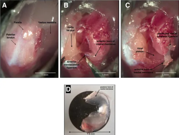
Osteoarthritis was induced by medial meniscectomy and medial collateral ligament transection, as previously described by Kamekura (Kamekura et al., 2005). Briefly, ten
week-old male mice were anaesthetised with a combination of ketamine (1mg/10g body weight , i.p., Merial France), xylazine (0.1mg/10g body weight, i.p., Bayer, Germany) and acepromazine (0.03mg/10g body weight, i.p., Vétoquinol, France)(Buitrago et al.,
2008). Using a stereomicroscope (Nikon SMZ1500) and microsurgical instruments a medial knee arthrotomy was performed and the patella was externally subluxated, the right knee medial collateral ligament was transected and the medial meniscus removed (MNX) (Figure 3.2). The left knee was sham-operated (SHAM), i.e., the arthrotomy and external subluxation of the patella were performed but no ligament transection or
Figure 3.1: Flowchart describing the allocation of animals in the different experiments
3.1. PAPER I 25
All surgical procedures were performed by the author, who is credited by the Por-tuguese National Authority for Animal Health as competent to perform experiments with laboratory animals (DGAV ref. 0421/000/000/2013). The procedures were carried out during the morning and the surgeon was blinded to the strain of the animals. Animals were euthanized 8 weeks after surgery and bilateral knee joints were collected and pro-cessed as described in the next sections. There were no surgery-induced mortality nor adverse effects and all of the operated animals were included in the analysis.
3.1.2 Assessment of iron accumulation
Unless noted otherwise all the reagents used were purchased from Sigma-Aldrich. For the assessment of iron accumulation in the liver and blood, samples of 6 animals of each strain were used. Hepatic non-heme iron concentration in µg of iron per mg of wet liver weight was determined as previously described (Rebouche et al., 2004). Briefly, a sample of liver was collected, homogenized in a 1:10 dilution (weight/volume), then equal volumes of the tissue homogenates and protein precipitation solution (1 mol L−1 HCl and 10% trichloroacetic acid) were mixed in “boil-proof” tubes and heated to 95ºC for 1h. After centrifuging for 10min the supernatant was collected and an aliquot was mixed with an equal volume of chromogen solution (0.508 mmol L−
1 of ferrozine,
1.5 mol L−
1 sodium acetate and 10% L-ascorbic acid). After 30min at room temperature the absorbance at 562 nm was measured and the concentration calculated using a calibration curve prepared on the same day from FeCl3 stock solution [1 mg mL−
1] and diluted to 5, 10, 15, 25 and 50µg mL−1 of iron. At the time of euthanasia blood was collected by intra-cardiac puncture, in tubes containing Heparin-Lithium, and sent to an external certified commercial laboratory (DNAtech, Lisbon, Portugal) for determination of serum iron, serum ferritin and also serum transferrin saturation.
3.1.3 Grading of osteoarthritic changes
Bilateral knee joints from 10 animals in each group were isolated, cleaned of adher-ent soft tissues in ice-cold phosphate buffer saline (PBS) and fixed for 24hrs in 4%
paraformaldehyde in PBS pH7.4, followed by decalcification with 0.5 mol L−
1 ethylene-dinitrilotetraacetic acid (EDTA) in PBS pH 7.4 for 3 weeks. After dehydration through graded alcohols and inclusion in paraffin, 5µmsagittal sections were cut from the medial compartment of the joints, three sections per level with a 150µminterval between levels, on a rotary microtome (Microm HM340E, Germany) and stained with Safranin-O/Fast Green/Meyer’s Hematoxylin. Two separate observers, without knowledge of the strain and intervention, graded the cartilage lesions using the semi-quantitative scoring system proposed by Glasson et al. (Glassonet al., 2010), also known as the OARSI scoring
sys-tem for murine osteoarthritis. This syssys-tem is based on the cartilage destruction extent, graded 0-6 depending on the depth of the lesion and on the percentage of the articular surface affected. The medial tibial plateau and the medial femoral condyle were graded
3.1. PAPER I 27
for that joint.
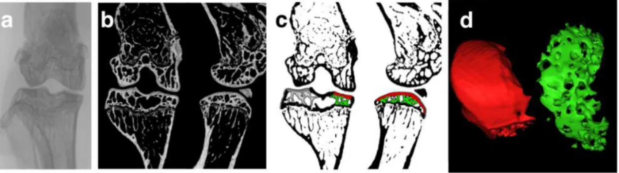
3.1.4 Morphological characterization of the knee joint by micro-CT
Micro-CT was performed on bilateral knee joints of 5 animals in each group with a Skyscan 1172 X-ray computed microtomograph (Bruker, Belgium) prior to inclusion in methyl methacrylate (MMA). The samples were wrapped in laboratory film (Parafilm) and placed inside an empty 1.5ml tube to prevent desiccation. The tubes were fixed on the sample holder with adhesive plasticine. For the image acquisition the following pa-rameters were used: X-ray tube potential 70 kVp, X-ray tube current 100µA, 0.5mmAl filter, rotation step 0.4º, isotropic voxel size 5µm3, integration time 500