(1) Faculdade de Medicina, Univ. Federal de Minas Gerais, Depto. Clínica Médica, Av. Alfredo Balena 190, sala 246, 30130-100 Belo Horizonte, MG, Brasil.
(2) Faculdade de Medicina, Univ. Federal de Minas Gerais, Depto. Medicina Preventiva e Social, Av. Alfredo Balena 190, sala 825, 30130-100, Belo Horizonte, MG, Brasil. (3) Centro de Pesquisas René Rachou, Fundação Oswaldo Cruz (FIOCRUZ), Av. Augusto de Lima 1715, 30190-002 Belo Horizonte, MG, Brasil.
(4) Faculdade de Medicina, Univ. Federal de Minas Gerais, Av. Alfredo Balena 190, 30130-100 Belo Horizonte, MG, Brasil.
Correspondence to: Silvana de Araújo Silva, R. Cássia 349/502, Bairro Prado, 30411-140 Belo Horizonte, MG, Brasil. Phones: +55 31 9119-8139, +55 31 3281-3115, +55 31 3248-9808. E-mail:
PREDICTIVE FACTORS FOR THE PROGRESSION OF CHRONIC CHAGAS CARDIOMYOPATHY IN
PATIENTS WITHOUT LEFT VENTRICULAR DYSFUNCTION
Silvana de Araújo SILVA(1), Eliane Dias GONTIJO(2), João Carlos Pinto DIAS(3), Camila Gomes de Souza ANDRADE(4) & Carlos Faria Santos AMARAL(1)
SUMMARY
The identification of predictors for the progression of chronic Chagas cardiomyopathy (CCC) is essential to ensure adequate patient management. This study looked into a non-concurrent cohort of 165 CCC patients between 1985 and 2010 for independent predictors for CCC progression. The outcomes were worsening of the CCC scores and the onset of left ventricular dysfunction assessed by means of echo-Doppler cardiography. Patients were analyzed for social, demographic, epidemiologic, clinical and workup-related variables. A descriptive analysis was conducted, followed by survival curves based on univariate (Kaplan-Meier and Cox’s univariate model) and multivariate (Cox regression model) analysis. Patients were followed from two to 20 years (mean: 8.2). Their mean age was 44.8 years (20-77). Comparing both iterations of the study, in the second there was a statistically significant increase in the PR interval and in the QRS duration, despite a reduction in heart rates (Wilcoxon < 0.01). The predictors for CCC progression in the final regression model were male gender (HR = 2.81), Holter monitoring showing pauses equal to or greater than two seconds (HR = 3.02) increased cardiothoracic ratio (HR = 7.87) and time of use of digitalis (HR = 1.41). Patients with multiple predictive factors require stricter follow-up and treatment.
KEYWORDS: Chagas cardiomyopathy; Clinical progression; Prognosis; Cohort studies.
INTRODUCTION
Although it was first described over a century ago, Chagas disease (CD) still remains a relevant endemic ailment in Latin America as it threatens some 16 million people in the continent. As the transmission via vectors has been controlled in several countries, the clinical follow-up of the millions still infected remains an important challenge. Chronic Chagas cardiomyopathy (CCC) is the main morbidity resulting from CD10. Left ventricular dysfunction is the strongest predictive factor for
morbidity and mortality in CCC8,9. The identification of markers for
disease progression before the occurrence of ventricular dysfunction may allow for earlier treatment and better prognosis. Although this disease has been extensively studied, the natural history of CCC and its independent predictive factors in outpatients – examined through the most sophisticated non-invasive cardiovascular methods such as echo-Doppler cardiography (ECHO), Holter monitoring, and exercise testing (ET) – are not completely understood. Most previous studies resorted to simpler risk stratification methods, such as electrocardiography (ECG) and chest radiography, and were conducted based on small selected heterogeneous groups including Chagas patients with varied prognoses, some of them for a short period of time23. Some authors26,31 have looked
at earlier stage CCC patients, with differing findings on the ECG, no left ventricular dysfunction, and unnoticeable symptoms. Very few studies
have measured prognostic factors of Chagas cardiomyopathy among asymptomatic Trypanosoma cruzi-infected persons26.
This study aimed to identify the predictive factors for the progression of CCC in patients with no left ventricular dysfunction and to check if this progression is different in patients who have an ECG with abnormalities indicative of CCC when compared to one who have unspecific ECG abnormalities.
MATERIALS AND METHODS
Patients: This non-concurrent cohort study covered adult CCC
Enrollment criteria: proven diagnosis for CD by two positive conventional serologic test results for T. cruzi infection32 having
completed the initial assessment protocol (iteration 1) comprised of an interview, physical examination, chest radiography, ECG, and ECHO; being diagnosed with CCC (abnormal ECG); having undergone clinical assessment, ECG, and ECHO at least two years since the initial assessment (iteration 2); having the tests at iteration 1 done within ≤ 12 months.
Exclusion criteria: having other heart conditions (ischemic,
hypertensive, congenital or valvular heart disease, alcoholic cardiomyopathy) referred or investigated through clinical and/or complementary tests, found at any stage of the follow-up; having left ventricular systolic or diastolic dysfunction on the ECHO on iteration 1; using a pacemaker (PM) or having ventricular tachycardia (VT) - defined as three or more consecutive premature ventricular complexes with a heart rate of more than 100 beats per minute - on the ECG at the initial assessment; patients with time intervals between tests > 12 months.
Cardiac non-invasive studies: They were all carried out as per the standard routines at HC-UFMG, from the preparation of the patients up to the interpretation of their results. All of them were conducted by personnel with previous experience of CCC and blind interpreted in relation to the clinical form of the disease. The date on which the first tests were done on the patients was considered as the patient’s date of entry into the study.
Conventional resting 12-lead ECG:interpreted by two examiners
using the diagnostic criteria for CCC3 accepted by the World Health Organization.
ECHO: Abnormal test results were characterized by LV (left
ventricular) dysfunction and/or anomalous segmental contractility, in addition to the presence of apical aneurysm. Systolic dysfunction was considered when EF < 54% and classified as mild to moderate (≥ 40%) and severe (< 40%), whereas diastolic disorder was characterized for dysfunction stages ≥ II (patients were classified according to diastolic function patterns: normal, impaired relaxation known as stage I, pseudonormal pattern known as stage II and restrictive pattern known as stage III). Segmental disorder was defined as the presence of akinesia, hypokinesia or dyskinesia in a defined area.
Chest radiography: with images taken from two views:
posteroanterior and lateral – using the cardiothoracic ratio (CTR) as reference and those patients with a CTR ≥ 0.50 were deemed as abnormal.
Exercise Testing (ET): It was completed by patients for whom the test was not contraindicated. They were considered abnormal when any of the following were observed: ventricular arrhythmia, blood pressure alterations, chronotropic response, myocardial ischemia criteria - namely J-point depression (the point at which the QRS complex meets the ST segment) ≥ 1 mm, with a horizontal or downsloping ST segment with duration ≥ 0.80 seconds (sec); Y-point depression 80 milliseconds (msec) after point J ≥ 1.5 mm with an upsloping ST segment; J-point elevation ≥ 1 mm. 24-hour Holter monitoring: They were considered abnormal when any of the following were found: arrhythmias with complexity ≥ Lown 218, intra or atrioventricular conduction disorders,
pauses of ≥ 2.0 seconds and changes in the ST segment matching myocardial ischemia criteria.
Categorization: After cardiac studies results had been analyzed,
patients were divided into two groups at iteration 1: group 1 (G1): ECG with at least two unspecific abnormalities; and group 2 (G2): ECG with abnormalities indicative of CCC. Further on, considering ECG and ECHO, patients were independently categorized for CCC8, taking
the most abnormal test result into account, thus describing four stages: 1- ECG: at least two unspecific abnormalities (G1) – sinus bradycardia (HR > 40 bpm), low voltage, incomplete right bundle branch block (RBBB), left anterior hemiblock (LAHB), first-degree atrioventricular block (AV block), unspecific ST-T alterations. ECHO: normal. 2- ECG: abnormalities indicative of CCC (G2) – complete RBBB in association or not with LAHB, isolated monomorphic ventricular extrasystoles (VES), sinus bradycardia (HR ≤ 40 bpm), second-degree AV block, T primary alterations. ECHO: abnormal, but no ventricular dysfunction. 3- ECG: abnormalities indicative of CCC (G2) – polymorphic or sustained VES, electrically inactive area, sinus node dysfunction. ECHO: diastolic ventricular dysfunction or abnormal EF, however ≥ 40%. 4- ECG: abnormalities indicative of CCC (G2) – atrial fibrillation (AF), complete AV block, left bundle branch block (LBBB), non-sustained ventricular tachycardia (NSVT) and PM at time 2. ECHO: diastolic ventricular dysfunction or EF < 40%.
Outcomes or response variables: worsening of the CCC scores and onset of left ventricular dysfunction.
Analyzed independent explanatory variables:
Social, demographic and epidemiologic variables: age; gender; ethnicity; intensity and duration of physical effort in previous and current occupations2; time spent in rural (RA) and CD endemic areas (EA); location and time spent at current residence; family history (FH) for CD, heart disease and sudden death – the last two occurring in family members aged 40 or less; drinking – weekly alcohol intake (in grams29) and period of abuse (in years); smoking (pack-year). Clinical
variables: symptoms, thromboembolism, comorbidities, systemic hypertension, body mass index (BMI), specific complete etiologic treatment with benznidazole, CCC score, regular and continuous use (in years) of cardiovascular drugs for at least two years including: loop diuretics, hydrochlorothiazide (HCTZ), beta blockers, spironolactone, amiodarone, angiotensin converting enzyme inhibitors (ACE inhibitors) or angiotensin II receptor blockers (ARBs). Cardiac non-invasive studies variables: abnormalities on ECG, chest radiography, ECHO, 24-hour Holter monitoring and on exercise testing.
Data collection tools and analysis: A structured questionnaire
was completed during first and return visits and all codified responses were entered into Microsoft Access. Statistical packages MINITAB for Windows 14.10, nQuery Advisor 4.0, SPSS 15.0 and EXCEL were used for data analysis purposes.
at the beginning and end of the study were considered when calculating the worsening of the CCC score. CCC score worsening was defined as having a greater score at the end of the study than the one collected at the beginning. Patients categorized as stage 4 were excluded from the aforementioned analysis because it was impossible for them to receive a worse score at the end of the study. Univariate analysis the Kaplan-Meier estimator was used to build survival curves, alongside Cox’s univariate model and the differences in survival between groups were assessed by the log-rank test. Multivariate analysis: The Cox regression model was used and a p-value of 0.20 was used to enter predictive variables into the Cox model and a 5% significance level was adopted as a cutoff threshold for the variables to be considered in the model. A new final model using only variables obtained in univariate analysis with a p- value of 0.5 was performed in order to avoid “overfitting phenomena”. A final model was considered adequate to be interpreted when cox proportional risk was tested by using a logarithm of cumulative risk function against time (in months) for each covariate.
Observations: A hazard ratio (HR) with a CI of 95% was calculated. A 5% significance level was adopted in all analyses. Some data sets were stratified in accordance with the literature in order to explore them better.
Analysis of the sample stratified into ECG groups: In order to assess whether the intensity of the initial abnormality of the ECG could predict CCC progression, groups G1 and G2 were analyzed solely in relation to the onset of left ventricular dysfunction – as the ECG accounted for part of the CCC score, thus the outcome “worsening of the CCC scores” should not be considered. The detection power of the sample was calculated using a 95% CI27.
RESULTS
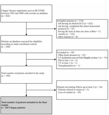
Patients were considered as lost when there was a failure to establish communications with patients after at least three phone calls on different days and at different times, sending a letter, and attempting to contact the patient’s neighbors (Fig. 1). The lost group was similar to the excluded and studied ones, there was no statistically significant difference between these groups.
Descriptive analysis:
Social, demographic and epidemiologic profile of the patients: the mean age of patients was 44.8 years (20-77 years) (SD = 10.6); mainly born in RA (97.0%) and residing in CD EA (88.5%) for a mean of 16 years (SD = 8.7). Most patients have lived away from RA and EA for a mean of 23.4 years (SD = 11.1). At the start of the study, patients had been involved in intense (49%) occupational physical effort for a mean of 14.6 years. Mean alcohol intake was 194.9 grams/week for 21.5 years (SD = 474.1 g/week), median 60 grams/week for 20 years; smoking history was quantified at a mean of 18.4 pack-years (SD = 16.3).
Clinical profile: Most patients were asymptomatic (63.6%). Systemic hypertension was the most prevalent comorbidity (21.8%) in iteration 1; incidence increasing to 28.5% in iteration 2, an increase which was also seen in the use of cardiovascular drugs, going from 30.3 in iteration 1 to 43.6% in iteration 2. The increase was statistically significant in both cases (McNemar’s: p < 0.01). Prevalence rates of the stages of CCC reduced from 49.7% (iteration 1) to 39.4% (iteration 2) in stage 1; from
42.4% to 40.6% in stage 2; while an increase from 6.7% to 13.3% was noticed in stage 3 and from 1.2% to 6.7% in stage 4.
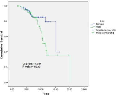
Cardiac non-invasive studies: ECG (Table 1) and ECHO (Table 2) explanatory variables were described and compared in both iterations of the study. They show a statistically significant (p-value Wilcoxon Test < 0.01) increase in both PR interval (56.4% of the patients) and QRS duration (40.8%), and in the reduction of heart rate (57.6%) all in iteration 2 of the study.
One-hundred patients underwent ET (61%), and 142 (86%) the 24-hour Holter monitoring.
Follow-up: More than 91% of the patients survived until the end of the study, while seven deaths were observed (4.5% lethality rate), two of which (28.6%) were due to sudden death, two (28.6%) due to decompensated CCC and three (42.8%) due to causes unrelated to CD. Eight (4.8%) patients were lost for over a year after completing the protocol in iterations 1 and 2 of the study. The minimum follow-up time in the study was two and the maximum was 20 years (mean = 8.2; DP = 3.2). The worsening of the CCC score was observed in 37 (22.7%) patients, while five (3.1%) improved their scores and 121 (74.2%) remained stable.
Analysis of the outcome “worsening of the CCC scores”: In
Table 1
Descriptive comparative analysis of ECG variables. CD patients. HC-UFMG
Variable Time 1 Time 2
Occurrences (n) Percentage (%) Occurrences (n) Percentage (%)
1st degree AV block*
No < 230 msec ≥ 230 msec
150 14 1 90.9 8.5 0.6 136 17 12 82.4 10.3 7.3
Sinus bradycardia* < 60 bpm 62 37.6 69 41.8
Complete RBBB No
yes 113 52 68.5 31.5 108 57 65.5 34.5
Inomplete RBBB No
yes 153 12 92.7 7.3 152 13 92.1 7.9
Isolated VES No
yes 150 15 90.9 9.1 154 11 93.3 6.7 LAHB No yes 117 48 70.9 29.1 117 48 70.9 29.1
Unspecific ST alterations No
yes 142 23 86.1 13.9 137 28 83.0 17.0 Rhythm sinus multifocal atrial AF PM 164 1 99.4 0.6 158 -2 5 95.8 -1.2 3.0
2nd degree AV block - grade I No
yes 163 2 98.8 1.2 164 1 99.4 0.6 LBBB No yes 163 2 98.8 1.2 162 3 98.2 1.8
Low voltage yes 3 1.8 3 1.8
NSVT yes 1 .6
* p-value McNemar’s test < 0.05.
Table 2
Descriptive comparative analysis of ECHO variables. CD Outpatient ward/HC-UFMG
Variable Time 1 Time 2
Occurrences (n) Percentage (%) Occurrences (n) Percentage (%)
LV final diastol.a diameter normal 151 91.5 137 83.0
altered 14 8.5 28 17.0
LV*c final systol.b diamater normal 155 93.9 134 81.2
altered 10 6.1 31 18.8
Ejection fraction
normal 165 100.0 144 87.3
53 to 40% 17 10.3
<40% 4 2.4
Shortening fraction normal 144 87.3 136 82.4
altered 21 12.7 29 17.6
Contractility alterations
absent 148 89.7 131 79.4
global dysf.d 1 0.6 9 5.5
segmental dysf.d 16 9.7 25 15.2
Diastolic function alteration
absent 133 80.6 75 45.7
grade I dysf.d. 32 19.4 85 51.8
grade II dysf.d - - 4 2.4
Degenerative* valve disorders no 158 95.8 140 84.8
yes 7 4.2 25 15.2
Table 3
Univariate analysis for outcome ‘worsening of CCC scores’. CD Outpatient ward/HC-UFMG
Variable Absolute # (n) / relative # (%) or
stratification
Worsening
incidence (%) p-value
a HR 95% CI for HR
Lower limit Upper limit
Female gender yes (103/62.4) 13.7 1.00
no (62/37.6) 37.7 0.010 2.59 1.25 5.36
Age < 50 years (112/67.8) 21.9 1.00
≥ 50 years (53/32.2) 24.5 0.024 2.18 1.11 4.26
Caucasian ethnicity no (112/67.9) 21.8 1.00
yes (53/32.1) 24.5 0.615 1.19 0.60 2.36
Permanence at RA <10 years (41/24.8) 31.1 1.00
≥ 10 years (124/75.2) 19.5 0.079 0.55 0.28 1.07
Permanence at EA < 10 years (34/20.6) 24.4 1.00
≥ 10 years (131/79.4) 15.6 0.363 0.64 0.25 1.66
Currently resides at RA no (160/97) 22.2 1.00
yes (05/3) 40.0 0.012 6.54 1.50 28.50
Currently resides at EA no (140 /84.8) 20.3 1.00
yes (25/15.2) 36.0 0.002 3.39 1.56 7.40
Remains currently at RA or EA < 10 years (163/98.2) 10.7 1.00
≥ 10 years (2/1.2) 25.2 0.071 2.99 0.91 9.77
FH of Chagas’ disease yes (127/77) 21.6 1.00
no (38/23) 23.0 0.912 1.05 0.47 2.32
FH of heart disease yes (74/44.8) 22.2 1.00
no (91/55.2) 23.3 0.656 0.86 0.45 1.66
FH of sudden death yes (59/35.8) 22.1 1.00
no (106/64.2) 24.1 0.839 1.05 0.63 1.77
Physical effort at current job mild (36/21.8) + moderate(48/29.1) 25.3 1.00
intense (67/40.6)+ VIb (14/8.5) 20.0 0.183 0.64 0.33 1.24
Time at current job < 10 years (42/25.5) 19.1 1.00
≥ 10 years (123/74.5) 25.3 0.318 1.42 0.72 2.80
Physical effort previous job mild (11/8.9) /moderate (33/26.8) 18.2 1.00
intense (49/39.8) / VIb(30/24.4) 22.1 0.362 1.48 0.64 3.45
Time at previous job < 10 years (112/67.9) 19.5 1.00
≥ 10 years (53/32.1) 24.2 0.359 1.48 0.64 3.44
Drinking present/past yes (75/45.5) 18.0 1.00
no (90/54.5) 28.4 0.438 1.30 0.67 2.51
Smoking present/past yes (58/35.2) 23.4 1.00
no (107/64.8) 21.4 0.639 0.84 0.41 1.72
Thrombo-embolics no (159/96.4) 22.8
yes (6/3.6) 20.0 0.564 0.556 0.08 4.08
Comorbidities no (117/70.9) 25.2
yes (48/29.1) 16.7 0.941 0.97 0.44 2.14
Systemic hypertension no (129/78.2) 23.6
yes (36/21.8) 19.4 0.617 1.24 0.54 2.84
BMI
↓ weight (9/5.5) 12.5 0.714
normal (92/55.8) 22.8 0.845 1.22 0.16 9.20
↑ weight (47/28.5) 21.7 0.784 1.33 0.17 10.51
Variable Absolute # (n) / relative # (%) or stratification
Worsening
incidence (%) p-value
a HR 95% CI for HR
Lower limit Upper limit
CTR normal (144/87.3) 16.8
altered (21/12.7) 65.0 0.000 6.419 3.213 12.823
Ventricular arrhythmia grade on ET
Lown 0 (63/63) 17.5
Lown 1- 4 (37/37) 36.1 0.041 2.36 1.04 5.39
Top HR on ET normal (81/81) 26.3
altered (19/19) 15.8 0.418 0.61 0.18 0.418
Blood pressure on ET normal (26/26) 24.7
altered (74/74) 23.1 0.587 0.77 0.30 1.98
AHA functional class on ET
I (27/27) 25.9 0.651
II (44/44) 25.6 0.513 0.68 0.21 2.17
III+IV (29/29) 20.7 0.871 1.09 0.40 2.96
cond.c dis.d AV/ IV Holter no (79/55.6) 26.6
yes (63/44.4) 18.0 0.111 0.55 0.26 1.15
pauses ≥ 2 sec. on Holter no (133/93.7) 21.4
yes (9/6.3) 44.4 0.043 2.99 1.04 8.64
SVe arrhythmia on Holter no (107/75.4) 22.9
yes (35/24.6) 22.9 0.384 1.46 0.62 3.40
Ventricular arrhythmia complexity on Holter
0 and 1 (74/52.1) 16.4
2 to 4 (68/47.9) 29.9 0.056 2.05 0.98 4.28
Sustained VAf (pairs) on Holter no (100/70.4) 16.2
yes (42/29.6) 39.0 0.004 3.09 1.42 6.72
NSVT on Holter no (124/87.3) 18.7
yes (18/12.7) 52.9 0.003 1.47 1.14 1.90
a *: Cox’s univariate model; b: VI: very intense; c: cond.: conduction; d: dis.: disorders; e: SV: supraventricular; f: VA: ventricular arrhythmia. Table 3
Univariate analysis for outcome ‘worsening of CCC scores’. CD Outpatient ward/HC-UFMG (cont.)
Table 4
Univariate analysis for outcome ‘worsening of CCC scores’. CD Outpatient ward/HC-UFMG. Drinking (weekly alcohol intake and time of abuse) and smoking (pack-year)
Worsening of CCC scores Estimation Weekly alcohol intake (in grams)
Time of drinking abuse (in years)
Smoking (pack-year)
No
Quartile 1 22.0 12.0 458.5
Median 58.0 21.0 1160.0
Quartile 3 157.5 27.5 4042.5
Yes
Quartile 1 20.0 13.5 603.0
Median 66.0 20.0 1020.0
Quartile 3 197.0 27.0 4028.0
p - value* 0.979 0.959 0.598
HR 1.00 1.00 1.01
95% CI to HR [0.99; 1.01] [0.96; 1.04] [0.97; 1.05]
increased CTR, and time of use of digitalis (Table 6). The Median survival time to CCC score worsening was 15.15 (8.91-21.39) years (standard error = 3.18; 95% CI).
Analysis of the outcome “onset of left ventricular dysfunction”:
Even after including the variables that had p < 0.20 in the univariate analysis (Table 7), an adequate final regression model was not attained for multivariate analysis.
Analysis of sample stratified into ECG groups: 88 (53%) patients were categorized into group 1 (G1) and 77 (47%) into group 2 (G2). Left ventricular dysfunction was seen in 11 (12.5%) patients in G1 and in 13 (16.9%) in G2. Univariate analysis did not reveal a statistically significant difference between both groups (HR = 0.89) for the onset of left ventricular dysfunction (p = 0.788; 95% CI: 0.39-2.02). Relative risk (RR) was 1.4 and calculated sample detection power found to be 12.23%.
DISCUSSION
Chagas disease is a complex heterogeneous illness with wide variation in clinical course and prognosis. The advantages of the present study include the homogeneous sample of patients at earlier stages of CCC with no left ventricular dysfunction, having minimal abnormalities on ECG, examined through noninvasive risk markers that can be routinely measured, the duration of follow-up (mean 8.2 years), and the use of multivariate methods of statistical analysis. This study demonstrated that the variables finally found to be predictive factors for CCC progression for both outcomes - worsening of the CCC scores and the onset of left ventricular dysfunction – were male gender, living in rural areas, time of use of digitalis and increased cardiothoracic ratio.
Fig. 4 - Kaplan-Meier estimator for time (in years) until the worsening of CCC scores for those currently residing in rural areas, CD Outpatient ward/HC-UFMG.
Fig. 3 - Kaplan-Meier estimator for time (in years) until the worsening of CCC scores by age range, CD Outpatient ward/HC-UFMG.
Fig. 2 - Kaplan-Meier estimator for time (in years) until the worsening of CCC scores by gender, CD Outpatient ward/HC-UFMG.
Table 5
Univariate analysis of time (in years) patients have taken cardiovascular drugs for outcome ‘worsening of CCC scores’. CD Outpatient ward/HC- UFMG
Variable p-value*a HR 95% CI to HR
Lower limit Upper limit
Loop diuretics 0.036 1.17 1.01 1.36
HCTZb 0.276 0.92 0.79 1.07
Digitalis 0.000 1.47 1.23 1.77
B-blockers 0.449 0.45 0.06 3.52
Spironolactone 0.004 1.42 1.12 1.80
ACEi/ARBs 0.030 1.13 1.01 1.26
Amiodarone 0.320 1.08 0.93 1.25
Fig. 6 - Kaplan-Meier estimator for time (in years) until the worsening of CCC scores based on CTR. CD Outpatient ward/HC-UFMG.
Fig. 5 - Kaplan-Meier estimator for time (in years) until the worsening of CCC scores for those currently residing in endemic areas, CD Outpatient ward/HC-UFMG.
Table 6
Multivariate analysis of time until the worsening of CCC scores. CD Outpatient ward/HC-UFMG
Variable Ratio Standard
error Wald’s test p-value HR
95% CI to HR
Lower limit Upper limit
Male gender 1.03 0.39 6.99 0.008 2.81 1.31 6.03
Pauses ≥ 2 seconds on Holter 1.11 0.55 4.07 0.044 3.02 1.03 8.83
CTR ≥ 0.50 2.06 0.42 24.51 <0.001 7.87 3.48 17.82
Time taking digitalis (years) 0.35 0.11 10.15 0.001 1.41 1.14 1.75
Resorting to univariate analysis, this paper – as did other important longitudinal studies – has revealed the following as statistically significant variables found to be predictive factors for CCC worsening as an outcome: male gender12,14, age > 50 years20,25,28; CTR ≥ 0.5013,14,22,25;
time of use of digitalis30, identification of ventricular arrhythmias in
the ET24 categorized from Lown 1 to 4; Holter monitoring showing
pauses equal to or greater than two seconds, and ventricular arrhythmia – sustained and non-sustained VT22,24. A statistically significant finding
– time taking cardiovascular medication – may be related to more severe cases of CCC (patients who needs treatment) as well as the variable “time of use of digitalis”. However, this study was not designed to discuss such findings.
Upon looking into patients’ current residential addresses in RA and CD EA two interesting issues surface: (1) the impact of exposure to reinfection in rural and endemic areas upon the deterioration of the patient’s clinical status. The progressive decrease in morbidity and mortality seen in CD in all controlled endemic areas was clearly predicted by DIAS11, who observed this in Bambuí, Minas Gerais, Brazil, where
a pioneering effort was made to systematically eradicate the vector insect. The author attributed the decrease to the cessation of exogenous reinfections, probably by the same strain, responsible for the more severe
cases of CD. And (2): access to healthcare services is more precarious in rural areas. It is clear that patients followed up at CD specialized care centers or at cardiology outpatient wards have greater access to therapy, enhanced compliance to treatment, better quality of life, and live longer, as the therapeutic arsenal used to treat heart failure from Chagas disease is highly effective and beneficial for the patient’s survival and quality of life, in addition to reducing hospitalization rates.
It was observed that patients with abnormal CTR have almost a 13-fold risk of this outcome happening, a very high risk, confirming the important predictive value of chest radiography. When evaluating a patient with abnormal CTR, attention should be turned to their adequate treatment as soon as possible. Less than one fourth of the patients with CCC had abnormal CTR, stressing the diagnostic value of ECG, due to its significant sensitivity in detecting abnormalities in early stage CCC8,28.
Although ECG unspecific abnormalities can commonly occur in the elderly as a result of atherosclerosis, hypertension, ischemia etc., the presence of electrocardiographic repolarization abnormalities increase sensitivity for the diagnosis of acute Chagas disease1. Those abnormalities
that ECG unspecific abnormalities are prevalent from acute phases of the disease. In the present study, a statistically significant difference between both groups (G1 and G2) for the onset of left ventricular dysfunction was not found, which can be explained by two ideas: the possibility that both groups were not really particularly different, thereby not allowing the authors to lessen the risk of patients with more than one unspecific abnormalities in the ECG progress CCC, and the low calculated sample detection power found to be 12.23% in the present study.
In the present study, the most prevalent abnormalities found in ECG are in accordance with the literature19 as well as the presence of ECG
alterations being an independent predictive factor for poor prognosis of CCC14,20,22,25: QRS width was associated with mortality, as seen in the
papers by RIBEIRO et al.24. A PR interval ≥ 0.16 seconds was shown
to be an independent predictive factor15. Some papers have indicated
that increased – and not reduced – heart rates are a predictive factor for mortality, but such a finding, albeit not correlated to the worst prognosis in several longitudinal studies, has been frequently reported in clinical settings in association with heart disease progression and severe sinus bradycardia (HR under 40 bpm).
Concerning ECHO, stage I diastolic dysfunction was the most
prevalent abnormality found in both iterations of the study, further supporting the finding seen in the literature that it may occur even in Chagas patients without heart disease and precede systolic dysfunction in CCC4. Some authors13 have described increased LV diastolic diameter
as a predictive factor for poor prognosis of CCC, while PETTI et al.20
correlated increased final LV diastolic diameter to death.
The authors’ data show that occupational reorientation is needed as the disease progresses to cardiac involvement, but many factors act as barriers to successfully finding a new career: the limits imposed upon the patient by the disease’s clinical manifestations and complications8,16,
the lack of specialized training seen among CD patients due to adverse socioeconomic circumstances, the migration from rural areas to urban regions – where the types of jobs available differ tremendously, the stigma that haunts those diagnosed with CD – even when the diagnosis is not accompanied by occupational disability.
In the present study, a significant share of the patients were overweight (BMI > 25) or obese (BMI > 30) due to their lifestyle and a third of them had comorbidities, with systemic hypertension ranking highest. However, the prevalence rate of systemic hypertension in the studied population, made up exclusively of CD patients, was lower than Table 7
Univariate comparison of ECG and CTR variables in both iterations of the study for outcome ‘onset of ventricular dysfunction on ECHO’. CD Outpatient ward/HC-UFMG
Variable Incidence of ventricular
dysfunction on ECHO (%) p-value*
a HR 95% CI to HR
Lower limit Upper limit
TIME 1
PR interval* (msec) ≤ 200 14.02
> 200 & < 230 21.4 0.772 1.20 0.36 4.02
HRate* (bpm) < 60 17.5
≥ 60 & < 100 9.7 0.321 0.62 0.25 1.58
QRS duration* (msec) < 120 14.4
≥ 120 14.8 0.324 0.64 0.26 1.55
RBBB + CRBBB no 13.9
yes 15.6 0.352 0.67 0.28 1.57
CRBBB no 15.0
yes 13.5 0.212 0.56 0.22 1.40
TIME 2
PR interval* (msec)
≤ 200 11.8 0.169
> 200 & < 230 17.6 0.871 1.11 0.32 3.82
≥ 230 41.7 0.061 2.62 0.96 7.19
HRate* (bpm) < 60 14.7
≥ 60 & < 100 14.5 0.612 0.81 0.36 1.83
QRS duration* (msec) < 120 11.3
≥ 120 19.1 0.852 1.08 0.47 2.49
RBBB + CRBBB no 13.7
yes 15.7 0.586 0.79 0.34 1.83
CRBBB no 14.8
yes 14.0 0.225 0.57 0.23 1.41
CTR normal 8.3
altered 57.1 0.000 12.73 5.34 30.33
that found for the Brazilian population in general within the same age range7. In spite of some controversy, there are publications to further
support this finding17. This study, however, was not designed to this
aim. The statistically significant increase in the incidence of systemic hypertension and in the use of cardiovascular drugs between iterations 1 and 2 of the study supports the verification of its increased prevalence rates as patient age increases while CCC progresses, giving both diseases a summational character. Some patients took inadequately prescribed digitalis, despite the authors’ efforts to convince patients and doctors of other services to stop it once it was not indicated. Increased CTR can result in a lack of opportunity of ready access to ACE inhibitor (this drug was first prescribed about ten years after the beginning of the study).
Low lethality rates in this paper relate to the overall status of the patients in the study. Mean follow-up was long enough to ensure that time was plenty for events to unfold, with regard to the onset of left ventricular dysfunction and CCC progression. Four patients died due to Chagas disease during the follow-up in this study, two due to sudden death and two due to heart failure requiring internation. Nowadays, there is a noticeable displacement of deaths to older patients, mainly among those from 50 to 79 years old21. More than 50% of the internations due
to Chagas disease in Brazil occurred among this population. Heart failure is the main cause of hospitalization (approximately 50%) of the South American population and it presents high mortality especially in patients with Chagas etiology6.
The limitations of the present study: selection bias might have occurred because patients with missing data were excluded and the multivariate analysis was restricted to the 165 patients who had complete data on all the variables. Although the authors believe that the patients in this cohort were representative of outpatients with Chagas heart disease in other areas of Brazil, further investigations involving this population are desirable. Patients with minimal abnormalities in their ECG were studied, thus it was necessary to define as G1 (group 1) and stage 1 those patients with two or more unspecific abnormalities, despite the low calculated sample detection power hindering the authors’ ability to draw certain conclusions. The authors could not assess the effect of therapy because treatment was not controlled in the study. The loss of follow-ups was more than 5%, although these patients and those excluded were statistically similar.
Conclusion: The identification of factors associated with the progression of CCC is essential to adequate patient management. In the present study the most important predictors were: male gender, Holter monitoring showing pauses equal to or greater than two seconds, time of use of digitalis and increased cardiothoracic ratio. Several other potential prognostic factors were pointed out in the univariate analysis, which will also contribute to the improvement in CCC patients care.
AUTHORS CONTRIBUTIONS
Silvana de Araújo Silva, Eliane Dias Gontijo and Carlos Faria Santos Amaral: Study design; collection, analysis, and interpretation of data; writing of the paper; and decision to submit it for publication. João Carlos Pinto Dias: interpretation of data; writing of the paper; and decision to submit it for publication. Camila Gomes de Souza Andrade: collection, writing of the paper.
RESUMO
Preditores da evolução da cardiopatia chagásica crônica em pacientes sem disfunção ventricular esquerda
A identificação de preditores da progressão da cardiopatia chagásica crônica (CCC) é essencial ao manejo adequado do paciente. Estudo coorte não concorrente de 165 pacientes portadores de CCC entre 1985-2010 quanto a preditores independentes da evolução da CCC. Os desfechos foram piora da classificação da CCC e surgimento de disfunção ventricular esquerda ao ecoDopplercardiograma. Variáveis sócio-demográficas, epidemiológicas, clínicas e propedêuticas foram estudadas e realizadas análise descritiva, análise de sobrevida com análise univariada (Kaplan-Meier e modelo univariado de Cox) e multivariada (modelo de regressão de Cox). O seguimento foi de dois a 20 anos, com média de 8,2 anos. A média de idade dos pacientes foi de 44,8 anos (20-77 anos). Comparando ambos os tempos do estudo, no tempo 2 houve significância estatística do aumento do intervalo PR e da duração do QRS, além da redução da frequência cardíaca (Wilcoxon < 0,01). Os preditores da evolução da CCC no modelo final de regressão foram sexo masculino (HR = 2,81), pausas iguais ou maiores que dois segundos ao Holter (HR = 3,02), aumento do índice cardiotorácico (HR = 7,87) e tempo de uso de digital (HR = 1,41), destacando-se necessidade de seguimento e tratamento mais rigoroso para os chagásicos que cumulam estes fatores.
REFERENCES
1. Alvarado-Tapias E, Miranda-Pacheco R, Rodríguez-Bonfante C, Velásquez G, Loyo J, Gil-Oviedo M, et al. Electrocardiography repolarization abnormalities are characteristic signs of acute chagasic cardiomyopathy. Invest Clin. 2012;53:378-94.
2. Andersen KL, Masironi R, Rutenfranz J, Seliger V. Habitual physical activity and health. Copenhagen: WHO;1979. (Regional Publications. European series n. 6).
3. Argentina. Ministerio de la Salud e Desarrolo. Programa de Salud Humana. Criterios de diagnostico eletrocardiografico en la cardiopatia chagasica crónica. Buenos Aires: Ministerio de la Salud e Desarrolo; 1985.
4. Barros MV, Rocha MO, Ribeiro AL, Machado FS. Tissue Doppler imaging enables the identification of diastolic dysfunction of pseudonormal pattern in Chagas’ disease. J Am Soc Echocardiogr. 2001;14:353-9.
5. Bestetti RB, Dalbo CM, Freitas OC, Teno LA, Castilho OT, Oliveira JS. Noninvasive predictors of mortality for patients with Chagas’ heart disease: a multivariate stepwise logistic regression study. Cardiology. 1994;84:261-7.
6. Bocchi EA. Heart failure in South America. Curr Cardiol Rev. 2013;9:147-56. 7. Brasil. Ministério da Saúde. Secretaria Executiva Subsecretaria de Planejamento e
Orçamento. Plano Nacional de Saúde/PNS 2008/2009-2011. Brasília: Ministério da Saúde; 2009.
8. Brasil. Ministério da Saúde. Secretaria de Vigilância em Saúde. Brazilian consensus on Chagas disease. Rev Soc Bras Med Trop. 2005;38(Supl 3):7-29.
9. Carrasco HA, Parada H, Guerrero L, Duque M, Durán D, Molina C. Prognostic implications of clinical, electrocardiographic and hemodynamic findings in chronic Chagas’ disease. Int J Cardiol. 1994;43:27-38.
10. Coura JR, Dias JC. Epidemiology, control and surveillance of Chagas disease: 100 years after its discovery. Mem Inst Oswaldo Cruz. 2009;104(Suppl 1):31-40.
12. Dias JC, Kloetzel K. The prognostic value of the electrocardiographic features of chronic Chagas’ disease. Rev Inst Med Trop Sao Paulo. 1968;10:158-62.
13. Espinosa RA, Pericchi LR, Carrasco HA, Escalante A, Martínez O, González R. Prognostic indicators of chronic chagasic cardiopathy. Int J Cardiol. 1991;30:195-202. 14. Garzon SAC, Lorga AM, Nicolau JC. Eletrocardiografia na cardiopatia chagásica. Rev
Soc Cardiol Est Sao Paulo. 1994;4:133-43.
15. Gonçalves JG, Dias Silva VJ, Calzada Borges MC, Prata A, Correia D. Mortality indicators among chronic Chagas patients living in an endemic area. Int J Cardiol. 2010;143:235-42.
16. Gontijo ED, Rocha MO, Torquato de Oliveira U. Perfil clínico-epidemiológico de chagásicos atendidos em ambulatório de referência e proposição de modelo de atenção ao chagásico na perspectiva do SUS. Rev Soc Bras Med Trop. 1996;29:101-8. 17. Guariento ME, Orosz JE, Gontijo JA. Interação clínica entre moléstia de Chagas e
hipertensão arterial primária em um serviço de referência ambulatorial. Arq Bras Cardiol. 1998;70:431-4.
18. Lown B, Wolf M. Approaches to sudden death from coronary heart disease. Circulation. 1971;44:130-42.
19. Macêdo VO. Inquérito eletrocardiográfico nacional para doença de Chagas. Rev Soc Bras Med Trop. 1993;26(Suppl 2):12-3.
20. Petti MA, Viotti R, Armenti A, Bertocchi G, Lococo B, Alvarez MG, et al. Predictores de insuficiencia cardiaca en la miocardiopatía chagásica crónica con disfunción asintomática del ventrículo izquierdo. Rev Esp Cardiol. 2008;61:116-22. 21. Ramos AN Jr, Carvalho DM. Doença de Chagas: passado, presente e futuro. Cad Saúde
Colet. 2009;17:787-94.
22. Rassi A Jr, Rassi A, Little WC, Xavier SS, Rassi SG, Rassi AG, et al. Development and validation of a risk score for predicting death in Chagas’ heart disease. N Engl J Med. 2006;355:799-808.
23. Rassi Jr A, Rassi A, Marin-Neto JA. Chagas heart disease: pathophysiologic mechanisms, prognostic factors and risk stratification. Mem Inst Oswaldo Cruz. 2009;104(Suppl 1):152-8.
24. Ribeiro AL, Cavalvanti PS, Lombardi F, Nunes M do C, Barros MV, Rocha MO. Prognostic value of signal-averaged electrocardiogram in Chagas disease. J Cardiovasc Electrophysiol. 2008;19:502-9.
25. Rodriguez-Salas LA, Klein E, Acquatella H, Catalioti F, Davalos V, Gomez-Mancebo JR, et al. Echocardiographic and clinical predictors of mortality in chronic Chagas’ disease. Echocardiography. 1998;15:271-8.
26. Sabino EC, Ribeiro AL, Salemi VM, Di Lorenzo Oliveira C, Antunes AP, et al. Ten-year incidence of Chagas cardiomyopathy among asymptomatic Trypanosoma cruzi
seropositive former blood donors. Circulation. 2013;127:1105-15.
27. Sahai H, Khurshid A. Formulae and tables for the determination of sample sizes and power in clinical trials for testing differences in proportions for the two- sample design: a review. Stat Med. 1996;15:1-21.
28. Silva SA, Gontijo ED, Amaral CF. Case-control study of factors associated with chronic Chagas heart disease in patients over 50 years of age. Mem Inst Oswaldo Cruz. 2007;102:845-51.
29. Skinner HA, Holt S, Schuller R, Roy J, Israel Y. Identification of alcohol abuse using laboratory tests and a history of trauma. Ann Intern Med. 1984;101:847-51. 30. Theodoropoulos TA, Bestetti RB, Otaviano AP, Cordeiro JA, Rodrigues VC, Silva AC.
Predictors of all-cause mortality in chronic Chagas’ heart disease in the current era of heart failure therapy. Int J Cardiol. 2008;128:22-9.
31. Viotti RJ, Vigliano C, Laucella S, Lococo B, Petti M, Bertocchi G, et al. Value of echocardiography for diagnosis and prognosis of chronic Chagas disease cardiomyopathy without heart failure. Heart. 2004;90:655-60.
32. World Health Organization. Control of Chagas disease: report of a WHO Expert Committee. World Health Organ Tech Rep Ser. 1991;811:1-95.