Case 4 - A 78-Year-Old Obese Woman with Sudden-Onset Dyspnea
Bruno Ceotto, Júlio César Vieira de Sousa, Luiz Alberto Benvenuti Heart Institute (Incor), University of São Paulo Medical School, Brazil
&RUUHVSRQGHQFH9HUD'$LHOOR
InCor – Av. Dr. Enéas de Carvalho Aguiar, 44 – 05403-000 – São Paulo, SP - Brazil E-mail: [email protected]
Key Words:
Obesity; dyspnea; hypertrophy, ventricular.
Section Editor: Alfredo José Mansur ([email protected])
Associated Editors: Desidério Favarato ([email protected]) Vera Demarchi Aiello ([email protected])
A 78-year-old woman sought medical attention for severe dyspnea. She had morbid obesity, hypothyroidism, and long-standing hypertension, and was taking captopril 150 mg, furosemide 40 mg, digoxin 0.25 mg, and levothyroxine 75µg daily.
For a long time she had dyspnea on heavy exertion, which became worse during the previous week, progressing to dyspnea at rest, orthopnea, nonproductive cough, and lower extremity edema. The patient had been diagnosed with heart failure at another medical institution. She was then medicated and discharged from hospital; nevertheless, her dyspnea worsened, and she sought emergency care at InCor.
Physical examination (April 28, 2007) revealed tachypnea (36 breaths/min), heart rate of 120 bpm, blood pressure of 150/80 mm Hg, and body mass index of 46,9 kg/m². She weighted 275.5 pounds and was 5 feet and 3 inches tall. Chest auscultation revealed bilateral rales in the lower lung fields and occasional wheezing. Examinations of the heart and abdomen were unremarkable. Bilateral lower-extremity edema was noted, greater in the right leg. Oxygen saturation (measured percutaneously) was 88%.
Laboratory data were as follows: urea 85 mg/dL, creatinine 1.8 mg/dL, blood glucose 116 mg/dL, hemoglobin 18 g/dL, hematocrit 57%, platelet count 228,000/mm³, troponin 2.8 ng/ml, CK-MB 10.8 Pg/L, fibrin D-dimer > 5000 Pg; and activated partial thromboplastin time (APTT) ratio 1.41.
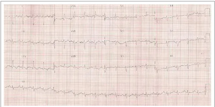
The electrocardiogram (ECG) showed sinus rhythm with a heart rate of 114 bpm, PR interval of 175 msec, QRS duration of 94 msec, QT interval of 282 msec, QRS axis +150 oriented posteriorly, S1Q3T3 pattern, and right bundle
branch block (Figure 1).
The echocardiogram yielded the following measurements: aortic diameter of 32 mm, left atrial diameter of 32 mm, interventricular septal thickness of 10 mm and free wall thickness of 9 mm. The right atrium was markedly dilated, and the right ventricle was dilated and hypokinetic, with a systolic pressure of 85 mm Hg. The left ventricle was found to be normal.
The diagnosis of pulmonary thromboembolism was proposed, and furosemide, 40 mg IV, was prescribed. The patient became hemodynamically unstable, requiring intravenous dopamine and orotracheal intubation for ventilatory support. Two hours of intravenous thrombolysis was performed with 100 mg of recombinant tissue plasminogen activator (rTPA).
On the night of the day after admission she went into cardiorespiratory arrest with pulseless electrical activity and died despite resuscitative efforts.
Clinical features
This is the case of a 78-year-old obese female patient with hypothyroidism, hypertension, and dyspnea on heavy exertion of one year duration that had progressed to dyspnea at rest during the previous week, associated with orthopnea, nonproductive cough, and lower-extremity edema.
Dyspnea is a symptom that results from the processing of multiple stimuli by the body1. It may therefore differ
from patient to patient, making an accurate diagnosis more challenging when assessed separately. A wide range of differential diagnoses, highly variable in severity, should be considered in a patient with dyspnea, including cardiac, pulmonary, neurological, muscular and even psychogenic causes2. A thorough and detailed clinical examination,
followed by appropriate selection and accurate interpretation of ancillary tests, may lead to the correct diagnosis. Even in the presence of obesity, which notoriously makes physical examination more difficult3, clinical symptoms play a decisive
role in the differential diagnosis.
Dyspnea of cardiac origin is a clinical expression of pulmonary venous and capillary hypertension. In the case described herein, the abnormal findings on chest auscultation probably prompted the diagnosis of heart failure made at another institution. The presence of systemic hypertension, grade III obesity (BMI > 40 kg/m2), and chronic dyspnea on
heavy exertion supports the hypothesis of heart failure, either systolic or diastolic, the latter being more common in elderly, obese4, and female patients, as was the case in this patient.
Nevertheless, in the absence of triggering factors, such as acute myocardial infarction or acute valvular dysfunction, the clinical course of heart failure is generally slow and progressive. The patient’s condition worsened relatively quickly over a one-week period, which argues against the diagnosis of heart failure.
Figure 1 – ECG - sinus tachycardia, right bundle branch block, right axis deviation, and a S1Q3T3 pattern.
during sleep, resulting in hypoventilation and hypoxemia, which leads to pulmonary hypertension, polycythemia and ultimately to heart failure, particularly right-sided heart failure, and impaired functional capacity5. A number of other
conditions can also cause pulmonary hypertension, such as chronic obstructive pulmonary disease (COPD), chronic pulmonary thromboembolism, congenital heart diseases, and schistosomiasis, among others.
The presence of asymmetrical lower-extremity edema together with dyspnea and hypoxemia led us to the diagnosis of acute pulmonary thromboembolism (PTE). Even though PTE is associated with a broad spectrum of clinical syndromes, they can be divided into three major groups6:
(1) pulmonary infarction, characterized by pleuritic pain and hemoptysis, (2) isolated dyspnea, which appeared to be the initial clinical presentation of our patient, and (3) circulatory collapse, defined as loss of consciousness or systolic blood pressure less than 90 mm Hg. Lung examination is abnormal in 29% to 37% of the patients with PTE, depending on whether or not prior cardiopulmonary disease is present7.
Fifteen-day mortality rates in patients with isolated dyspnea and circulatory collapse (6.2% e 6.5%, respectively) are higher than those found in patients with pulmonary infarction (2.5%)8.
Venous thromboembolism (VTE) results from an imbalance in the coagulation system. The cardinal factors that contribute to intravascular coagulation have been known for more than a century, being originally described as a triad by Rudolf Virchow9
as follows: (1) endothelial injury, (2) hypercoagulability, and (3) blood stasis. These risk factors may be primary (inherited) or secondary (acquired) in nature.
The secondary risk factors are the most frequently found and thus more easily identified, such as prolonged immobilization, surgery in the previous three months, stroke, and malignancy10.
However, in some cases, known as idiopathic or primary venous thromboembolism, these risk factors are not present. Under these circumstances, particular attention must be given to the investigation of genetic factors. The most common genetic defect in these patients is a mutation in factor V Leiden, accounting for up to 40% of the cases11. The patient described
herein was obese and hypertensive, and both conditions are major risk factors for venous thromboembolism in women12.
Her oxygen saturation, measured by percutaneous oximetry, was 88%. Such hypoxia associated with tachypnea (36 breaths/min) and tachycardia (120 bpm) reflects both the presence of respiratory failure and severity of the clinical picture. Hypoxemia in PTE may be explained by numerous mechanisms, the most important of which are changes in the pulmonary ventilation-perfusion ratio. While the presence of hypoxemia is not helpful in establishing the diagnosis13,
it plays a key role as a predictor of death. Pulse oximetry, widely available in emergency departments, is a quick and easy-to-perform test. Even in patients who initially are neither hypotensive nor requiring mechanical ventilation, a pulse oximetry reading < 95% at hospital admission is associated with greater 30-day mortality (20% compared with only 2%
LQSDWLHQWVZLWKSXOVHR[LPHWU\14.
The patient’s ECG showed sinus tachycardia, right axis deviation and a S1Q3T3pattern. This pattern is traditionally
considered as suggestive of pulmonary embolism, and for decades has been known to reflect acute cor pulmonale15.
Care must be taken when interpreting this electrocardiogram. The S1Q3T3 pattern is not specific for PTE, occurring in 13.5%
of the patients in whom this diagnosis was initially suspected but subsequently ruled out16. Its absence is not sufficient to
The electrocardiogram may be completely normal in patients with PTE, the most common rhythm disturbance being sinus tachycardia, which pathophysiologically reflects an increase in cardiac output to improve oxygen delivery17.
Plasma fibrin D-dimer was > 5000 µg, an elevated level that shows activation of the endogenous fibrinolytic system, since D-dimer is a fibrin degradation product. Its use in the diagnosis of PTE has been extensively studied. The D-dimer assay has high sensitivity and negative predictive value but poor specificity18. Therefore, it is best used to exclude the diagnosis
of PTE in patients with low or intermediate probability. Plasma D-dimer levels are elevated postoperatively and in patients with acute myocardial infarction, sepsis, malignancies, and other systemic diseases.
This patient showed increased CK-MB and troponin levels, which initially could suggest acute myocardial infarction. Although cardiac troponins have been shown to be highly specific markers of myocardial injury, we must bear in mind that an elevation in serum levels of troponin does not mean that the mechanism of such elevation was atherosclerotic plaque instability, which is found in AMI patients. It is known that troponin elevation may be secondary to acute right ventricular overload, with increased metabolic demand and thereby ischemia19; this may have been the mechanism of
troponin elevation in our patient. Kinetics of troponin release may be useful in the differential diagnosis between acute myocardial infarction and PTE, since in the latter condition troponin serum levels usually return to normal levels within the first 40 hours, unlike in acute myocardial infarction20.
Elevated levels of cardiac troponin have proven to be of prognostic value in patients with PTE. Mortality rates are substantially higher in patients with high troponin levels than in those with normal levels (19.7% and 3.7%, respectively)21.
Even in a subgroup of hemodynamically stable patients, elevated troponin levels were associated with a mortality rate 5.9 times higher21.
The echocardiogram confirmed the suspicion raised by clinical and electrocardiographic findings, together with elevated troponin levels, severe pulmonary hypertension, and right ventricular overload. Pulmonary artery systolic pressure was 85 mm Hg, and the right ventricle was dilated and hypokinetic. The echocardiogram also showed normal left ventricular function, an important finding as it rules out heart failure, either systolic or diastolic, as the cause of the patient’s clinical condition. Only 40% of PTE patients have suggestive changes on echocardiogram, but if present these changes are helpful in establishing the presumptive diagnosis and constitute an excellent prognostic tool22. Mortality rates
are twice as high in patients with right ventricular hypokinesis as in those without hypokinesis22.
The diagnosis of pulmonary thromboembolism is difficult. It is usually based on the clinical probability of each patient, followed by identification of the thrombus or its repercussion in lung perfusion imaging. A number of diagnostic algorithms for PTE have been proposed, but none has offered substantial benefit over the others23.
Their differences are based primarily on the availability of the ancillary test chosen and the experience of each institution with it.
Scores were created in an attempt to standardize the risk stratification of PTE. The most widely used are those developed by Geneva and Wells, both of which are very helpful and share operating characteristics24. The modified
Wells criteria include clinical signs of deep venous thrombosis, absence of a more likely diagnosis than PTE, tachycardia, prolonged immobilization, previous thrombotic event, hemoptysis, and malignancy25. After clinical
stratification, if suspicion of PTE still remains it should be confirmed by diagnostic imaging using two main strategies. One of them is helical CT pulmonary angiography, which may confirm the diagnosis and has been the method of choice of centers experienced with this technique. Despite the disadvantage of using iodinated contrast medium, it has sufficient sensitivity to safely exclude the diagnosis of clinically significant pulmonary embolism25. The second
strategy is based on ventilation/perfusion lung scans, which can confirm the diagnosis of PTE in patients with high clinical probability. However, in some patients invasive pulmonary angiography is needed to rule out the disease10.
Lower-extremity venous Doppler ultrasound may also be used, and has sufficient diagnostic sensitivity for patients in whom therapies associated with greater morbidity, such as thrombolysis or surgery, will not be considered26.
The patient progressed to hemodynamic instability, requiring intravenous dopamine and orotracheal intubation for ventilatory support.
The shock associated with pulmonary thromboembolism results from right ventricular dilation and dysfunction, with a reduction in cardiac output associated with a leftward shift of the interventricular septum, thereby decreasing LV preload and output.
This causes both a decrease in coronary perfusion and right ventricular ischemia, further impairing RV function and creating a vicious circle that ultimately leads to tissue hypoperfusion4.
At that point, we were faced with an extremely difficult clinical decision. There was no time to make a definite diagnosis of PTE, which could have been obtained using imaging studies. The patient’s clinical instability did not allow any ancillary test to be made outside the confines of the intensive care unit, hence the presumptive diagnosis of PTE, which was based mostly on clinical and echocardiographic findings. A fibrinolytic agent was then administered.
The aim of the fibrinolytic therapy was threefold: to improve RV dysfunction by dissolving the thrombus and thus reduce pulmonary arterial obstruction; to prevent the continuous release of serotonin and other neurohumoral factors that may exacerbate pulmonary hypertension; and perhaps to dissolve thrombi in other sites, such as lower extremities and the pelvic region. It is especially indicated for patients with massive or submassive pulmonary thromboembolism. The fibrinolytic approved by the FDA was t-PA (Alteplase) 100 mg, in continuous infusion, followed by heparinization26. Patients
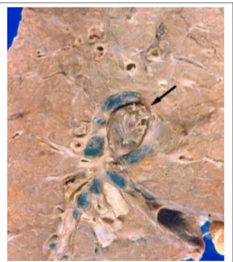
Figure 3 - Longitudinal section of the right lung at the hilar level showing occlusion of the pulmonary artery by a fresh thrombus (arrow).
Figure 2 - Cross-section of the heart showing left ventricular (LV) and right ventricular (RV) hypertrophy, greater in the latter.
reduction in 90-day mortality and recurrence of pulmonary thromboembolism13. In patients with massive or submassive
PTE in whom fibrinolysis is contraindicated or failed, surgical or catheter embolectomy must be considered26.
On the night of hospital day 2 the patient went into cardiorespiratory arrest with pulseless electrical activity, which may have been due to pulmonary thromboembolism, myocardial ischemia, hypoxia, electrolyte disorders (hypo- and hyperkalemia), hypovolemia, acidosis, or hypothermia. The cardiorespiratory arrest was refractory to cardiopulmonary cerebral resuscitation.
%UXQR&HRWWR0'-~OLR&pVDU9LHLUDGH6RXVD0'
Diagnostic hypothesis - acute massive pulmonary thromboembolism
Other diagnoses - morbid obesity, hypertension, hypothyroidism
%UXQR&HRWWR0'-~OLR&pVDU9LHLUDGH6RXVD0'
Autopsy
Postmortem examination showed a severely obese woman. The heart weighed 640 g and showed biventricular hypertrophy, greater in the right ventricle, which was slightly dilated.
The pulmonary artery was dilated, and a fresh, massive, bilateral pulmonary thromboembolism was found occluding first-generation branches of the left and right pulmonary arteries at the hilar level (Figure 3). Histological examination of several organs confirmed the above findings and showed in the thyroid gland, which was normal in size and not adhered to neighboring structures, a severe lymphocytic inflammatory infiltrate, without formation of lymphoid follicles, accompanied by fibrosis (Figure 4).The kidneys were found to have a finely granular surface, hyaline arteriolosclerosis, plus rare foci of interstitial fibrosis and glomerulosclerosis. The gallbladder was small and contained a single large stone. The aorta was mildly atherosclerotic. No internal genital organs were found (probably due to prior radical hysterectomy).
/XL]$OEHUWR%HQYHQXWL0'
Figure 4 - Cross-section of the thyroid gland showing the presence of
PRQRQXFOHDU LQÀDPPDWRU\ FHOO LQ¿OWUDWH ¿EURVLV DQG DWURSK\ RI WK\URLG
follicles, characterizing chronic lymphocytic thyroiditis. There are no lymphoid follicles. Hematoxylin-eosin staining.
Pathologic Diagnoses
Morbid obesity; right and left ventricular hypertrophy; chronic lymphocytic thyroiditis; chronic calculous cholecystitis; and acute massive pulmonary thromboembolism (cause of death).
/XL]$OEHUWR%HQYHQXWL0'
Comments
Referências
1. ATS Board of Directors. Dyspnea. Mechanisms, assessment, and management: a consensus statement. Am J Respir Crit Care Med. 1999; 159: 321-40.
2. Michaelson E, Hollrah S. Evaluation of the patient with shortness of breath: an evidence based approach. Emerg Med Clin North Am. 1999; 17: 221-37.
3. National Task Force on the Prevention and Treatment of Obesity. Medical care for obese patients: advice for health care professionals. Am Fam Physician. 2002; 65: 81-8.
4. Poirier P, Giles TD, Bray GA, Hong Y, Stern JS, Pi-Sunyer FX, et al. Obesity and cardiovascular disease: pathophysiology, evaluation, and effect of weight loss: an update of the 1997 American Heart Association Scientific Statement on Obesity and Heart Disease From the Obesity Committee of the Council on Nutrition, Physical Activity, and Metabolism. Circulation. 2006; 113: 898-918.
5. Flemons WW. Obstructive sleep apnea. N Engl J Med. 2002; 347: 498-504.
6. Stein PD, Willis PW, de Mets DL. History and physical examination in acute pulmonary embolism in patients without preexisting cardiac or pulmonary disease. Am J Cardiol. 1981; 47: 218-23.
7. Stein PD, Beemath A, Matta F, Weg JG, Yusen RD, Hales CA, et al. Clinical characteristics of patients with acute pulmonary embolism: data from PIOPED II. Am J Med. 2007; 120: 871-9.
8. Lobo JL, Zorrilla V, Aizpuru F, Uresandi F, Garcia-Bragado F, Conget, F, et al. Clinical syndromes and clinical outcome in patients with pulmonary embolism: findings from the RIETE registry. Chest. 2006; 130: 1817-22.
9. Virchow R. Phlogose und thrombose im Gefasssystem. Gesammelte Abhandlungen zur Wissenschaftlichen Medizin. Berlin: Verlag; 1862.
10. Value of the ventilation/perfusion scan in acute pulmonary embolism: results of the prospective investigation of pulmonary embolism diagnosis (PIOPED). The PIOPED Investigators. JAMA. 1990; 263: 2753-9.
11. Rosendaal, FR. Venous thrombosis: a multicausal disease. Lancet. 1999; 353: 1167-73.
12. Goldhaber SZ, Grodstein F, Stampfer MJ, Manson JE, Colditz GA, Speizer FE, et al. A prospective study of risk factors for pulmonary embolism in women. JAMA. 1997; 277: 642-5.
13. Rodger MA, Carrier M, Jones GN, Rasuli P, Raymond F, Djunaedi H, et al. Diagnostic value of arterial blood gas measurement in suspected pulmonary embolism. Am J Respir Crit Care Med. 2000; 162: 2105-8.
14. Kline JA, Hernandez-Nino J, Newgard CD, Cowles DN, Jackson RE, Courtney M. Use of pulse oximetry to predict in-hospital complications in normotensive patients with pulmonary embolism. Am J Med. 2003; 115: 203-8.
15. Mcginn S, White PD. Acute cor pulmonale resulting from pulmonary embolism. JAMA. 1935; 104: 1473.
16. Rodger M, Makropoulos D, Turek M, Quevillon J, Raymond F, Rasuli P, et al. Diagnostic value of the electrocardiogram in suspected pulmonary embolism. Am J Cardiol. 2000; 86: 807-9.
17. Ullman E, Brady WJ, Perron AD, Chan T, Mattu A. Electrocardiographic manifestations of pulmonary embolism. Am J Emerg Med. 2001; 19: 514-9.
18. Tapson VF, Carroll BA, Davidson BL, Elliot CG, Fedullo PF, Hales CA, et al. The diagnostic approach to acute venous thromboembolism: clinical practice guideline. American Thoracic Society. Am J Respir Crit Care Med. 1999; 160: 1043-66.
19. Meyer T, Binder L, Hruska N, Luthe H, Buchwald AB. Cardiac troponin I elevation in acute pulmonary embolism is associated with right ventricular disfunction. J Am Coll Cardiol. 2000; 36: 1632-6.
20. Muller-Bardorff M, Weidtmann B, Giannitsis E, Kurowsky V, Katus HA. Release kinetics of cardiac troponin T in survivors of confirmed severe pulmonary embolism. Clin Chem. 2002; 48: 673-5.
21. Becattini C, Vedovati MC, Agnelli G. Prognostic value of troponins in acute pulmonary embolism: a meta-analysis. Circulation. 2007; 116: 427-33.
22. Kucher N, Rossi E, De Rosa M, Goldhaber SZ. Prognostic role of echocardiography among patients with acute pulmonary embolism and a systolic arterial pressure of 90 mm Hg or higher. Arch Intern Med. 2005; 165: 1777-81.
23. Roy PM, Colombet I, Durieux P, Chatellier G, Sors H, Meyer G. Systematic review and meta-analysis of strategies for the diagnosis of suspected pulmonary embolism. BMJ. 2005; 331: 259-68.
24. Chagnon I, Bounameaux H, Drahomir A, Roy PM, Gourdier AL, Cornuz J. Comparision of two clinical prediction rules and implicit assessment among patients with suspected pulmonary embolism. Am J Med. 2002; 113: 269-75.
25. van Belle A, Büller HR, Huisman MV, Huisman PM, Kaasjager K, Kamphuisen PW, et al. for Christopher Study Investigators. Effectiveness of managing suspected pulmonary embolism using an algorithm combining clinical probability, D-dimer testing, and computed tomography. JAMA. 2006; 295: 172-9.
26. Arcasoy SM, Kreit JW. Thrombolytic therapy of pulmonary embolism: a comprehensive review of current evidence. Chest. 1999; 115: 1695-707.
27. Sugerman HJ. Pulmonary function in morbid obesity. Gastroenterol Clin North Am. 1987; 16: 225-37.
28. Blaszyk H, Wollan PC, Witkiewicz AK, Björnsson J. Death from pulmonary thromboembolism in severe obesity: lack of association with established genetic and clinical risk factors. Virchow Arch. 1999; 434: 529-32.
29. Bray GA. Health hazards of obesity. Endocrinol Metab Clin North Am. 1996; 25: 907-19.
30. Michalaki MA, Vagenakis AG, Leonardou AS, Argentou MN, Habeos IG, Makri MG, et al. Thyroid function in humans with morbid obesity. Thyroid. 2006; 16: 73-8.
31. Portmann L, Giusti V. Obesity and hypothyroidism: mith or reality? Rev Med Suisse. 2007; 3: 859-62.
revealed not only mild left ventricular hypertrophy, which could be explained by systemic hypertension, but also moderate right ventricular hypertrophy. The latter is most probably due to chronic pulmonary hypoventilation and/or obstructive sleep apnea secondary to morbid obesity, which may cause chronic pulmonary hypertension and right ventricular overload27.
It is important to note that in addition to right ventricular hypertrophy, other changes in this case are closely related to severe obesity, such as gallstones, the systemic hypertension itself, and pulmonary thromboembolism28,29.
Therefore, we believe that morbid obesity was the
primary disease of the patient.
The morphological change found in the thyroid gland, that is, chronic lymphocytic thyroiditis, may explain the thyroid dysfunction. Indeed, hypothyroidism (either clinical or subclinical) is also associated with morbid obesity, and is found in 19.5% of the patients30.
Interestingly, even though hypothyroidism has been traditionally considered as one of the causes of obesity, no consensus exists on the actual causal relationship between both entities31.