Hospital Espírita de Marília
Mailing address: Paulo Borini - Rua Gabriel Monteiro da Silva, 40 Cep 17501-150 - Marília, SP, Brazil - E-mail: [email protected] Received 11/25/02
Accepted 4/14/03
English version by Stela Maris C. e Gandour
Arq Bras Cardiol, volume 81 (nº 5), 512-7, 2003
Paulo Borini, José Henrique Imaca Terrazas, Arlindo Ferreira Júnior, Romeu Cardoso Guimarães,
Sabrina Bicalho Borini
Marília, SP - Brazil
Female Alcoholics. Electrocardiographic Changes and
Associated Metabolic and Electrolytic Disorders
In men and women, abusive ingestion of alcohol is as-sociated with arterial hypertension 1-3, cardiac arrhythmias4,
and cardiac muscle impairment with several structural and functional abnormalities 5- 8.
Myocardial lesions are expected to occur when the daily alcohol consumption is greater than 80g for an appro-ximate period of 10 years 9. However, lower daily doses
(around 60g) for longer periods (approximately 25 years) may cause dilated cardiomyopathy, arrhythmias, and car-diac hypertensive disease 5,10. A subgroup of chronic
alco-holics with a greater sensitivity to development of hepato-pathy, probably due to genetic reasons, exists. As such, vulnerability to cardiac changes may also depend on cons-titutional predisposition 11.
The following mechanisms have been cited as partici-pating in the physiopathogeny of heart disease due to alco-hol: direct action of alcohol or its metabolites on the cardiac muscle causing inflammation and fibrosis 12, macro- and
mi-croangiopathic changes 13,14, electrolytic changes,
partici-pation of metabolic and nutritional factors 15,16, and the
toxic effect of the additives present in alcoholic beverages17.
Deficiency in thiamin itself does not seem to be the cause of alcoholic cardiomyopathy 18, because it is associated with
low cardiac output and systemic vasoconstriction, while, in the cardiomyopathy of beriberi, an elevated cardiac output occurs with reduced peripheral vascular resistance 4.
The increasing prevalence of female alcoholism among us 19,20 will certainly be a serious public health
pro-blem in the near future because of the increase in morbidity and mortality resulting from abusive alcohol consumption. If the cardiovascular problems of male alcoholism have not been sufficiently studied in Brazil, even less has been done in regard to female alcoholism. Women have been reported to be more susceptible than men are to diseases related to chronic alcoholism, even when ingesting a lower amount of alcohol. The difference in susceptibility between the sexes has been related to different proportions of body water and fat 21 and different rates of alcohol metabolism 22. Therefore,
women would be more susceptible than men in regard to
Objective - To identify the electrocardiographic changes and their associations with metabolic and elec-trolytic changes in female alcoholics.
Methods - The study comprised 44 female alcoholics with no apparent physical disorder. They underwent the following examinations: conventional electrocardiogra-phy; serologic tests for syphilis, Chagas’ disease, and hepa-titis B and C viruses; urinary pregnancy testing; hematime-tric analysis; biochemical measurements of albumin, fibri-nogen, fasting and postprandial glycemias, lipids, hepatic enzymes, and markers for tissue necrosis and inflammation.
Results - Some type of electrocardiographic change was identified in 33 (75%) patients. In 17 (38.6%) patients, more than one of the following changes were present: pro-longed QTc interval in 24 (54.5%), change in ventricular repolarization in 11(25%), left ventricular hypertrophy in 6 (13.6%), sinus bradycardia in 4 (9.1%), sinus tachycardia in 3 (6.8%), and conduction disorder in 3 (6.8%). The pa-tients had elevated mean serum levels of creatine phospho-kinase, aspartate aminotransferases, and gamma glutamyl transferase, as well as hypocalcemia and low levels of total cholesterol and LDL-cholesterol. The patients with altered electrocardiograms had a more elevated age, a lower alco-hol consumption, hypopotassemia, and significantly eleva-ted levels of triglycerides, postprandial glucose, sodium and gamma glutamyl transferase than those with normal elec-trocardiograms. The opposite occurred with fasting glyce-mia, magnesium, and alanine aminotransferase.
Conclusion - The electrocardiographic changes found were prolonged QTc interval, change in ventricular repola-rization, and left ventricular hypertrophy. Patients with normal and abnormal electrocardiograms had different metabolic and electrolytic changes.
cardiomyopathy and myopathy 5. Asymptomatic alcoholic
females have a lower ejection fraction and greater ventricu-lar mass than do nonalcoholic females 5.
The medical problems caused by alcoholism are usual-ly preceded by social and psychic problems for several years. Consequently, alcoholics admitted to specialized units for the treatment of addiction comprise a group diffe-rent from those admitted to clinical hospitals for the treat-ment of physical problems. Most studies on the cardiac changes in chronic alcoholics assess the latter group of al-coholics, ie, patients with evident clinical manifestations. The information about the changes occurring when the symptoms are not evident or are very mild is scarce.
This study aimed at assessing the frequency and type of electrocardiographic changes in chronic alcoholic pa-tients with no manifestation of previous heart disease and at detecting the metabolic and electrolytic changes associa-ted with them.
Methods
According to the principles of the Helsinki Declara-tion and after informed written consent, female patients diagnosed with alcoholism (IDC-10 1997) 23 and admitted to
a psychiatric hospital (Hospital Espírita de Marília, HEM) from October 1993 to September 1994 were studied.
The patients came from the Center of First Assistance and Triage of the Hospital de Clínicas of the Medical School of Marília, where they underwent clinical and psychiatric examinations and were considered suitable for the psychia-tric treatment at the HEM, because they had no relevant or-ganic problems. Within the first 24 hours after hospital ad-mission, the patients once again underwent a complete clini-cal examination and were considered cliniclini-cally asymptoma-tic with no evidence of hepaasymptoma-tic, renal, cerebral, or cardiac dysfunction. Information was gathered after the phase of al-coholic intoxication through an interview based on a medi-cal form comprising, among other items, psychiatric and cli-nical anamnesis 24. At the time of hospital admission, the
patients were using alcohol exclusively, and none of them had used any medication or other licit or illicit drug in the 30 days preceding hospitalization.
Blood for laboratory tests was collected within the first 24 hours of admission after a 10- to 12-hour fast and no pharmacological therapy. The following laboratory tests were performed: qualitative serology for syphilis, Chagas’ disease, and hepatitis B and C viruses; serum levels of he-moglobin, albumin, fibrinogen, lipids, fasting and postpran-dial glycemias, electrolytes, creatine phosphokinase, lactic dehydrogenase, and hepatic enzymes; and the urinary pregnancy test.
Conventional electrocardiograms at rest were recorded with the FUNBEC - ECG 4 device and were interpreted by 2 cardiologists. When they disagreed, a third cardiologist was consulted. The QT interval was measured from the beginning of the QRS complex to the end of the T wave. When the U wave was present, the QT interval was measured at the nadir
of the curve between the T and U waves. The duration of the QT interval was calculated using the Bazett formula in the D2 lead and was considered altered for women when the value found for QTc was greater than 440 ms 25,26.
Systemic blood pressure was measured on hospital admission and on subsequent days with a mercury sphyg-momanometer on the left upper limb with the patient seated. The first and last Korotkoff sounds were used to determine systolic and diastolic blood pressure, respectively. Three measurements were taken at 1-minute intervals in the mor-ning, afternoon, and evening periods for 5 consecutive days. The mean of the 3 measurements in each period was considered the blood pressure value in the period, and the mean of the values obtained in the 3 periods was considered the patient’s blood pressure. Arterial hypertension was diagnosed according to the criteria of the Joint National Committee (JNC VI) 27 when systolic blood pressure was
> 140 mmHg and diastolic blood pressure was = 90 mmHg. The patients whose blood pressure levels normalized in the first 5 days subsequent to hospital admission were called hypertensive/normotensive, and those who remained hy-pertensive beyond that period were called hyhy-pertensive/ hypertensive.
Of the 100 patients hospitalized in the period studied, 17 did not take part in the study for different reasons (symptoms or signs of the syndrome of dependence on the occasion of clinical examination, early hospital discharge, patient’s transferences, and evasions) and the following were excluded from the study: patients with positive serolo-gy for syphilis (n = 4), for Chagas’ disease (n = 8), and for he-patitis B and C (n = 6), patients with a positive pregnancy test (n = 4), patients who had received hypertonic glucose, those who had received fluid and electrolyte replenishment and other medications during hospitalization (n = 12), and patients with incomplete laboratory tests (n = 5).
The sample of this comparative cross-sectional study comprised 44 patients divided into 2 groups based on the presence or absence of electrocardiographic changes.
Data are presented as mean ± standard deviation. The statistical comparisons were performed with the chi-square test with the Yates correction or the 2-tailed Fisher test for qualitative variables and the Student t test for quantitative variables. The 95% confidence intervals (CI) are shown for some data. The significance level of 5% was adopted for statistical analysis. Data were processed and analyzed using Epi-Info 6.0 statistical software.
Results
ventricular repolarization changes (25%), and left ventricu-lar hypertrophy (13.6%). The prolonged QTc interval was associated with ventricular repolarization changes in 8 pa-tients (18.2%) and left ventricular hypertrophy in 5 (11.4%) (tab. II). Arterial hypertension did not influence the preva-lences of prolonged QTc intervals and ventricular repolari-zation changes.
The patients with and without electrocardiographic changes had hypocalcemia and mean serum levels of creati-ne phosphokinase, aspartate aminotransferase, and gamma glutamyl transferase above the normal limits.
Hypopotassemia was observed only in patients with
abnormal electrocardiograms, and elevation in alanine ami-notransferase was found in those with normal electrocardio-grams (tab. III).
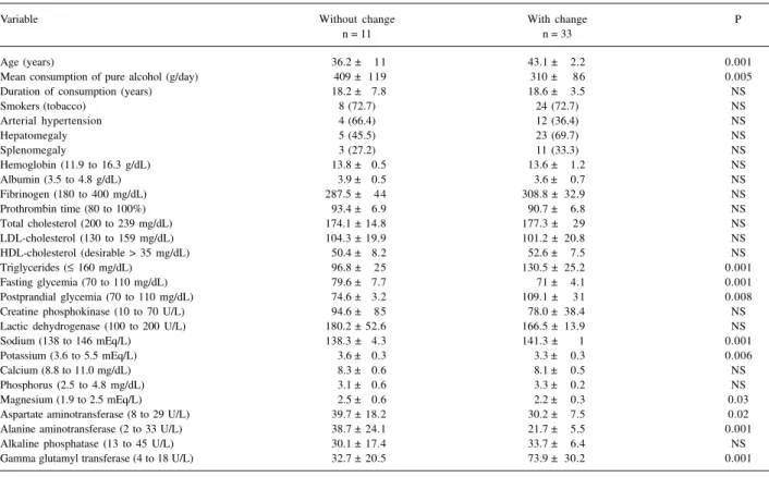
The patients with abnormal electrocardiograms com-pared with those with normal electrocardiograms had a more elevated mean age (P = 0.001; 95% CI of 2.89 - 10.91); a lower daily alcohol consumption (P = 0.005; 95% CI of 32.32 -165.68); and more elevated serum levels of triglycerides (P = 0.001; 95% CI of 16.03 – 51.37), postprandial glycemia (P=0.001; 95% CI of 15.45 – 53.54), and sodium (P=0.001; 95% CI of 1.4 – 4.6). The opposite occurred with the follo-wing levels: fasting glycemia (P=0.001; 95% CI of 4.95 – 12.25), magnesium (P=0.034; 95% CI of 0.02 – 0.58), aspartate aminotransferase (P=0.018; 95% CI of 1.75 – 17.25), alanine aminotransferase (P=0.001; 95% CI of 8.08 – 25.92), and gamma glutamyl transferase (P=0.001; 95% CI of 21.39 – 61.01). No correlation was observed between the serum le-vels of gamma glutamyl transferase and daily alcohol con-sumption (r = 0.15; F = 0.85, 95% CI of -0.29 to 0.33) (tab. III). The relations between total cholesterol and HDL-cho-lesterol were 3.45 (174.1/50.4) in the group with normal elec-trocardiographic findings and 3.37 (177.3/52.6) in the group with abnormal electrocardiographic findings.
Smoking was not a significant influence in the occurren-ce of electrocardiographic findings. The similarity between the levels of hemoglobin and albumin in patients with abnor-mal and norabnor-mal electrocardiographic findings suggest that nutritional factors played no significant role in the occurrence of the electrocardiographic abnormalities (tab. III).
Discussion
Electrocardiographic abnormalities are common in alcoholism. Atrial fibrillation followed by atrial flutter and ventricular extrasystoles are the most frequently found ab-normalities. In addition, atrioventricular conduction disor-ders, mainly first-degree atrioventricular block, bundle-branch block, left ventricular hypertrophy, and repolariza-tion abnormalities are common electrocardiographic findings 12, 28,29.
In this study, the most frequent electrocardiographic changes were prolonged QT interval, ventricular repolariza-tion changes, and signs of left ventricular hypertrophy, follo-wed by rhythm abnormalities and conduction disorders of the cardiac stimulus. Unlike other studies, we found no ar-rhythmias. Even not considering the QT interval change, an abnormality not usually assessed on routine readings of elec-trocardiograms, the rate of electrocardiographic abnorma-lities was greater in the alcoholic patients (61% x 45%) than that observed in an epidemiological study conducted in an open population comprising individuals aged between 18 and 65 years, individuals with heart disease not excluded30.
The observation that the mean ages of the female pa-tients with abnormal electrocardiographic findings, particu-larly those with a prolonged QT interval and changes in ventricular repolarization, were significantly greater than those of the patients with normal electrocardiographic
fin-Table I - Demographic characteristics, pattern of alcoholism and smoking, and characteristics of systemic arterial hypertension of
female alcoholics with no previous cardiovascular disease
%
Mean age (years) 41.4 ± 3.4 Color
White 30 68.2
Black 9 20.5
Mulatto 5 11.3
Mean pure alcohol 334 ± 81 consumption (grams/day)
Mean duration of 18 ± 3.9
consumption (years) Smoking
No 12 27.3
Yes 32 72.7
Systemic arterial blood pressure
Normotensive patients 22 50
Systolic blood pressure 11.3 ± 0.6 Diastolic blood pressure 7.4 ± 0.2
Hypertensive/normotensive patients 12 27.3 Systolic blood pressure 15.2 ± 0.5
Diastolic blood pressure 9.6 ± 0.3
Hypertensive/hypertensive patients 10 22.7 Systolic blood pressure 16.4 ± 0.6
Diastolic blood pressure 10.6 ± 0.5 Electrocardiogram
Without changes 11 25
With changes 33 75
Table II – Electrocardiographic changes in female alcoholics with no clinical manifestations of cardiovascular disease
Electrocardiographic changes nº %
I - Changes considered in isolation
Prolonged QT interval 24 54.5
(QTc > 440 milliseconds)
Changes in ventricular repolarization (CVR) 11 25 Left ventricular hypertrophy (LVH) 6 13.6
Sinus bradycardia (SB) 4 9.1
Sinus tachycardia (ST) 3 6.8
Left anterior superior 2 4.5
hemiblock (LASH)
Nodal inferior rhythm (NIR) 1 2.3
II - Association of changes
Prolonged QTc + CVR 8 18.2
Prolonged QTc + LVH 5 11.4
Prolonged QTc + ST 2 4.5
Prolonged QTc + SB 1 2.3
gregation, increased serum fibrinolytic activity, and a possi-ble increase in insulin sensitivity. The major protective fac-tor could be the increase in HDL-cholesterol levels 33. High
HDL-cholesterol levels (50 to 60 mg/dL) are associated with a low risk for coronary artery disease, and a 1% reduction in this lipid fraction increases the risk for the disease by 3% 34.
In our study, the patients with and without electrocardio-graphic changes had mean HDL-cholesterol levels above 50 mg/dL. In addition, the mean levels of total cholesterol and LDL-cholesterol remained within the normal range and below the levels considered risky for artery disease. A higher risk for coronary artery disease is estimated to occur when the ratio between total cholesterol and HDL-choleste-rol is greater than 5 35. In our study, that ratio was similar for
the groups with and without electrocardiographic abnorma-lities (3.37 and 3.45, respectively), far below the value of risk. The differences observed in fasting and postprandial glycemias according to the presence or absence of electro-cardiographic abnormalities are difficult to analyze. Glyce-mia control does not only depend on insulin and glucagon secretion, but also on adrenergic, cholinergic, and possibly peptidergic mechanisms 36, during the phase of alcohol
withdrawal, when plasma concentrations of aldosterone, norepinephrine, epinephrine, and cortisol increase 37, and
manifestations of increased sympathetic nervous system activity occur 38.
The prevalence of hypocalcemia, a condition in which increased muscle irritability occurs, was similar in patients dings, is in accordance with that observed in nonalcoholic
persons. The prevalence of depolarization and repolariza-tion changes increases with age, independently of alcohol consumption. As the mean age of the patients with electro-cardiographic abnormalities was relatively low, it seems un-likely that age was the factor responsible for the changes.
The fact that the alcohol intake of patients with abnor-mal electrocardiographic findings is lower than that of pa-tients with normal electrocardiographic findings during ap-proximately equal periods suggests that individual differen-ces exist in the vulnerability to the noxious effects of alco-hol. This impression was reinforced by the different beha-viors of several substances, such as triglycerides, glucose, hepatic enzymes, and some electrolytes in patients with normal and abnormal electrocardiograms. The serum levels of triglycerides in patients with electrocardiographic abnor-malities were greater than those in patients with normal elec-trocardiograms. Prospective studies have established that hypertriglyceridemia is a risk factor for vascular disease, mainly coronary heart disease, in both sexes 31. In our study,
except for the changes related to ventricular repolarization, no other electrocardiographic abnormality suggestive of myocardial ischemia was found. It is worth noting that elec-trocardiography at rest is a procedure with low specificity for the diagnosis of ischemic heart disease. The protective effect provided by alcohol ingested in the form of alcoholic beverages 32 could depend on several factors, such as an
in-crease in HDL-cholesterol levels, inhibition of platelet
ag-Table III - Comparisons between variables of chronic female alcoholics with and without electrocardiographic changes
Variable Without change With change P
n = 11 n = 33
Age (years) 36.2 ± 11 43.1 ± 2.2 0.001
Mean consumption of pure alcohol (g/day) 409 ± 119 310 ± 86 0.005
Duration of consumption (years) 18.2 ± 7.8 18.6 ± 3.5 NS
Smokers (tobacco) 8 (72.7) 24 (72.7) NS
Arterial hypertension 4 (66.4) 12 (36.4) NS
Hepatomegaly 5 (45.5) 23 (69.7) NS
Splenomegaly 3 (27.2) 11 (33.3) NS
Hemoglobin (11.9 to 16.3 g/dL) 13.8 ± 0.5 13.6 ± 1.2 NS
Albumin (3.5 to 4.8 g/dL) 3.9 ± 0.5 3.6 ± 0.7 NS
Fibrinogen (180 to 400 mg/dL) 287.5 ± 44 308.8 ± 32.9 NS
Prothrombin time (80 to 100%) 93.4 ± 6.9 90.7 ± 6.8 NS
Total cholesterol (200 to 239 mg/dL) 174.1 ± 14.8 177.3 ± 29 NS
LDL-cholesterol (130 to 159 mg/dL) 104.3 ± 19.9 101.2 ± 20.8 NS
HDL-cholesterol (desirable > 35 mg/dL) 50.4 ± 8.2 52.6 ± 7.5 NS
Triglycerides (≤ 160 mg/dL) 96.8 ± 25 130.5 ± 25.2 0.001
Fasting glycemia (70 to 110 mg/dL) 79.6 ± 7.7 71 ± 4.1 0.001
Postprandial glycemia (70 to 110 mg/dL) 74.6 ± 3.2 109.1 ± 31 0.008
Creatine phosphokinase (10 to 70 U/L) 94.6 ± 85 78.0 ± 38.4 NS
Lactic dehydrogenase (100 to 200 U/L) 180.2 ± 52.6 166.5 ± 13.9 NS
Sodium (138 to 146 mEq/L) 138.3 ± 4.3 141.3 ± 1 0.001
Potassium (3.6 to 5.5 mEq/L) 3.6 ± 0.3 3.3 ± 0.3 0.006
Calcium (8.8 to 11.0 mg/dL) 8.3 ± 0.6 8.1 ± 0.5 NS
Phosphorus (2.5 to 4.8 mg/dL) 3.1 ± 0.6 3.3 ± 0.2 NS
Magnesium (1.9 to 2.5 mEq/L) 2.5 ± 0.6 2.2 ± 0.3 0.03
Aspartate aminotransferase (8 to 29 U/L) 39.7 ± 18.2 30.2 ± 7.5 0.02
Alanine aminotransferase (2 to 33 U/L) 38.7 ± 24.1 21.7 ± 5.5 0.001
Alkaline phosphatase (13 to 45 U/L) 30.1 ± 17.4 33.7 ± 6.4 NS
Gamma glutamyl transferase (4 to 18 U/L) 32.7 ± 20.5 73.9 ± 30.2 0.001
with and without electrocardiographic abnormalities. Hypo-potassemia was observed exclusively in patients with elec-trocardiographic abnormalities, but it was only significant in patients with prolonged QT or ventricular repolarization changes. Only patients with prolonged QT had hypomag-nesemia, but in alcoholics a depressed T wave and prolon-ged QT interval may be related to hypopotassemia and hy-pomagnesemia. Although low levels of potassium and mag-nesium have been associated with a greater prevalence of ventricular arrhythmias 39, this was not observed in our
study.
Aminotransferases and gamma glutamyl transferase have different behaviors according to the presence or ab-sence of electrocardiographic changes. While the serum le-vels of both aminotransferases were significantly lower in patients with electrocardiographic changes, the level of gamma glutamyl transferase was more elevated. It is worth emphasizing that no correlation was observed between the serum levels of gamma glutamyl transferase and daily alco-hol consumption.
The prevalences of hepatomegaly and splenomegaly were similar in women with and without electrocardiogra-phic changes, assuming that the extension of the QT inter-val in alcoholics does not depend on the autonomic cardiac function, but is related to the severity of the hepatopathy 40
or to the simple presence of portal hypertension, regardless of the degree of hepatic dysfunction 41. In our study, the
ab-sence of symptoms and signs of hepatic dysfunction and portal hypertension, the absence of abnormalities in the biochemical tests assessing liver function (albumin, fibrino-gen, and prothrombin time), as well as the presence of
nor-mal serum levels of alanine aminotransferase (the enzyme that most reliably indicates hepatic lesions) in patients with abnormal electrocardiograms suggest that the change in the QT interval depends on factors other than the existence of hepatopathy and portal hypertension.
Approximately 50% of the patients in our study had ar-terial hypertension on hospital admission, but only 22.7% remained hypertensive in the subsequent days. An electro-cardiographic change suggestive of ventricular hypertro-phy was present in 50% of the hypertensive/hypertensive patients and in only 8.3% of the hypertensive/normotensi-ve patients.
Creatine phosphokinase, the enzyme that is altered in lesions of several tissues, especially the skeletal and smooth muscles, was elevated in patients with and without electrocardiographic changes. In alcoholics, these enzyme changes have been attributed to changes in skeletal muscles 42,43.
The association between smoking and alcoholism is very frequent 44,45. Alcohol and tobacco may interact in the
production of hepatic changes 46. However, in this study, no
influence of smoking in the prevalence of electrocardiogra-phic changes was observed.
The results of our study suggest that electrocardio-graphy, a noninvasive method available in almost all medi-cal facilities, should be part of the preliminary assessment of alcoholics.
Acknowledgments
We thank CNPq and FAPEMIG for their support.
1. Oparil S. Arterial Hypertension. In: Wyngaarden JB, Smith Jr. LH, Bennett JC, eds. Cecil Textbook of Medicine. 19th ed. Philadelphia, Saunders; 1992; 258-74. 2. Puddey IB, Beilin LJ, Vandongen R, Rouse IL, Rogers P. Evidence for a direct ef-fect of alcohol consumption on blood pressure in normotensive men: a randomi-zed controlled trial. Hypertension 1985; 7: 707-13.
3. Klatsky AL, Friedman GD, Siegelaub AB, Gérard MJ. Alcohol consumption and blood pressure: Kaiser - permanents multiphasic health examination data. N Engl J Med 1977; 296:1194-200.
4. Wynne J, Braunwald E. As miocardiopatias e as miocardites. In: Braunwald E, Is-selbacher KI, Petersdorf RG, Wilson JD, Martin JB, Fauci AS eds. Harrison- Me-dicina Interna, 11ª ed, vol 1. Rio de Janeiro: Guanabara Koogan, 1988; 927-33. 5. Urbano-Márquez A, Estruch R, Fernández-Solá J et al. The greater risk of alcoho-lic cardiomyopathy and myopathy in women compared with men. JAMA 1995; 274: 149-54.
6. Kasper EW, Agema WRP, Hutchins GM et al. The cause of dilated cardiomyopa-thy: a clinicopathologic review of 673 consecutive patients. J Am Coll Cardiol 1994; 23: 586-90.
7. Teragaki M, Takeuchi K, Takeda T. Clinical and histological features of alcohol drinkers with congestive heart failure. Am Heart J 1993; 125: 808-17. 8. Piano MR, Schwertz DW. Alcoholic heart disease: a review. Heart-Lung 1994;
23: 3-17.
9. Burch GE, Gilles TD. Alcoholic cardiomyopathy: concept of disease and its treatment. Am J Med 1971; 101: 461-4.
10. Gillet C, Julliere Y, Pirollet P et al. Alcohol consumption and biological markers for alcoholism in idiopathic dilated cardiomyopathy: a case-controlled study. Alcohol Alcohol 1992; 27: 353-8.
References
11. Fischbein L, Sachs RN, Geay D et al. Study of HLA system A and B antigens in dilated cardiomyopathy associated with alcoholism. Arch Mal Coeur 1987; 80: 1171-5. 12. Moreira DAR, Reyes CAS. Bases eletrofisiológicas da fibrilação atrial - fatores
de-sencadeantes e de perpetuação. Rev Soc Cardiol Estado de São Paulo 1994; 3: 207-13. 13. Facour SM. Intramyocardial small vessels disease in chronic alcoholism. Am
Heart J 1976; 92: 561-75.
14. Pintar K, Woalnsky BM, Cubbay ER. Alcoholic cardiomyopathy. Can Med Assoc J 1969; 93: 103-7.
15. Neville JN, Eagles JA, Sampson G, Samson G, Olson RE. Nutricional status of al-coholics. Am J Clin Nut 1968; 21: 1.329-40.
16. Pinn G, Bovet P. Alcohol-related cardiomyopathy in Seychelles. Med J Aust 1991; 155: 529-32.
17. Walsh TK, Vacek JL. Ethanol and heart disease: an underestimated contributing factor. Postgrad Med 1986; 79: 60-3.
18. Regan TJ. Alcoholic cardiomiopaty. Prog Cardiovasc Dis 1984; 27: 141-52. 19. Hochgraf PB, Zilberman ML, Andrade AG. Women alcoholics - social,
demogra-phic and clinical characteristics in a Brasilian sample. Alcohol Alcohol 1995; 30: 427-32.
20. Pechansky F, Soibelman M, Gus G. Consumo de álcool em mulheres: preconcei-to? Revista ABP-APAL 10: 87-90.
21. Marshall AW, Kingstone D, Boss M, Morgan MY. Ethanol elimination in males and females: relationship to menstrual cycle and body composition. Hepatology 1983; 3: 701-6.
23. Classificação de Transtornos de Doenças Mentais e de Comportamento da CID 10 (1997): Descrições Clínicas e Diretrizes Diagnósticas. Organização Mun-dial de Saúde - Genebra, Artes Médicas, Porto Alegre.
24. Borini P. Anamnese psiquiátrica e clínica, exame físico e exames complementares es-truturados e informatizados para aplicação em alcoolistas. J Bras Psiq 1990; 39: 250-65.
25. Fisch C. Eletrocardiografia e vetocardiografia. In: Braunwald W ed. Tratado de Medicina Cardiovascular, vol 1, 3ª ed. São Paulo, Roca, 1991; 189-233. 26. Tranchesi J. Eletrocardiograma Normal e Patológico - Noções de
vectocardiogra-fia. São Paulo: Atheneu, 1975; 63-92
27. Joint National Commitee on Detection, Evaluation, and Treatment of High Blood Pressure. The sixth report of the Joint National Commitee on Detection, Evaluation, and Treatment of High Blood Pressure (JNC VI). Arch Intern Med 1997; 157: 2413-46.
28. Regan TJ. Alcoholic cardiomyopathy. In: Zipes DP, Rowlands DJ eds. Progress in Cardiology. Philadelphia, Lea and Fibiger, 1989; 129.
29. McCall D. Alcohol and the cardiovascular system. Curr Probl Cardiol 1987; 12: 1-414.
30. Johnson JL, Heineman EF, Heiss G, Hames CG, Tyroler HA. Cardiovascular di-sease risk factors and mortality among black women and white women aged 40-64 years in Evans County, Georgia. Am J Epidemiol 1986; 123:209-20. 31. Hokanson JE, Austin MA. Triglyceride is a risk factor of coronary disease in men
and women. A meta-analysis of population based prospective studies. Circula-tion 1993; 88: 510-4.
32. Klatsky AL, Armstrong Ma, Friedman GD. Red wine, white wine, liquor, beer, and risk for coronary artery disease hospitalization. Am J Cardiol 1997; 80: 416-20. 33. Rimm EB, Williams P, Fosher K, Criqui M, Stampfer MJ. Moderate alcohol intake
and lower risk of coronary heart disease: meta-analysis of effects on lipids and haemostatic factors. BMJ 1999; 319: 1523-8.
34. Consenso Brasileiro sobre Dislipidemia - Detecção, Avaliação e Tratamento. So-ciedade Brasileira de Cardiologia. Arq Bras Cardiol 1996; 67: 113-28.
35. Assmann G, Schulte H. The importance of triglycerides - results from The Prospec-tive Cardiovascular Munster (PROCAM) Study. Eur J Epidemiol 1992; 8: 99-103. 36. Patto RJ, Neves MM, Russo EK, Borges DR. A hipoinsulinemia do alcoólatra
in-depende do eixo enteroinsular.Rev Paul Med 1985,103: 15-18, 1985. 37. De Marchi S, Cecchin E, Basile A et al. Renal tubular dysfunction in chronic
alco-hol abuse - effects of abstinence. N Engl J Med 1993, 329: 1927-34. 38. Beevers DG, Bannan LT, Saunders JB, Paton A, Walters JR. Alcohol and
hype-tension. Contrib Nephrol 1982, 30: 92-7.
39. Tsuji H, Vendititti FJ Jr, Evans JC, Larson MG, Levy D. The associations of levels of serum potassium and magnesium with ventricular premature complexes (the Framingham Heart Study). Am J Cardiol 1994; 74: 232-5.
40. Puthumana L, Edwin D, Chaudhary V, Thuluvath PJ. Prolonged QTc interval seen in patients with cirrhosis is related to the severity of liver disease and not with autonomic cardiovascular reflex. Gastroenterology 1999; 116(4 - part 2): L0371, A 1264.
41. Valeriano V, Trevisani F, Merli M et al. Prolongation of the QT interval in cirrho-sis: role of portal hypertension. Gastroenterology 1999; 116(4 - part 2): L0377, A 1266.
42. Borini P, Guimarães RC. Indicators of inflammation and cellular damage in chro-nic asymptomatic or oligosymptomatic alcoholics: correlation with alteration of bilirubin and hepatic and pancreatic enzymes. Rev Hosp Clín Fac Med S Paulo 1999; 54: 53-60.
43. Urbano-Marquez A, Estruch R, Navarro-Lopez F et al. The effects of alcoholism on skeletal and cardiac muscle. N Engl J Med 1989; 320: 409-15.
44. Bobo JK, Gilchrist L. Urging the alcoholic client to quit smoking cigarettes. Addict Behav 1983; 8: 297-305.
45. Borini P, Silva CO. Aspectos demográficos, epidemiológicos e sociais do alcoo-lismo: uma análise de alcoolistas internados em hospital psiquiátrico. Rev ABP-APAL 1989; 11: 89- 96.