199
https://doi.org/10.1590/0004-282X20160196
IMAGES IN NEUROLOGY
Late radiation therapy brain abnormalities
that mimic leukoencephalopathy with anterior
temporal lobe cysts
Alterações encefálicas tardias pós radioterapia mimetizando leucoencefalopatia com
cistos temporais anteriores
Marcos Rosa Júnior
1,2, Antônio José da Rocha
3A 43-year-old man who had been treated for a
cra-niopharyngioma with surgery and radiotherapy (33 years
ago) was referred for evaluation of a sudden hemiparesis.
Neuroimaging documented a brainstem cavernoma
associ-ated with basal ganglia calciications and white matter hyper
-intensity near anterior temporal lobe cysts (Figures 1 and 2).
Late brain abnormalities are often observed after
radiotherapy for childhood intracranial tumors.
Microbleeds and cavernomas are well recognized in these
cases. However, bilateral anterior temporal lobe cysts are
uncommon, and it is crucial to recognize these to avoid
misdiagnoses, particularly when they are associated with
white matter abnormalities. Differential diagnoses include
hereditary disorders, infectious diseases, and dilated
peri-vascular spaces
1,2,3,4.
1Universidade Federal do Espírito Santo, Departamento de Radiologia, Vitória ES, Brasil; 2BRAEN, Laboratório de Pesquisa em Neurorradiologia, Vitória ES, Brasil;
3Santa Casa de Misericórdia de São Paulo, Seção de Neurorradiologia, São Paulo SP, Brasil.
Correspondence: Marcos Rosa Júnior; Universidade Federal do Espírito Santo – UFES, Centro de Ciências da Saúde; Av Marechal Campos, 1468; 29043-900
Vitória ES, Brasil; E-mail: marcosrosajr@hotmail.com
Conflict of interest: There is no conflict of interest to declare. Received 18 August 2016; Accepted 31 December 2016.
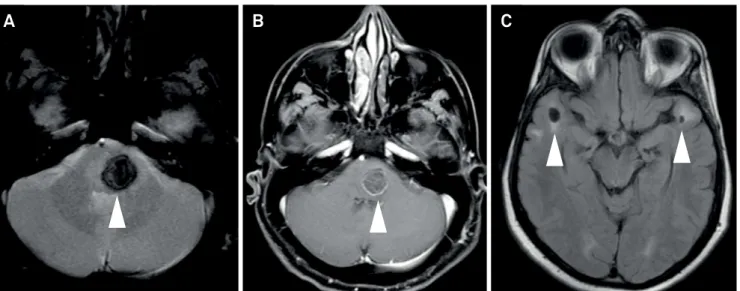
Figure 1.
Axial GRE (A) and T1 GD (B) showed a focal hemorrhage compatible with a radiation-induced cavernoma in the
brainstem. Axial FLAIR (C) showed white matter hyperintensity around anterior temporal lobe cysts.
200
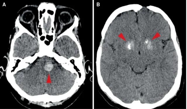
Arq Neuropsiquiatr 2017;75(3):199-200Figure 2.
Axial non-contrast CT confirmed a hemorrhagic lesion in the brainstem (A) and basal ganglia calcifications (B).
A
B
References
1. Nunes RH, Pacheco FT, Rocha AJ. Magnetic resonance imaging of anterior temporal lobe cysts in children: discriminating special imaging features in a particular group of diseases. Neuroradiology. 2014;56(7):569-77. https://doi.org/10.1007/s00234-014-1356-9
2. Cross NE, Glantz MJ. Neurologic complications of radiation therapy. Neurol Clin N Am. 2003;21(1):249-77. https://doi.org/10.1016/S0733-8619(02)00031-2
3. Osborn AG, Preece MT. Intracranial cysts: radiologic-pathologic correlation and imaging approach. Radiology. 2006;239(3):650-4. https://doi.org/10.1148/radiol.2393050823