Megalencephalic leukoencephalopathy with
subcortical cysts (MLC)
–
a case with clinical
and magnetic resonance imaging (MRI)
dissociation
Leucoencefalopatia megalencefálica com cistos subcorticais (MLC)
–
um caso com
dissociação clínica e de resonância magnética
Fabiano Reis
1, Ricardo Yoshio Zanetti Kido
1, João Amaral Mesquita
1, Mariana Mari Oshima
1,
Maria Augusta Montenegro
22Universidade Estadual de Campinas, Departamento de Neurologia, Campinas SP, Brazil.
Correspondence: Fabiano Reis; Universidade Estadual de Campinas, Hospital de Clínicas, Departamento de Radiologia; Cidade Universitária Zeferino Vaz s/n; 13025-080 Campinas SP, Brasil; E-mail: [email protected]
Conflict of interest:There is no conflict of interest to declare.
Received 28 August 2014; Received in final form 01 October 2014; Accepted 21 October 2014.
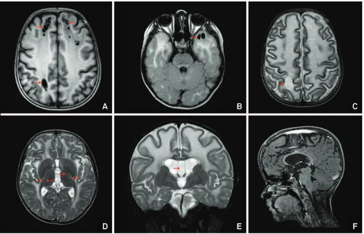
Figure 1.
(A), Axial T1-weighted image shows diffuse cerebral white-matter abnormalities with frontoparietal subcortical cysts
(arrows). (B), Axial FLAIR image exhibits subcortical cysts (arrows) in the anterior temporal lobe intermingled with diffuse
white-matter hyperintensity. (C) and (D), Axial T2-weighted images with diffuse white-white-matter abnormalities, the external and extreme
capsules are involved and the posterior limb of the internal capsule shows a double-line of abnormal intensity (arrows in D). (E),
Coronal T2-weighted image highlights a diffuse brain swelling with white-matter hyperintensity and persistence of the cavum
septum pellucidum (arrow). (F), Sagittal T1-weighted image with fat suppression and after endovenous gadolinium contrast, note
the cranio-facial disproportion without abnormal enhancement. The corpus callosum is relatively spared.
1Universidade Estadual de Campinas, Hospital de Clínicas, Departamento de Radiologia, Campinas SP, Brazil;
DOI:10.1590/0004-282X20140206
IMAGES IN NEUROLOGY
A ten-year-old boy with macrocephaly. His head
circumfer-ence soon after birth started growing above normal.
Developmental milestones were normal and neurological
exam-ination showed mild hypotonia in the first years of life. The only
complain is frequent falls, without cerebellar or motor
abnor-malities on neurological examination. MLC diagnosis was based
on MRI findings (Figures 1 and 2). The absence of cerebellar
abnormalities in this case is useful to rule out congenital
mus-cular dystrophy in patient with macrocephaly
1.
MLC is associated with MCL1 gene mutation in 70% of
the cases
2, leading to disbalanced intracelullar ion
concen-tration and astrogliosis
3,4.
References
1. Nunes RH, Pacheco FT, Rocha AJ. Magnetic resonance imaging of anterior temporal lobe cysts in children: discriminating special imaging features in a particular group of diseases. Neuroradiology. 2014;56:569-77. http://dx.doi.org/10.1007/s00234-014-1356-9
2. Olivier M, Lenard HG, Aksu F, Gärtner J. A new leukoencephlapathy with bilateral anterior temporal lobe cysts. Neuropediatrics.1998;29(5):225-8. http://dx.doi.org/10.1055/s-2007-973566
3. Knaap MS, Boor I, Estévez R. Megalencephalic leukoencephalopathy with subcortical cysts: chronic white matter oedema due to a defect in brain ion and water homoeostasis. Lancet Neurol. 2012;11(11):973-85. http://dx.doi.org/10.1016/S1474-4422(12)70192-8
4. Sinhal BS, Gorospe JR, Naidu S. Megalencephalic leukoencephalo-pathy with subcotical cysts. J Child Neurol. 2003;18(9):645-52. http:// dx.doi.org/10.1177/08830738030180091201