Arq Neuropsiquiatr 2008;66(2-A):261-263
261
CYSTIC LEUKOENCEPHALOPATHY
WITHOUT MEGALENCEPHALY
Eliete Chiconelli Faria
1, Juliana Harumi Arita
1, Mirella Maccarini Peruchi
2,
Jaime Lin
3, Marcelo Rodrigues Masruha
4, Luiz Celso Pereira Vilanova
5LEUCOENCEFALOPATIA CÍSTICA SEM MEGALENCEFALIA
Division of Child Neurology / Department of Neurology and Neurosurgery, Federal University of Sao Paulo, Sao Paulo SP, Brazil (UNIFESP-EPM): 1
Resi-dent, Department of Pediatrics, UNIFESP-EPM; 2Resident, Department of Radiology, Hospital Heliopolis, Sao Paulo SP, Brazil; 3Resident, Division of
Child Neurology / Department of Neurology and Neurosurgery, UNIFESP-EPM; 4Associated Physician, Division of Child Neurology / Department of
Neurology and Neurosurgery, UNIFESP-EPM; 5Professor and Chairman, Division of Child Neurology / Department of Neurology and Neurosurgery,
UNIFESP-EPM.
Received 14 September 2007, received in inal form 29 November 2007. Accepted 4 February 2008.
Dra. Eliete Chiconelli Faria – Rua Jacinto de Lima Santos 263 - 03738-090 São Paulo SP - Brasil. E-mail: [email protected]
The coming of magnetic resonance imaging (MRI) into clinical practice added advances in understanding the white matter diseases in children1. Previously, white
mat-ter and metabolic diseases were found to have a relative-ly speciic pattern that could help in differential diagnosis, and primary defects in many inborn leukoencephalopa-thies have been elucidated including disorders from lysos-somal storage, amino and organic acids, muscle and mito-chondrial dysfunction1,2. Recent reports have described an
apparently non-progressive condition characterized by se-vere psychomotor delay with variable degrees of tone and relex abnormalities, normo- or microcephaly, and MRI indings consisting of bilateral anterior temporal cystic le-sions with pericystic abnormal myelination and symmetric patchy lesions with increased signal in the frontal and oc-cipital periventricular white matter regions3. Few patients
with this condition have been described in literature1-4. In
some aspects, this condition may resembles other leuko-encephalopathies such as “megalencephalic leukoenceph-alopathy with subcortical cysts” and “leukoencephalopa-thy with vanishing white matter”, due to the presence of subcortical cysts and white matter abnormalities, howev-er, the clinical presentation and other neuroimaging fea-tures are distinct3.
We describe the clinical, laboratory and neuroimaging characteristics of a patient with this rare entity.
CASE
A one-year-old boy was born to healthy and non-consan-guineous parents at 36 weeks of gestation. There was no family history of neurological diseases. Birth weight was 2,2 kg (25–50th centile), length 45 cm (25–50th centile) and head circumference 31 cm (below 5th centile). Apgar indices were 6 and 9 (at one and ten min). Right after birth, he had transient respiratory discom-fort and hypomagnesaemia with no further complications and he was discharged at the third day of life. It was already
record-ed that this child was hypotonic, with poor movements and ad-ducted thumbs since the irst weeks of life. He also presented a delay in neuropsychomotor acquisitions. The infant was admit-ted in our service at the age of 12 months. On examination he presented good overall condition with a head circumference of 44 cm (below 5th centile). Neurological examination revealed a good contact with the examiner but he did not either ix on or catch an offered object. It was also observed increased limb muscle tone, brisk tendon relexes in contrast with axial and ap-endicular hypotonia. Evaluation of cranial nerves was normal.
Hematological tests and blood screening for metabolic dis-eases, including those for concentrations of very long-chain fat-ty acids, lactate and pyruvate, gave results in the normal ranges. Cerebrospinal luid lactate concentration was normal.
Arq Neuropsiquiatr 2008;66(2-A)
262
Encephalopathy without megalencephaly Faria et al.
ical markers for CMV, herpes virus, toxoplasmosis and syphilis were undetectable.
Brainstem auditory evoked potentials detected severe bilat-eral sensorineural deafness and electroencephalogram showed discrete disorganized background activity with an epileptiform activity in the median line.
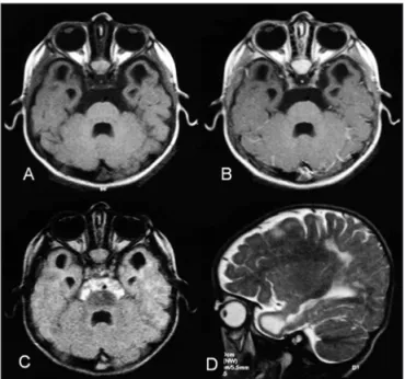
Noncontrast-enhanced cranial computed tomography (CT) revealed hipoatenuation of parietal white matter and well-cir-cumscribed temporal subcortical cysts bilaterally (Fig 1). MRI showed bilateral lesions with signal intensity that is isointense to the one of cerebrospinal luid in both temporal lobes, compati-ble with cysts (Fig 2) and patchy areas of high signals intensity in the white matter of temporal, frontal and parietal lobes (Fig 3).
Although the patient presented severely handicapped he al-so continued to make slow developmental progress in motility, speech, and cognitive function.
The hospital ethic commission approved this case report and the parents gave informed consent for publication.
DISCUSSION
Cystic leukoencephalopathy without megalencephaly was irst described by Olivier et al. in 1998 and since then, fewer than 30 cases have been described4. The distinctive
indings of this rare condition are bilateral temporal lobe cysts combined with a speciic pattern of multifocal white matter lesions, normo- or microcephaly, and severe psy-chomotor retardation in a non-progressive clinical course2,3.
Considerable effort has been done into distinguishing these patients from another leukoencephalopathy known as “megalencephalic leukoencephalopathy with subcorti-cal cysts” (MLC)3,5. Firstly described in 1995, this entity is
characterized by dramatic MRI changes including a typical pattern of diffuse white matter lesions and swelling with invariable frontoparietal and anterotemporal subcortical cysts, but mild clinical manifestations initially, develop-ment of macrocephaly and functional deterioration later in course2,3,6. Mutations in MLC1 gene have been shown to
cause MLC, although there are patients with the condition in whom mutations of MLC 1 cannot be found2-5.
In our patient, we did not perform MLC1 testing be-cause of its non-availability; however, clinical and neuro-imaging features of cystic leukoencephalopathy without megalencephaly are clearly distinct from MLC. Patients with cystic leukoencephalopathy without megalencephaly are severely involved from the beginning with a non-pro-gressive course, they have normal or small head circumfer-ence and MRI indings contrast to the patients with MLC. Another recently described cystic leukoencephalopa-thy called “childhood ataxia with central nervous system hypomyelination” (CACH) or “leukoencephalopathy with vanishing white matter” supericially resembles cystic
leu-Fig 2. MRI, serial T1-weighted (A), T1-weighted after gadolinium injec-tion (B), luid-attenuated inversion recovery (FLAIR) sequence (C) ax-ial images and T2-weighted sagital image (D) show bilateral lesions with signal intensity that is isointense to the one of cerebrospinal lu-id in both temporal lobes, compatible with cysts.
Arq Neuropsiquiatr 2008;66(2-A)
263
Encephalopathy without megalencephaly Faria et al.
koencephalopathy without megalencephaly7. Patients with
CACH syndrome have extensively dramatic cystic changes in the cerebral white matter; however, the temporal lobes are not preferentially involved7. Furthermore, the clinical
picture in CACH syndrome is characterized by a mild ini-tial course, with later deterioration and bouts of sudden decline in function associated with minor head trauma7.
It was proposed that the MRI abnormalities found in cystic leukoencephalopathy without megalencephaly could be the result of congenital CMV infection and not a leukodystrophy8. Late CMV infection is one important
condition to be considered in the differential diagnosis of cystic leukoencephalopathy without megalencephaly, once it could lead to microcephaly, retarded psychomo-tor development in a clinically static encephalopathy, and sensorial deafness1,8,9. Neonatal signs of intrauterine
CMV infection include systemic signs of jaundice, hepato-splenomegaly and petechiae. Most commonly neurologi-cal impairment includes sensorineural hearing deicits and decreased vision caused by chorioretinitis. The severity of the neurological impairment may vary from mild learning, behavioral, and motor coordination problems to serious mental deiciency and motor handicap1,9. In CMV
congeni-tal infection, MRI studies can be normal or demonstrate several abnormal indings such as cerebellar hypoplasia, disorders of neuronal migration and white matter abnor-malities including hemorrhage, calciications and the pres-ence of anterior temporal lobe cysts. Those indings as a result of late infection (third trimester) can be regarded as a result of infection at a time when neuronal migration and cortical plate formation had already occurred result-ing in a more selective lesion of glial cells1,9,10.
It is important to note that, in this case, both mother and the patient were tested for CMV with negative results. Additionally, in our patient neither signs of systemic CMV
infection nor signs of calciications, which are commonly associated with congenital CMV infection, were found. Therefore, according to serological, clinical, and neuroim-aging indings, CMV infection is extremely unlikely.
In conclusion, we have reported a patient affected by non-progressive cystic leukoencephalopathy without megalencephaly. The identiication of patients with this new white matter disease depends on the recognition of the characteristic of MRI indings of bilateral anterior temporal lobe cystic lesions, with abnormal myelynation in the pericystic region, and patchy symmetric white-mat-ter lesions. It is important to remember that this clinical condition, the neuroimaging conirmation is necessary to avoid mistaken with cerebral palsy.
REFERENCES
1. Gomes AL, Vieira JP, Saldanha J. Non-progressive leukoencephalopa-thy with bilateral temporal cysts. Eur J Ped Neurol 2001;5:121-125. 2. Henneke M, Preuss N, Engelbrecht V, et al. Cystic
leukoencephalop-athy without megalencephaly: a distinct disease entity in 15 children. Neurology 2005;64:1411-1416.
3. Bodensteiner JB, Kerrigan JF, Johnsen SD. Leukoencephalopathy with bilateral anterior temporal lobe cysts. J Child Neurol 2006;21:419-422. 4. Olivier M, Lenard HG, Aksu F, Gärtner J. A new leukoencephalopathy
with bilateral anterior temporal lobe cysts. Neuropediatrics 1998;29: 225-228.
5. Grosso S, Cerase A, De Stefano N, et al. Non-progressive leukoenceph-alopathy with bilateral anterior temporal cysts: a case report and re-view of the literature. Brain Dev 2005;27:73-77.
6. Van der Knaap M, Barth P, Stroink H. Leukoencephalopathy with swell-ing and discrepantly mild clinical course in eight children. Ann Neurol 1995;37:324-334.
7. Schiffman R, Moller J, Trapp B. Childhood ataxia with diffuse central nervous system hypomyelination. Ann Neurol 1994;35:331-340. 8. Tatli B, Ozman M, Aydinli N, Caliskan M. Not a new leukodistrophy but
a congenital cytomegalovirus infection. J Child Neurol 2005;20:525-527. 9. Van der Knaap M, Vermeulen G, Barkhof F, Hart AA, Loeber JG, Weel
JF. Pattern of white matter abnormalities at MR imaging: use of poly-merase chain reaction testing of Guthrie cards to link pattern with con-genital cytomegalovirus infection. Radiology 2004;230:529-535. 10. Barkovich A, Lindan C. Congenital cytomegalovirus infection of the