Malformations of cortical development
Current concepts and advanced neuroimaging review
Celi Santos Andrade1, Claudia da Costa Leite2
ABSTRACT
Malformations of cortical development (MCD) result from disruptions in the complex process of the human brain cortex formation and are highly associated to severe epilepsy, neurodevelopmental delay and motor dysfunction. Nowadays, magnetic resonance imaging (MRI) is the cornerstone of the work-up of patients with epilepsy and modern advanced imaging techniques have improved not only our ability to detect and characterize cortical malformations, but also in identifying associated functional abnormalities that are far beyond the structural visualized lesions. Herein, we address the most currently used classifications of MCD and make a concise review of the embryological process of cortical development. Our main goal is to summarize recent advances and new trends in diagnostic imaging techniques concerning MCD. Thereafter, follows a brief discussion of specific disorders and their radiological features.
Key words: malformations of cortical development, epilepsy, magnetic resonance imaging, diagnostic imaging.
Malformações do desenvolvimento cortical: conceitos atuais e revisão de neuro-imagem avançada
RESUMO
As malformações do desenvolvimento cortical (MDC) resultam de distúrbios no complexo processo do desenvolvimento do córtex cerebral humano e estão comumente associadas a epilepsia severa e disfunções neuropsicomotoras. Atualmente, as imagens por ressonância magnética (RM) são a pedra angular no manejo de pacientes com epilepsia e modernas técnicas avançadas de imagem melhoraram não só a nossa capacidade de detectar e caracterizar as malformações corticais, mas também levaram ao reconhecimento de anomalias funcionais associadas que estão muito além das lesões estruturais visibilizadas. Abordaremos as classificações mais utilizadas de MDC e revisaremos a embriologia do desenvolvimento cortical. Nosso principal objetivo é destacar os avanços recentes e as novas tendências em técnicas de diagnóstico por imagens relacionadas às MDC. Em seguida, faremos uma breve discussão sobre alguns transtornos específicos e suas características radiológicas.
Palavras-chave: malformações do desenvolvimento cortical, epilepsia, imagem por ressonância magnética, diagnóstico por imagem.
Correspondence
Claudia da Costa Leite Instituto de Radiologia Setor de Ressonância Magnética Av. Dr. Enéas de Carvalho Aguiar 255 Portaria 5 / 3º andar
05403-001 São Paulo SP - Brasil E-mail: [email protected]
Support
We are very grateful to FAPESP (ClnAPCe project 05/56464-9) and to CNPq for the funding and support. Da. Claudia da Costa Leite was supported by CNPq (Grant 308267/008-7)
Received 20 July 2010
Received in final form 5 August 2010 Accepted 12 August 2010
Radiology Department, School of Medicine, University of São Paulo, São Paulo SP, Brazil: 1MD, Post-Graduate Radiologist of
the Radiology Department, Clinics Hospital of University of São Paulo, School of Medicine, São Paulo SP, Brazil; 2MD,PhD,
Associate Professor of the Radiology Department of the University of São Paulo, School of Medicine, São Paulo SP, Brazil. Chief of the Neuroradiology Group and of the Magnetic Resonance Section, Clinics Hospital of University of São Paulo, School of Medicine, São Paulo SP, Brazil.
Malformations of cortical development (MCD) are highly epileptogenic lesions and some cases also present in childhood with early encephalopathy, neurodevelop-mental delay, motor deicits and learning diiculties1-5. Neuroimaging is an
met-abolic and functional abnormalities beyond the visible structural lesions, shedding light to the pathophysiologi-cal basis of human epileptogenesis.
he aim of this review is to address the current con-cepts of MCD and recent advances in modern imaging techniques concerning the diagnosis and understanding of these epileptogenic disorders. Before that, we will make a concise revision of the normal human brain cortex for-mation. For each subtype of MCD, we will state its his-topathological, clinical and imaging characteristics with a pertinent diferential diagnosis. Because of its medical and radiological signiicance, the following MCD will be addressed in this article: focal cortical dysplasia, hemi-megalencephaly, heterotopia, lissencephaly, polymicro-gyria, and schizencephaly.
Classiication of MCD and normal cortical development
he study and classiication of MCD are complicat-ed by the fact that two malformations are virtually never completely identical and often combinations of diferent subtypes of malformations occur together in the same pa-tient. Indeed, MCD have a wide spectrum of histomor-phological alterations, heterogeneous clinical presenta-tions and highly variable neuroradiological manifesta-tions, that makes this group of diseases even more in-triguing and somewhat challenging.
he most currently adopted categorization is the clas-siication scheme of Barkovich et al.2,6,7, which is based
on three embryological processes of brain development: neuronal and glial proliferation, and diferentiation, and eventual apoptosis; neuronal migration; and cortical orga-nization. hese embryological steps should not be viewed as separate sequential processes, but as a complex mecha-nism that ultimately results in brain formation. he clas-siication is based on the earliest stage at which the ab-normality occurs, together with pathological, genetic and imaging aspects2,3.
he description of radial glia dates from the 19th
cen-tury and has strongly inluenced the history of neurosci-ence8. he normal human brain cortical development is
a so extraordinary and complex biological process, with so many molecular requirements, signaling cascades and precise expression patterns of key genes9-12, that one may
wonder how is it possible not to have more commonly malformations and misdevelopments.
Neurons originate from stem cells of the subependy-mal layer of the wall of the ventricles (the so-called ger-minal matrix). he gerger-minal matrix cells with neuronal diferentiation (neuroblasts), when mature, begin a pro-cess of migration. he migration of neurons in the peri-ventricular region to the deep nuclei and to the surface of the cerebral cortex is driven by radial ibers in the eighth
week of gestation. he cerebellar nuclei and cortex are also formed in this period. he migration occurs in an or-derly manner, so that the cells that will form the irst layer of the cortex are the irst to migrate and thereafter cells that will form the layers 6, 5, 4, 3 and 2. Cortical neurons migrate for varying distances until they reach the cor-tex in their inal position, forming the hexalaminar cyto-architecture of the adult neocortex9,10. After being
orga-nized in several layers, the neurons separate from the ra-dial ibers and initiate the formation and growth of den-drites and synapses. he radial ibers are separated from the neurons after the migration and originate glial cells8.
Most of the process of neuronal migration occurs around the 16th week of gestation; however, this process will only
end in about the ifth month of life. he cortical connec-tions are formed very early, and continue to develop in an intricate mechanism that leads to the development of gray matter and white matter.
Brain sulcation and gyration are directly related to neuronal migration and are responsible for the increased cortical surface, without increasing its volume.
Disturbances in several molecular pathways11-15 that
regulate these complex mechanisms may have a genetic basis3,10, although exogenous insults, such as
cytomega-lovirus infection, can also cause some types of MCD; the nature and timing of the insult in relation to the devel-opment process are the factors that determine the type of MCD.
Modern neuroimaging techniques
he diagnosis of any MCD ideally requires high reso-lution multiplanar magnetic resonance imaging (MRI)6,16.
his is by all means the method of choice for evaluation of the MCD because it demonstrates excellent contrast between white matter and gray matter, gives exact top-ographic characterization of the development of corti-cal gyri and sulci, and allows more accurate analysis of the formation of white matter and stages of myelination. Computed tomography (CT) scans can fail to make the diagnosis in more than 30% of afected patients2, but it
is crucial to remember that this may be the only meth-od available in some cases. hus, it is important to rec-ognize that CT changes, although subtle, are similar to those found in MRI studies.
New advanced MRI techniques have gradually im-proved image quality and reinforced our ability to iden-tify, delineate and characterize brain lesions. According to some authors, the gain in spatial resolution with in-creasing magnetic ield strength - moving from 1.5 T to 3.0 T, 7.0 T and greater - and subsequent increase in sig-nal to noise ratio, can potentially bring more anatomi-cal details in complex brain malformations16. Advances
sur-face coils, have increased resolution and contrast of the acquired images16, with maximum gains in the cortical
surface, which is especially useful in the detection of mild cortical dysplasias.
Some recent MR advanced sequences may also im-prove image quality and might be helpful in characteriza-tion of MCD. he introduccharacteriza-tion of volumetric T1-weight-ed sequences with high spatial resolution (SPGR - spoilT1-weight-ed gradient recalled echo, MPRAGE - magnetization pre-pared rapid gradient echo) that allow high quality mul-tiplanar reconstructions favors a more accurate diagno-sis. Volumetric FLAIR at 3 Tesla can be very sensitive for characterizing subjacent white matter involvement16.
Sus-ceptibility weighted imaging (SWI), due to its increased contrast and resolution, has a potential role in delineat-ing cortical lesions17.
Computer-assisted methods, such as voxel-based morphometry (VBM)18, surface reconstructions and
seg-mentation techniques19,20 may aid in detection of subtle
lesions, but these automatic approaches still need to sur-pass intrinsic inaccuracies and remain to be proven help-ful to the unassisted human eye in prospective studies16,18.
Nevertheless, clinical information is still absolutely im-portant in guiding the intensive and time-consuming vi-sual search that is required in the evaluation of milimetric volume acquisitions with multiplanar reformats.
Improvements in fetal MRI with rapid pulse sequenc-es, parallel imaging, more sophisticated coils and motion correction tools are also optimizing the early in utero
de-tection of MCD16,21,22. he prenatal appearance of MCD
is similar to that seen in the child and fetal MRI may de-tect malformations not visualized in sonography22.
Be-sides that, the lack of myelination at this age may po-tentially make subtle cortical dysplasias easier to identify. Although conventional MRI with high spatial resolu-tion readily identify the malformed cortex in most cases, recent investigations have begun to look into the associ-ated abnormalities in the white matter and cerebral con-nectivity by difusion tensor imaging (DTI) and tractog-raphy23. Some studies have shown a reduction in the
vol-ume of white matter in the afected cerebral hemispheres containing the MCD and more widespread alterations in difusivity and anisotropy, that could be of potential clin-ical importance if surgery is considered, and also in the investigation of cryptogenic epilepsy16,24.
Magnetoencephalography (MEG) may detect interic-tal epileptiform activity in up to one-third of electroen-cephalogram (EEG)-negative patients, by measuring very weak magnetic ields induced by neuronal discharges16,25.
MEG seems to be useful in identifying cortical lesions and also mapping normally functioning eloquent cortex, thus improving overall surgical outcome26.
In the last decade, functional neuroimaging studies
have also led to a better understanding of the pathophys-iology of symptomatic epilepsy. Available techniques in-clude single photon emission computed tomography (SPECT), positron emission tomography (PET), and func-tional magnetic resonance imaging (fMRI). SPECT scans showing changes in regional cerebral blood low (rCBF) can predict the localization of an epileptogenic focus as-sociated with an underlying abnormality, which may be particularly useful in the absence of obvious structural ab-normality on MRI27. PET shows hypometabolism in the
interictal phase, which can aid to lateralize the epilepto-genic focus16. Studies using PET and fMRI demonstrated
functional cortex located in MCD, conirming indings of invasive EEG and warning of the extreme care required when surgical resection is contemplated27. Despite
wide-spread use of these diagnostic techniques in the presur-gical evaluation, there are few studies speciically con-cerning MCD.
Leite and colleagues, in a study of multivoxel proton magnetic resonance spectroscopy (MRS) showed reduced levels of N-acetylaspartate/creatine (NAA/Cr) in both the MCD and on the normal appearing contralateral side in patients with unilateral lesions, warning the fact that the metabolic abnormalities in MCD extend far beyond fo-cal lesions evident on structural tests28,29. Others authors
have also demonstrated metabolic abnormalities in MCD, such as reduced NAA and increased choline (Cho), as well as in the macroscopically normal perilesional zone, usually decreased NAA and Cr30-32.
Currently, our group is developing experiments with phosphorus MRS that can potentially bring more infor-mation on the phospholipid metabolism and brain en-ergetic status in patients with MCD. Preliminary results have shown elevation of phosphocreatine (PCr) levels, which may correlate to hypometabolism in the interic-tal phase, and alterations in phosphomonoesthers (PME) and phosphodiesthers (PDE), which indicate a breakdown in the membrane synthesis. We also could note abnor-malities in the contralateral normal appearing cerebral tissue33.
he idea that in MCD the visible lesion represents only the “tip of the iceberg” has been widely debated. he presence of perilesional or contralateral metabolic abnor-malities might be of clinical interest, particularly in pa-tients in whom a surgical procedure is being considered. his could explain why surgical resection does not bring good results for the control of epilepsy in some patients with focal MCD34.
Focal cortical dysplasia
Palmini et al. published a panel consensus on the termi-nology and classiication of cortical dysplasias35.
FCD is an important cause of unfamiliar, non-syn-dromic, intractable epilepsy, with early onset in child-hood. here are no features on physical examination sug-gestive of the diagnosis, although some reports of asso-ciated indings, such as linear epidermal nevus36. FCD is
a major cause of focal motor epilepsy or continuous par-tial epilepsy that can be life threatening, so that surgical treatment is a therapeutic modality considered in many of these patients2,37,38.
Researchers have identified a variety of cytolog-ic and histomorphologcytolog-ic abnormalities: giant pyrami-dal neurons, disorganized dysplastic neurons, “balloon” cells (large round cells with eosinophilic cytoplasm re-sembling a balloon), disordered lamination and cluster-ing of neurons1,2,36,37,39-42. Evidences from in vitro studies
show that cytomegalic neurons are primary epileptogen-ic cells, whereas “balloon” cells are electrophysiologepileptogen-ical- electrophysiological-ly inactive38,43.
FCD are probably not a single entity. Indeed, the his-tological appearance is not uniform, ranging from molec-ular phenotypes, cytological components, organizational disturbances, genetic disorders, mechanisms of activation and appearance on MRI15,17,40.
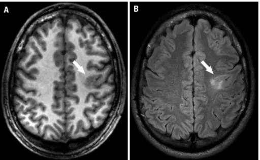
FCD are characterized on MRI (Fig 1) as focal cor-tical thickening with abnormal signal intensity, blurring of the gray-white matter interface, sometimes with sig-nal intensity abnormalities in the underlying white matter mostly hyperintense signal in T2 and FLAIR (luid-atten-uated inversion recovery) and hypointense signal in T1, with the aspect of a wedge-shaped tail extending radially to the ventricle (“transmantle” dysplasia)5,16,36,37,39,44.
Oth-er charactOth-eristic imaging indings are focal white mattOth-er atrophy, a deep sulcus and broadening of the gyri adja-cent to the FCD.
hese changes are not always present together, nor are pathognomonic in isolation. FCD is usually found in fron-tal or temporal lobes, although it can occur in any loca-tion. Electroencephalogram (EEG) may show particular ictal changes in individuals. Detailed pre-surgical evalu-ation is mandatory, since FCD can occur in areas of elo-quent cortex and the epileptogenic zone may be more ex-tensive than the lesion visualized on MRI6,25,26,36.
his lesion can occasionally be quite subtle, and anal-ysis of MR images together with clinical data, EEG and possibly nuclear medicine tests such as SPECT is essen-tial for a correct assessment of the case and valorization of more subtle indings6. Sometimes supericial
neuro-nal or neuroglial tumors like dysembryoplastic neuroep-ithelial tumor (DNET) and ganglioglioma can be a rele-vant consideration in the diferential diagnosis, especial-ly in less typical cases.
Hemimegalencephaly
Hemimegalencephaly or unilateral megalencephaly is related to the unilateral hamartomatous excessive growth of all or part of one cerebral hemisphere, with defects in neuronal proliferation, migration and cortical organization.
Afected patients typically have macrocephaly at birth, and can present early in childhood with refractory epi-lepsy, neurological development delay and hemiparesis45.
Across this spectrum, milder cases may present with dis-crete seizures, completely controlled. he brain may be afected in isolation, or there may be hypertrophy of part or all of the ipsilateral body.
Some syndromes may be associated: neuroibromato-sis type I, tuberous scleroneuroibromato-sis, epidermal nevus syndrome, Proteus syndrome, ipsilateral hypomelanosis of Ito, Klip-pel-Trenaunay-Weber syndrome, focal alopecia, and dys-embryoplastic neuroepithelial tumor1,2,14,44.
Anatomic or functional hemispherectomy may be in-dicated for clinically intractable epilepsy, if contralater-al hemisphere is normcontralater-al. he transfer of functions to the contralateral hemisphere is greater in younger children, due to brain plasticity.
Microscopically, laminar cortical organization is com-pletely changed, with the presence of giant neurons in the cortex and in the underlying white matter. Histopatholog-ical studies usually demonstrate indings similar to those observed in cortical dysplasias14,40, with the presence of
“balloon” cells in some cases1.
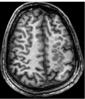
MRI may show moderate to severe increase of the cerebral hemisphere, with increased volume of white matter, which may show in some cases abnormal signal. here is generally cortical thickening, with shallow sul-ci resembling agyria/pachygyria (Fig 2). he distinction of the interface between white matter and gray matter is usually blurred2,5. he lateral ventricle on the afected side
is usually irregularly shaped and enlarged, in proportion to the increased cerebral hemisphere. However, in a few cases, the ipsilateral ventricle may be small2.
he main clue to the diagnosis of unilateral megalen-cephaly is the increase of the lateral ventricle on the af-fected side, contrary to what occurs in expansile or inil-trative lesions with mass efect, which compress and shift the ventricular system. In hemimegalencephaly, there is usually an association with other MCD, such as lissen-cephaly and polymicrogyria5.
Heterotopia
Heterotopia, one of the most commonly MCD re-vealed on MRI, occurs when neurons originating in the periventricular region fail to migrate, leaving tracks or nodules of neurons in heterotopic location, adjacent to the ependymal line or in subcortical topography. Based on MR images, there are three types of heterotopia:
peri-ventricular nodular heterotopia, focal subcortical hetero-topia, and band heterotopia.
he typical patient with periventricular nodular het-erotopia (Fig 3) is most commonly female, during or af-ter the second decade of life that presents with epilepsy, refractory or not to drug treatment. Usually there is no cognitive impairment and there are rare associations with disturbances outside the central nervous system, such as vasculopathy, patent ductus arteriosus or coagulopathy36.
Most cases of familial periventricular nodular hetero-topia afect predominantly women and are caused by mu-tations in FLNA. However, mutations in this gene can
also cause sporadic periventricular heterotopia in wom-en as well as familial and sporadic cases in mwom-en. Other genes also appear to be involved, such as DCX, ARFGEF2
and a locus on chromosome 5p36,46.
Periventricular nodular heterotopia can be associated with mesial temporal sclerosis (dual pathology). In these cases, surgical resection of the afected hippocampus in-variably brings no beneits and should be avoided36.
Patients with periventricular nodular heterotopia as-sociated with other MCD compared to those with isolat-ed periventricular heterotopia, have more severe clinical course, with more frequent seizures, failure to drug treat-ment and increased risk of treat-mental retardation. he extent and distribution of subependymal nodules contraindicate surgery in most cases. Only a few patients were treated surgically, with varying results36.
he nodules of heterotopic gray matter can also oc-cur in subcortical white matter (focal subcortical hetero-topia) with distortion of the ventricle, unilaterally or bilat-erally (Fig 4). here are reduction in the volume of white matter and thinning of overlying cortex. In these cases, more extensive clinical and cognitive deicits may ensue.
In band heterotopia (Fig 5), layers of gray matter are found in subcortical white matter, and are separated from
the cortex by a thin layer of white matter. he thickness of the bands varies among patients and also along the fron-to-occipital axis in the same individual. he thickness of the band is also related to the severity of the clinical pic-ture, being more severe when there are thicker bands of gray matter36,45.
Histological specimens show heterotopic neurons with normal morphology in isolation or in clusters, but lacking appropriate synaptic connections. he pathophysiologi-cal basis is not completely understood and it seems that the gray matter nodules are intrinsically epileptogenic36.
Imaging diagnosis is usually obvious, showing peri-ventricular or subcortical tissue isointense to gray matter on all sequences evaluated, that does not enhance after intravenous injection of paramagnetic contrast agent. In cases of band heterotopia, there is a characteristic conig-uration of “double cortex”. he thickness of the bands var-ies, with blurring of the interface between the white mat-ter and gray matmat-ter. Diferential diagnosis includes sub-ependymal nodules that occur in tuberous sclerosis, but these may enhance after contrast injection and may be calciied. he identiication of the signal intensity similar to the gray matter on all MR sequences is the key to the correct diagnosis of gray matter heterotopia.
Lissencephaly
Lissencephaly is a rare cause of severe epilepsy, men-tal retardation and early death. However, MRI has shown that many patients survive into adulthood.
In lissencephaly, migration of all neurons is severe-ly afected and the brain surface is smooth, with cortical
disruption and reduced white matter volume. he term agyria deines the complete absence of cerebral gyri on the cerebral surface and is synonymous of complete lis-sencephaly. Pachygyria represents a few large and thick gyri, with shallow sulci, and corresponds to incomplete lissencephaly2,5.
Studies in relatives with subcortical band heterotopia and others with lissencephaly have led to the discovery
Fig 3. Periventricular nodular heterotopia. Volumetric T1 FFE (fast ield echo) sequence performed at a 3 Tesla scanner with axial reconstruction in thin sections (1 mm thick) shows subependy-mal heterotopic gray matter adjacent to the right lateral ventri-cle (arrow).
Fig 4. Subcortical heterotopia. T1 SPGR (spoiled gradient recalled echo) in the axial plane demonstrates multiple nodules isointense to gray matter in subcortical location in the left cerebral hemi-sphere. There can be also seen reduction in the volume of white matter, and thinning of the overlying cortex.
of new genes, DCX on chromosome X and LIS1 on
chro-mosome 17. Mutations in any of these genes can lead to any of these disorders or combinations of them, in men or women. Other genes also seem involved in the gener-ation of this phenotype, such as YWHAE, ARX, TUBA1A
and RELN2,3,5,36,41.
Lissencephaly may occur in isolation or as part of oth-er syndromes. “Cobblestone” cortical malformations are mainly related to muscular dystrophy syndromes, clinical-ly heterogeneous, but usualclinical-ly manifesting with hypotonia at birth, generalized muscle weakness and joint contrac-tures in varying degrees. he diagnosis may be obtained by muscle biopsy, allied to characteristic imaging ind-ings in the central nervous system. he three main dis-turbances in this group are Walker-Warburg syndrome, muscle-eye-brain disease, and Fukuyama congenital mus-cular dystrophy5,41,44.
he Walker-Warburg syndrome shows characteristic imaging indings: hydrocephalus with marked ventriculo-megaly, hypogenesis of corpus callosum, hypoplasia, and dysmorphism of brain stem and cerebellum, microph-thalmia, and severe hypomyelination. Occipital cephalo-celes are present in half of the cases2,5. he cortex is thick,
with few shallow sulci and irregularity of gray-white matter junction, relecting the extent of bundles of cortical neurons to the underlying white matter, hence the charac-teristic imaging appearance of the “cobblestone” cortex. he Fukuyama congenital muscular dystrophy is a ge-netic condition, seen primarily in Japanese descendants with mutations in the FCMD gene on chromosome
9q31-332. hese patients have two main types of cortical
anom-alies: polymicrogyria in the frontal lobes, and “cobble-stone” cortex with shallow sulci predominantly in the posterior temporal, and occipital lobes. here are also cerebellar dysplasias with subcortical cysts, and delayed myelination of encephalic white matter5.
Muscle-eye-brain disease also features “cobblestone” cortex with irregular gray-white matter interface, corti-cal thickening (most pronounced anteriorly in the frontal lobes than posteriorly), with a reduction in number and depth of the sulci. here is severe hypomyelination, and the brain stem and cerebellum are dysmorphic. he pons is small, with an anterior slit, and there may also be fusion of the colliculi and presence of cerebellar cysts. he oc-ular globes are small, with subretinal luid collections2,5.
here is no single efective drug treatment in patients with lissencephaly, and the epilepsy caused is usually re-fractory. Surgery in these patients is usually inefective, but sometimes callosotomy may be considered.
Microscopically, there is disorganization of the lis-sencephalic cortex, and blurring of the interface with the white matter. If there is association with band hetero-topia, layers of subcortical gray matter are composed of neurons similar to those found in cortical gray matter2.
In MRI, the cortical surface is smooth, the gyri may be scarce and lat (pachygyria) or absent (agyria) (Fig 6). Incomplete opercularization with shallow and vertical-ized sylvian issures give an aspect of brain in “eight” or also called “snowman” or “hourglass” brain in the axial plane44. In Walker-Warburg synd rome, the appearance
of lissencephaly is typical, with the characteristic image of the “cobblestone” cortex.
Classic lissencephaly usually presents no great diag-nostic challenge; however, pachygyria may sometimes be more subtle, close to the standard brain conigura-tion usually found.
Polymicrogyria
Polymicrogyria is the presence of too many small ab-normal gyri that produce an irregular cortical surface. Polymicrogyria is not invariably associated with epilepsy, and may present as neurological development delay, oro-motor apraxia or congenital hemiparesis36,47,48.
Several patterns of inheritance have been described. A mutation in MECP2 has been found in a man with peri-sylvian syndrome and severe neonatal encephalopathy. A recent study involving hybrid genetic and MRI identiied
PAX6 as a gene in which mutations can cause
unilater-al polymicrogyria36.
It has been reported clinical association with congen-ital arthtogryposis2,5,47 and with some speciic syndromes,
such as Delleman syndrome, Fukuyama congenital mus-cular dystrophy, Neu-Laxova syndrome, hypomelanosis of Ito, Aicardi syndrome, and diGeorge syndrome2.
Several particular patterns of polymicrogyria have been described; the main one is bilateral perisylvian poly-microgyria, clinically characterized by a combination of pseudobulbar palsy, spastic tetraparesis, learning diicul-ties and epilepsy. Some speciic combinations of periven-tricular nodular heterotopia with overlying polymicrogy-ria have also been described3,46,48,49.
Moreover, the fact that there is necrotic tissue with-in the malformed cortex, that most changes occur with-in the middle cerebral artery territory, and the existence of poly-microgyria adjacent to the borders of schizencephaly and porencephaly, suggest that hypoxic-ischemic insults may, at least in some cases, be related to the genesis of polymi-crogyria5. here may also be a relation to congenital
infec-tions such as those caused by cytomegalovirus.
Some experimental models suggest that the epilep-togenic area extends far beyond the obviously visualized abnormality. Resection of an isolated area rarely is bene-icial in refractory epilepsy36.
At least two major histological types are recognized: the four-layered cortex with simpliied organization (most common), and the unlayered type, yet there are several histological intermediate subtypes and these two main types can coexist in adjacent areas36,46.
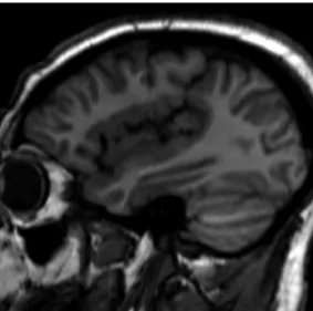
MRI shows excessive cortical gyration with an aspect of cortical thickening and shallow sulci, and irregularity of the gray-white matter interface. his entity may be focal, multifocal or difuse; unilateral or bilateral, symmetric or assymetric. In cases of perisylvian syndrome (Fig 7), there is polymicrogyria surrounding opercular portions with en-larged and vertically oriented sylvian issures, extending to the parietal lobes, well demonstrated in the sagittal plane2,47.
Polymicrogyria may, in some contexts, simulate pachygyria, since excessive cortical gyration, when very small, simulates a thickened cortex with smooth surface, but these entities may be more easily distinguished in vol-umetric MRI sequences with high resolution images.
Schizencephaly
Schizencephaly is deined by the presence of a trans-cortical cleft, extending from the ventricular surface to the pial surface, lined with gray matter, and often with polymicrogyria along its edges.
here may be a variety of associated abnormalities involving the septum pellucidum, optic nerves, the cor-pus callosum or the hippocampi5. Septo-optic dysplasia
(deMorsier syndrome) might be considered in a patient with schizencephaly, optic nerve hypoplasia and absence of septum pellucidum5,44. As in polymicrogyria, vascular,
infectious and genetic causes have been considered in the genesis of this malformation2,46, and perhaps the severity
of the insult diferentiates these two entities, being more severe in the case of schizencephaly.
here may be a wide spectrum of clinical presenta-tions. Patients with bilateral clefts usually have micro-cephaly and severe developmental delay, with spastic quadriparesis, while patients with unilateral clefts may have much less severe clinical phenotypes2. Surgical
treat-ment has been reported in cases of medically intractable epilepsy, but it is rarely considered.
Usually the cleft is lined by dysplastic gray matter, and there may be adjacent areas of polymicrogyria, pachygy-ria and heterotopic gray matter nodules46.
MRI shows a transcortical cleft which may be apposed
Fig 7. Perisylvian polymicrogyria. Imaging at 3 Tesla displays an operculum dysplastic and incomplete, with polymicrogyric cor-tex surrounding the sylvian issure, which is vertically oriented and extends posteriorly to the parietal lobe, well demonstrated in the sagittal reformat of a volumetric T1-FFE (fast ield echo) sequence.
together (closed lips, type I) or clearly unapposed (open lips, type II), communicating the ventricle with the peri-encephalic subarachnoid space, lined by gray matter5. he
lesions may be unilateral or bilateral (in this case, usually symmetrical in location but not in size). In a closed lips schizencephaly, the presence of an indentation in the ep-endymal surface may be the most signiicant clue to the correct diagnosis, the so-called nipple (Fig 8).
The greatest differential here is between open lips schizencephaly and porencephaly. Both are lesions with signal intensities similar to cerebrospinal luid, but the tissue that lines the cavity is the key to the correct diag-nosis - if it shows signal intensity characteristics similar to gray matter, the hypothesis of schizencephaly can be safely considered.
In summary, recognition of the main patterns of MCD in MR images is the mainstay in the work-up of these pa-tients, and advanced neuroimaging techniques may aid in the correct diagnosis, leading to a more accurate classii-cation of these complex and highly epileptogenic entities.
REFERENCES
1. Guerrini R, Sicca F, Parmeggiani L. Epilepsy and malformations of the cere-bral cortex. Epileptic Disord 2003;5(Suppl):S9-S26.
2. Kuzniecky RI, Jackson GD. Magnetic Resonance in Epilepsy. 2 ed. Oxford: Elsevier; 2005.
3. de Wit MC, Lequin MH, de Coo IF, et al. Cortical brain malformations: ef-fect of clinical, neuroradiological, and modern genetic classiication. Arch Neurol 2008;65:358-366.
4. Tinuper P, D’Orsi G, Bisulli F, et al. Malformation of cortical development in adult patients. Epileptic Disord 2003;5 (Suppl 2):S85-S90.
5. Barkovich AJ, Raybaud CA. Malformations of cortical development. Neuro-imaging Clin N Am 2004;14:401-423.
6. Colombo N, Salamon N, Raybaud C. Imaging of malformations of cortical development. Epileptic Disord 2009;11:194-203.
7. Barkovich AJ, Kuzniecky RI, Jackson GD. A developmental and genetic classiication for malformations of cortical development. Neurology 2005; 65:1873-1887.
8. Bentivoglio M, Mazzarello P. The history of radial glia. Brain Res Bull 1999; 49:305-315.
9. Sidman RL, Rakic P. Neuronal migration, with special reference to develop-ing human brain: a review. Brain Res 1973;62:1-35.
10. Valiente M, Marín O. Neuronal migration mechanisms in development and disease. Curr Op Neurobiol 2010;20:68-78.
11. Kriegstein A, Noctor S, Martínez-Cerdeño V. Patterns of neural stem and progenitor cell division may underlie evolutionary cortical expansion. Na-ture Rev 2006;7:883-890.
12. Kubo K, Nakajima K. Cell and molecular mechanisms that control cortical layer formation in the brain. Keio J Med 2002;52:8-20.
13. Battaglia G, Becker AJ, LoTurco J, et al. Basic mechanisms of MCD in animal models. Epileptic Disord 2009;11:206-214.
14. Crino PB. Focal brain malformations: seizures, signaling, sequencing. Epi-lepsia 2009;50 (Suppl 9):S3-S8.
15. D’Arcangelo G. From human tissue to animal models: insights into the pathogenesis of cortical dysplasia. Epilepsia 2009;50 (Suppl 9):S28-S33. 16. Madan N, Grant E. New directions in clinical imaging of cortical dysplasias.
Epilepsia 2009;50:9-18.
17. Jensen FE. Introduction - epileptogenic cortical dysplasia: emerging trends in diagnosis, treatment, and pathogenesis. Epilepsia 2009;50 (Suppl 9):S1-S2. 18. Bruggemann JM, Wilke M, Som SS, Bye AM, Bleasel A, Lawson JA. Voxel-based morphometry in the detection of dysplasia and neoplasia in child-hood epilepsy: limitations of grey matter analysis. J Clin Neurosci 2009; 16:780-785.
19. Dale AM, Fischl B, Sereno MI. Cortical surface-based analysis. I. Segmenta-tion and surface reconstrucSegmenta-tion. Neuroimage 1999;9:179-194.
20. Fischl B, Sereno MI, Dale AM. Cortical surface-based analysis. II: Inlation, lattening, and a surface-based coordinate system. Neuroimage 1999;9: 195-207.
21. Glenn OA, Barkovich AJ. Magnetic resonance imaging of the fetal brain and spine: an increasingly important tool in prenatal diagnosis: part 1. AJNR Am J Neuroradiol 2006;27:1604-1611.
22. Glenn OA, Barkovich J. Magnetic resonance imaging of the fetal brain and spine: an increasingly important tool in prenatal diagnosis: part 2. AJNR Am J Neuroradiol 2006;27:1807-1814.
23. Lim CCT, Yin H, Loh NK, Violet GE, Francis H, Barkovich AJ. Malformations of cortical development high-resolution MR and difusion tensor imaging of iber tracts at 3T. Am J Neuroradiol 2005;26:61-64.
24. Eriksson SH, Rugg-Gunn FJ, Symms MR, Barker GJ, Duncan JS. Difusion ten-sor imaging in patients with epilepsy and malformations of cortical devel-opment. Brain 2001;124:617-626.
25. Knake S, Halgren E, Shiraishi H, et al. The value of multichannel MEG and EEG in the presurgical evaluation of 70 epilepsy patients. Epilepsy Res 2006; 69:80-86.
26. Knowlton RC. Can magnetoencephalography aid epilepsy surgery? Epi-lepsy Curr 2008;8:1-5.
27. Cross JH. Functional neuroimaging of malformations of cortical develop-ment. Epileptic Disord 2003;5(Suppl):S73-S80.
28. Leite CC, Lucato LT, Sato JR, Valente KD, Otaduy MCG. Multivoxel proton MR spectroscopy in malformations of cortical development. Am J Neuro-radiol 2007;28:1071-1075.
29. Leite CC. Malformações do desenvolvimento cortical: avaliação através da espectroscopia de prótons com aquisição simultânea de múltiplos volumes de interesse: Faculdade de Medicina da Universidade de São Paulo. Tese. São Paulo; 2003.
30. Mueller SG, Laxer KD, Barakos JA, et al. Metabolic characteristics of cortical malformations causing epilepsy. J Neurol 2005;252:1082-1092.
31. Li LM, Cendes F, Bastos AC, Andermann F, Dubeau F, Arnold DL. Neuronal metabolic dysfunction in patients with cortical developmental malforma-tions: a proton magnetic resonance spectroscopic imaging study. Neurol-ogy 1998;50:755-759.
32. Kuzniecky R, Hetherington H, Pan J, et al. Proton spectroscopic imaging at 4.1 tesla in patients with malformations of cortical development and epi-lepsy. Neurology 1997;48:1018-1024.
33. Andrade CS, Otaduy MCG, Maia DF, Valente KDR, Leite CC. Phosphorus MR spectroscopy at high iled in patients with malformations of cortical development - preliminary results. In: XXXIII Brazilian Epilepsy Congress. Brasília; 2010.
34. Roulet-Perez E, Davidof V, Mayor-Dubois C, et al. Impact of severe epilep-sy on development: recovery potential after successful early epilepepilep-sy sur-gery. Epilepsia 2010;51:1266-1276.
35. Palmini A, Najm I, Avanzini G, et al. Terminology and classiication of the cortical dysplasias. Neurology 2004;62(Suppl 3):S2-S8.
36. Sisodiya SM. Malformations of cortical development: burdens and insights from important causes of human epilepsy. Lancet Neurol 2004;2:29-38. 37. Colombo N, Citterio A, Galli C, et al. Neuroimaging of focal cortical dysplasia:
neuropathological correlations. Epileptic Disord 2003;5 (Suppl 2):S67-S72. 38. Duchowny M. Clinical, functional, and neurophysiologic assessment of dys-plastic cortical networks: Implications for cortical functioning and surgical management. Epilepsia 2009;50 (Suppl 9):S19-S27.
39. Sisodiya SM, Fauser S, Cross JH, Thom M. Focal cortical dysplasia type II: bi-ological features and clinical perspectives. Lancet Neurol 2009;8:830-843. 40. Blumcke I, Vinters HV, Armstrong D, Aronica E, Thom M, Spreaico R. Malfor-mations of cortical development and epilepsies: neuropathological indings with emphasis on focal cortical dysplasia. Epileptic Disord 2009;11:181-193. 41. Crino PB, Miyata H, Vinters HV. Neurodevelopmental disorders as a cause of
seizures: neuropathologic, genetic, and mechanistic considerations. Brain Pathol 2002;12:212-233.
42. Chamberlain WA, Cohen ML, Gyure KA, et al. Interobserver and intraobserv-er reproducibility in focal cortical dysplasia (malformations of cortical de-velopment). Epilepsia 2009;50:2593-2598.
43. Mathern GW, Cepeda C, Hurst RS, Flores-Hernandez J, Mendoza D, Levine MS. Neurons recorded from pediatric epilepsy surgery patients with corti-cal dysplasia. Epilepsia 2000;41 (Suppl 6):S162-S167.
44. Leite CC, Amaro JE, Lucato LT. Neurorradiologia - Diagnóstico por imagem das alterações encefálicas. São Paulo: Guanabara Koogan; 2008. 45. Guerreiro MM. Malformations of cortical development. Arq Neuropsiquiatr
2009;67:570-574.
46. Guerrini R, Marini C. Genetic malformations of cortical development. Exp Brain Res 2006;173:322-333.
47. Poduri A, Chitsazzadeh V, D’Arrigo S, et al. The syndrome of perisylvian polymicrogyria with congenital arthrogryposis. Brain Dev 2009;32:550-555. 48. Leventer RJ, Jansen A, Pilz DT, et al. Clinical and imaging heterogeneity of
polymicrogyria: a study of 328 patients. Brain 2010;133:1415-1427. 49. Wieck G, Leventer RJ, Squier WM, et al. Periventricular nodular heterotopia
![Fig 2. Hemimegalencephaly. Axial T2 FSE (fast spin echo) [A] shows an enlarged right cerebral hemisphere](https://thumb-eu.123doks.com/thumbv2/123dok_br/15433550.595414/5.955.58.578.94.412/fig-hemimegalencephaly-axial-shows-enlarged-right-cerebral-hemisphere.webp)