1Assistente do Serviço de Neurocirurgia do Hospital da Força Aérea do Galeão, Rio de Janeiro RJ, Brasil (HFAG); 2Graduação em Medicina, Universidade Federal do Rio de Janeiro (UFRJ) Rio de Janeiro RJ, Basil; 3Chefe de Serviço de Neurocirurgia (HFAG) Received 23 June 2004, received in final form 27 October 2004. Accepted 8 December 2004.
Dr. José Alberto Landeiro - Rua Conde de Bonfim 211/310 - 20520-050 Rio de Janeiro RJ - Brasil. E-mail: jlandeiro@aol.com
INTERNAL STRUCTURE OF THE
CEREBRAL HEMISPHERES
An introduction of fiber dissection technique
Igor de Castro
1, Daniel de Holanda Christoph
2,
Daniel Paes dos Santos
2, José Alberto Landeiro
3ABSTRACT - The aim of this study is to introduce the fiber dissection technique and its importance in the comprehension of the three-dimensional intrinsic anatomy of the brain. A total of twenty brain hemispheres were dissected. Using Kingler’s technique we demonstrated the intrinsic structures of the brain. The supra lateral aspect of the brain as well as the medial aspect were presented. The most important fiber systems were demonstrated. The use and comprehension of new neuroimaging techniques demand a better standing of this fascinating anatomy. The knowledge acquired with this technique will improve our under-standing of critical pathways of the central nervous system.
KEY WORDS: brain, anatomy, white matter dissection.
Estrutura interna dos hemisférios cerebrais: introdução à técnica de dissecação de fibras
RESUMO - O objetivo é mostrar a técnica de dissecação de fibras e sua importância na compreensão da anato-mia tridimensional do cérebro. Um total de 20 hemisférios cerebrais foram dissecados. Usando a técnica de dissecação descrita por Kingler, pudemos demonstrar as estruturas que compõem a anatomia interna do cérebro. A anatomia da face súpero-lateral assim como da face medial foi apresentada. O uso e compreen-são de novas técnicas de neuroimagem requerem um melhor conhecimento desta anatomia. O conhecimen-to adquirido com essa técnica contribuirá para o melhor entendimenconhecimen-to de vias essenciais do sistema nervo-so central.
PALAVRAS-CHAVE: cérebro, anatomia, dissecação, substância branca.
Traditionally the brain sulci and gyri anatomy of the brain have been studied by anatomists and clinicians but the intrinsic anatomy of the complex fibers of the white matter has been somewhat ig-nored. Very few books or publications regarding this topic are available when compared to the exten-sive literature about the external structure of the brain. Recently in Rhoton’s masterpiece, “The Su-pratentorial Cranial Space: Microsurgical Anatomy and Surgical Approaches” - Supplement of Neuro-surgery, some beautiful dissections showing the in-ternal fasciculus can be appreciated1. With the most
recent advances in neuroimaging one can experien-ces full details pictures of the internal anatomy of the brain2-7.Therefore, there is an increasing
de-mand for knowledge of intrinsic brain anatomy. As radiological and surgical techniques become
in-creasingly precise, our knowledge of the superfi-cial anatomy and also the recognition of the inter-nal white matter tracts of the brain is essential. In the last years many studies using diffusion-weight-ed and diffusion tensor MR imaging were publis-hed. The new MRI devices which use high magnet-ic fields and techniques promoting superior qual-ity images can be used to show the full white mat-ter anatomy in detail4. The term so-called
tractogra-phy is becoming very popular. This analysis of tracts is essential for understanding and explaining the pathophysiologic patterns of certain disease states, especially intrinsic gliomas. In fact, in the studies of the intrinsic brain tumors such as the gliomas, the anatomy of the white matter tracts is more impor-tant than the anatomy of the sulci and gyri3.
the brain’s intrinsic anatomy is essential. The knowl-edge gained from this technique can be applied to microsurgical procedures. Not only the neurosur-geon who can benefit from a detailed description of this anatomy, but also the neurologist, neurop-athologist and the neuroradiologist. The precise diagnosis and treatment of central nervous system (CNS) lesions depends upon the comprehension of the whole anatomy itself. Our ability to think in tree-dimensions should be exercised. According with Ture et al.8the reestablishment of the fiber
dissec-tion of the white matter as a standard study method is recommended. The learning is essential and it should be mandatory in the continuing education of neurosurgeons in training.
METHOD
The human brains were harvested from autopsy pati-ents that had a non-neurological cause of death. The stu-dies were performed in the microsurgery laboratory of The Brazilian Air Force Hospital, Rio de Janeiro. Each spec-imens received careful postmortem attention, and those with gross defects were rejected from this study. We used the preservation method of Kingler, with minor adjust-ments9,10. In removing the brain from the skull, every effort
was made to minimize damage to the delicate surface. The organ was then suspended, by means of a ligature placed around the basilar artery, in a vessel containing 10% for-maldehyde solution. This is an essential maneuver to main-tain the brain in its normal contours. This fluid was repla-ced first after 24 hours and again after an interval of two weeks. After a total period of 4 weeks or longer in the for-maldehyde solution, the brain was washed for several hours in fresh cold water. The pia matter, arachnoid mem-brane, and vessels of the specimens were carefully remo-ved using the operative microscope. Subsequently the spec-imens were placed in a plastic vessel containing 10% formal-dehyde solution and stored for 8 days in a deep freeze at -10º C. At this point the brain was thawed under running cold water for 24 hours. Sometimes, repeating the deep fre-ezing procedure (in 10% formaldehyde solution) two or three times can facilitate the dissection. After the last freezing, the brains were kept in the 10% formaldehyde solution.
For making the dissections, simple anatomic instru-ments were found to be quite satisfactory. The main ins-trument utilized was the wooden spatula. To perform the dissections, it was necessary to use different sizes of spatulas tips. A fine forceps was used to execute the deli-cate fiber bundle preparations. It was also necessary on most occasions to use a dissecting microscope to magni-fy the specimen and provide better illumination.
RESULTS
The dissection begins at the supero-lateral surfa-ce of the surfa-cerebral hemispheres.
Supero-lateral aspect– The superolateral surfa-ce of each surfa-cerebral hemisphere is markedly convex and fits into the corresponding half of the skull vault. In this specimen, we removed the gray mat-ter of the right hemisphere. The gyri and sulci pat-terns are still the same and can be readily appre-ciated. The longest sulcus in this aspect is the cen-tral sulcus. The so-called “U” fibers connecting on gyri with another under their respective sulcus are revealed (Fig 1).
The insula is a substantial portion of the cere-bral cortex that forms the floor of the lateral sul-cus that can be opened up by removing the lips bordering the lateral sulcus (Sylvius) and its rami. These lips are known as the frontal, fronto pari-etal and temporal opercula. After their excision, the insula appears as a triangular eminence that is marked by a number of sulci and gyri. In this spe-cimen, the insular cortex was removed revealing a number of sulci, one of which the central sulcus of the insula is deepend more proeminent than the rest. Again the “U” fibers of the insula gyri can be visualized as well as the circular sulcus of the insu-la which surrounds the entire lobe. The inferior part of this triangular lobe is known as the limen insu-la. The entire cortex of the whole hemisphere was removed revealing the “U” fibers of the white mat-ter underneath (Fig 2).
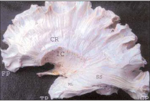
The lentiform nucleus has been removed to expose the lateral aspect of the internal capsule. The corona radiate and its continuity with the in-ternal capsule can be seen in this dissection, since the ends and upper margin of the putamen mark the junction of the internal capsule with the base of the corona. A contrasting appearance is afford-ed by the long, parallel, closafford-ed packafford-ed fibers of the sagittal stratum, the fibers of which remain rather discrete as they persue their long and wavy course toward the occipital cortex. The optic nerve, optic chiasma and optic tract can be traced backwards in this specimen. The optic tract terminates in the lateral geniculate body. The fibers of the optic radiation, or geniculostriate projection, emerge from the dorsal surface of the lateral geniculate body and can be traced as part of the sagittal stra-tum of white fibers that run anterior and inferior into the temporal lobe. Subsequently, they sweep backwards and terminate in the region of the cal-carine sulcus (Fig 3).
lateral geniculate nucleus is appreciated in this spe-cimen. The fibers of internal capsule condense to run down through the peduncular part of the mid-brain. A line of demarcation can be seen showing the original position of the globo pallidum. The an-terior comissure is also despicted. The hippocam-pus and the choroid plexum above it is also visua-lized (Fig 4).
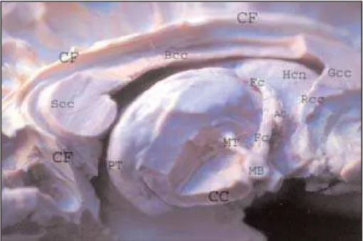
The medial aspect– Above the corpus callosum, the cingulum is seen. The cingulum is an associa-tion tract that commences below the rostrum of the corpus callosum (cc), in the region of the olfac-tory cortex, and arches around the entire cc. After curving round the splenium of the cc, the bundle proceeds forward within the parahippocampal gyrus to reach the uncus and nearby cortical areas of the temporal lobe. In this preparation, the cor-pus callosum was partially remove to show the cau-date nucleus (head).
The thalamus is a large nucleus in the depth of the brain. The constituent nuclei on its medial as-pect are displaced. The column of the fornix has been divided a short distance above the anterior comissure. The fibers of the mamillothalamic fasci-culus (bundle of Vicq d’Azir) arise from the mamil-lary body and travel upwards and backwards to the anterior, expanded part of the anterior nuclear group of the thalamus (Fig 5).
The cingulum and some fibers of the cc have
Fig 2. Insula region. FP, frontal pole; OP, occipital pole; TP, temporal pole; FO, frontal opercu-la; PO, parietal opercuopercu-la; TO, temporal opercuopercu-la; CSi, central sulcus of insuopercu-la; SG, short gyrus of insula; LG, long gyrus of insula; CiS, circular sulcus of insula; LI, limen insula.
Fig 1. Supero lateral surface of the brain. FP, frontal pole; OP, occipital pole; CS, central sulcus; LF, longitudinal fissure.
thala-Fig 3. Optic pathway. CR, corona radiate; IC, internal capsule; P, putamen impression; GP, globo pallidum impression; ON, optic nerve; OC, optic chiasm; OT, optic tract; SS, stratum sagittal; ML, meyer loop.
mus towards the habenular trigone. In addition to the habenular trigone, the following components of the epithalamus can be distinguished: the habe-nular comissure, the pineal body and the posteri-or comissure. The posteriposteri-or thalamic nucleus, known as pulvinar is demonstrated (Fig 6).
In this preparation the corpus callosum, cauda-tenucleus, and most of the brain stem structures
have been removed. The thalamus is shown in the center of the picture. The thalamus is an important integrating center which receives sensory signals of various modalities, and transmits impressions to appropriate areas of the cerebral cortex. An exten-sive accumulation of the axons connecting various thalamic nuclei to practically all cortical areas is seen in a fan-like array and this, in three dimensions,
flects the profusion of the thalamic radiations. For descriptive purposes, different parts of the thalam-ic radiations are grouped into four thalamthalam-ic pedun-cles. The anterior, superior, posterior and inferior pe-duncles are displayed (Fig 7).
The internal capsule and corona radiate have be-en exposed by removal of the corpus callosum,
cauda-te nucleus and diencephalon. The most striking figure of this preparation is the convergence of the great masses of corticofugal fibers from extensive areas of cerebral cortex into the relativily narrow, but thick, basis pedunculi. Some torn fibers of the thalamic peduncles can still be identified. The stratum sagit-tal is also visualized (Fig 8).
Fig 5. Medial aspect. CF, cingulate fascicullum; Rcc, rostrum of corpus callosum; Gcc, genu of cc; Bcc, body of cc; Scc, splenium of corpus callosum; PT, pulvinar thalamus; CC, crus cerebri; MT, mamilothalamic fascicullum; MB, mamilar body; Fc, fornix (columm); AC, anterior comissure.
Fig 7. Thalamic peduncles. A, anterior thalamic peduncles; S, superior; P, posterior; I, inferior; T, thalamus.
DISCUSSION
Using fiber dissection techniques some early ana-tomists such as Vieussens, Willis, Steno, Bell, Reil and Foville could demonstrate many tracts and fas-ciculi of the brain8,11-14. In fact, the fiber dissection
technique was one of the first methods used to de-monstrate the internal structures of the brain8. An
extensive historical review is beyond the scope of this paper and was made in an outstanding
man-ner by Ture et al.8, although we can not forget to
mention the work of Joseph Kingler (1888-19 63). He was an anatomist in Basel that made the greatest contribution to the fiber dissection techni-que. In 1935, he developed an improved method of brain fixation and a technique that now bears his name9,10(Kingler’s technique). Like others, he
dissected formalin-fixed brains with wooden spatu-las. However, he froze and thawed the brains
fore dissection. Freezing helps to separate the fi-bers. His superb atlas containing detailed anatom-ic studies of the brain was published in 1956. Al-though his studies were impressive, this technique never became widely used8-10.
While the freezing method is an aid to dissec-tion and generally increases the distincdissec-tion between the grey and white matter of the brain, it does not produce absolutely consistent results, as Kingler himself acknowledged9. As a rule, however, the
te-chnique makes it easier to prepare dissections of the both fiber tracts and nuclei.
Illustrations of the internal structures of the brain in current textbooks are usually pictures of sections or schematic drawings. Only few fiber dis-sections from earlier textbooks are still reprodu-ced8. In Rhoton’s masterpiece published in 2002 we
can appreciate some superb pictures of white mat-ter anatomy exposed. In this publication he descri-bed the white matter anatomy in detail. With the learning gained from the above mentioned dissec-tions, the younger generation is stimulated to per-form dissections on their own.
The evolution of neuroimaging techniques has imposed a greater demand of knowledge. Without the understanding of the three-dimensional intrin-sic anatomy of the brain one can not interpret pre-cisely the new neuroimaging studies such as “trac-tography”. Diffusion-tensor MR imaging is a promi-sing tool to evaluate white matter anatomy. The first attempt to visualize the white matter tract was made by Kinosada et al in 19932. With best MRI
de-vices using higher magnetic field such as 3.0 Tesla, better images are now available4,5,7. In the
litera-ture, one can find reports about the correlation of AVMs6or tumors3with the white matter tracts. We
project that in a few years, these images will be es-sential for the precise diagnosis or treatment plan-ning of many diseases of the central nervous sys-tem. According with Ture et al.8the fiber
dissec-tion technique is a demanding and time-consum-ing technique. The technique was somehow aban-doned after the introduction of the microtome and histological preparations. The histological sections are very important to the interpretation of bipla-nar MRI films, but our ability to think in three di-mensions should be stimulated. Therefore, the knowledge of intrinsic white matter anatomy is of great importance. The combination of histologi-cal techniques with fiber dissection technique
im-proves the understanding and prevents misinter-pretation of the complex anatomic features of struc-tures. The first surgeon that gave attention to this technique was Yasargil17.
After he gained knowledge with this technique, he applied it to all of his routine microsurgical pro-cedures15-17. The goal of this manuscript is to
stimu-late the younger generation of neurosurgeons to acquire proficiency in fiber dissection technique and become experts in surgical neuroanatomic features.
Acknowledgments- The authors would like to ex-press the deep appreciation to Evandro de Oliveira, M.D., Ph.D., who placed his laboratory facilities at our disposal. He stimulated the technique and introduced the fiber dissection technique in his Microsurgical Course of sulci and gyri at Benificiencia Portuguesa Hospital Mi-crosurgical Laboratory (Sao Paulo - Brazil).
REFERENCES
1. Rhoton, AL Jr. The supratentorial cranial space: microsurgical Anatomy and surgical approaches: Chapter 1. The Cerebrum. Neurosurgery 2002;51:(Suppl):1-52.
2. Kinosada Y, Ono M, Okuda Y, et al. MR tractography: visualization of structure of nerve fiber system from diffusion weighted images with maximum intensity projection method. Nippon Igaku Hoshasen Gakkai Zasshi 1993;53:171-179.
3. Holodny AI, Schwartz TH, Ollenschleger M, Liu WC, Schulder M. Tu-mor involvement of the corticospinal tract: diffusion magnetic resonance tractography with intraoperative correlation. J Neurosurg 2001;95:6:1082. 4. Baleriaux D, David P, Sadeghi N, Neugroschl C, Jissendi P, Metens T. Role of new MRI techniques in neuroradiologic practice. Rev Med Brux 2003;24:279-286.
5. Nguyen TH, Stievenart JL, Yoshida M, et al. Tractography of the visu-al pathways: routine examination in magnetic resonance imaging. Fr Ophtalmol 2003;26:941-951.
6. Yamada K, Kizu O, Ito H, Nishimura T. Tractography for an arteriove-nous malformation. Neurology 2004;24:62-69.
7. Toosy AT, Ciccarelli O, Parker GJ, Wheeler-Kingshott CA, Miller DH, Thompson AJ. Characterizing function-structure relationships in the human visual system with functional MRI and diffusion tensor imag-ing. Neuroimage 2004;21:1452-1463.
8. Ture U, Yasargil MG, Friedman AH, Al-Mefty O. Fiber dissection techni-que: lateral aspect of the brain. Neurosurgery 2000;47:417-426. 9. Klingler J. Erleichterung der makroskopischen Praeparation des Gehirns
durch den Gefrierprozess. Schweiz Arch Neurol Psychiatr 1935;36:247-256. 10. Klingler J, Gloor P. The connections of the amygdala and of the anteri-or tempanteri-oral canteri-ortex in the human brain. J Comp Neurol 1960; 115:333-369. 11. Vieussens R. Neurographia universalis. Lyons: Lugduni, Apud
Joan-nem Certe, 1685.
12. Bell C. The anatomy of the brain. London: Longman and Co., 1802. 13. Reil JC. Fragmente über die Bildung des kleinen Gehirns im Menschen.
Arch Physiol Halle 1807-1808;8:1-58.
14. Foville ALF. Traité complet de l’anatomie, de la physiologie et de la patho-logie du système nerveux cérébrospinal. Paris: Fortin, Masson et Cie, 1844. 15. Yasargil MG. Microneurosurgery: microsurgical anatomy of the basal
cisterns and vessels of the brain.Stuttgart: Georg Thieme, 1984. 16. Yasargil MG. Microneurosurgery: AVM of the brain - history,
embryo-logy, pathological considerations, hemodynamics, diagnostic studies, microsurgical anatomy. Stuttgart: Georg Thieme, 1987.