Arq Neuropsiquiatr 2008;66(1):108-110
108
Optic neuritis due tO sOlvent abuse
Carlos Alexandre Twardowschy
1, Hélio Afonso Ghizoni Teive
2, Fábio Siquineli
1,
Arthur Furlaneto Fernandes
1, Ismael Paulo Búrigo
1, Arnolfo Carvalho-Neto
3, Lineu César Werneck
4neurite óptica relaciOnada aO abusO de sOlvente
Neurology Service, Internal Medicine Department, Hospital de Clínicas, Federal University of Paraná, Curitiba PR, Brazil: 1Resident in Neurology; 2Adjunt
Professor of Neurology; 3Adjunt Professor of Radiology; 4Full Professor of Neurology.
Received 1 August 2007, received in inal form 1 November 2007. Accepted 4 December 2007.
Dr. Carlos Alexandre Twardowschy – Rua Dias da Rocha Filho 261 / 11 - 80040-050 Curitiba PR - Brasil. E-mail: [email protected] Solvent abuse is a public health problem in Brazil
par-ticularly among young adults and children. Inhalation of toluene-based products is popular with solvent sniffers because of the euphoric effect and easy availability of these substances. Chronic inhalation of toluene may re-sult in a variety of neurologic complications like cerebel-lar dysfunction, optic atrophy, pyramidal tract signs, cra-nial nerve abnormalities. Also, personality changes, emo-tional instability and general cognitive decline have been attributed to its abuse1.
We report an unusual case of acute optic neuritis in-duced by thinner snifing.
case
A 34-year-old man presented with an acute visual loss. He was admitted to the hospital reporting a sudden and progressive loss of visual acuity after snifing a hole can of thinner during uninterrupted 48 hours. This homeless patient had a history of chronic solvent abuse for ive years, moderate alcohol ingestion and tabagism, but denied other substances abuse. At the mo-ment of the admission, 4 days after the beginning of the symp-toms, he related to see only countenances. The physical exami-nation and vital data were normal. The neurological examiexami-nation was normal except for presenting bilateral mydriasis with severe
reduction of the photomotor and consensual responses. Visual acuity was worse than 20/200. Ophthalmological examination demonstred bilateral papilledema, with normal ocular pressure and ocular movements. Other cranial nerves were normal. Labo-ratory examinations are listed in Table. Lumbar puncture showed 130 mmH2O opening pressure, 27 red blood cells/mm³, 1
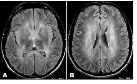
leu-cocyte/mm³, glucose concentration 75 mg/dL, protein 39 mg/ dL and VDRL was negative. Magnetic resonance imaging (MRI) showed bilateral symmetric hyperintense lesions in the deep white matter, corpus callosum (splenium), centrum semiovale (Fig 1) and pons (Fig 2). EEG was irregular without speciic path-ological meanings. The patient was treated with methylprednis-olone 1 gr/day for three days associated with replacement of B complex vitamins. Six days after the admission, the patient ran away from the hospital and one month later returned in the neu-rology clinic relating important improvement of the visual acuity.
discussiOn
Today, organic solvents contained in industrial and do-mestic products are the most commonly abused volatile substances. Formerly, benzene was the major organic sol-vent in paints, lacquers, and thinners. However, benzene is toxic to bone marrow and liver and has been replaced by n-hexane or toluene (methyl benzene), which are
Arq Neuropsiquiatr 2008;66(1)
109 Solvent abuse: optic neuritis Twardowschy et al.
Fig 2. MRI. Axial FLAIR image shows bilateral sym-metric hyperintense lesions in the base of the pons (arrows).
toxins. Toluene can cause multifocal neurologic disorders. Prior reports have been concerned with its acute effects1.
Because of its lipophilicity, toluene rapidly penetrates into the central nervous system (CNS) after inhalation. Toxic toluene inhalation is most commonly the result of occupational exposure or recreational abuse.
A multiinstitutional study of 138 outpatients and in-patients with solvent dependence reported the organic solvents abused were “thinner” (a word often used as a general term for organic solvents for abuse), 69.6%; pure toluene, 40.6%; and glues, 39%, with multiple replies2.
Gas chromatographic analysis of the volatile solvents revealed that the paint thinner consist almost entirely of toluene (methyl benzene), with traces of xylene (dimeth-ylbenzene). The aerosol products contain 59 to 61% tolu-ene, with traces of xylene and 10% methylene chloride (dichloromethane), a halogenated hydrocarbon. The re-mainder are propellants (butane-isobutane), which escape as gas when sprayed on the rag and are rarely inhaled. The solvent fractions that are inhaled consisted primarily of toluene and traces of methylene chloride1. Multiple
com-ponents in the mixtures may enhance the net toxicity in a synergistic or additive fashion. Inhaled volatile hydro-carbons are rapidly absorbed from the lungs. Being highly lipophilic, they most easily enter and are retained within the lipid-rich nervous system.
Toxic and deiciency optic neuropathies are due to toxic chemicals and nutritional deficiency. The visual symptoms (dyschromatopsia and progressive reduction of visual acuity) usually occur bilaterally, simultaneously and painlessly. Total blindness is unusual with the exception of that caused by methanol. Neurologic abnormalities were
Table. Patient’s laboratory exams results.
Exams Results Exams Results
Hemoglobin (g/dL) 12.8 HBsAG Not reagent
MCV 88 Anti-HBC total Not reagent
Leucocytes (per mm3) 5000 Anti-HBS Not reagent
Platelets (per mm3) 247000 Anti-HCV Not reagent
Creatinin (mg/dL) 0.6 VDRL Not reagent
Urea (mg/dL) 38 HIV Negative
Glucose (mg/dL) 132 PCR HSV Negative
INR 1.01 PCR CMV Negative
TSH (um/dL) 2.09 PCR VZV Negative
ESR 5 PCR EBV Negative
Total bilirubin (mg/dL) 1.0 Serum B12 Normal
AST (mg/dL) 22
ALT (mg/dL) 24
Serum Ca (mg/dL) 8.9
Serum Na (mmol/L) 143
Serum K (mmol/L) 3.5
Arq Neuropsiquiatr 2008;66(1)
110
Solvent abuse: optic neuritis Twardowschy et al.
seen in 65% of patients with a history of chronic solvent vapor abuse for 2 years or more1. The abnormalities were
cognitive (60%), pyramidal (50%), cerebellar (45%), cranial nerve brainstem (25%) and tremor (15%).
We found ive other reports of optic neuritis induced by thinner snifing3-6. Like in our case, after they became
conscious of their symptoms of slight visual disturbance, they continued to snifing until they lost their vision. An-other similar feature was lack of pain and response to steroids plus vitamin B complex3. The MRI exam did not
reveal any signs of optic nerve lesion (Figs 1A, 1B, 2). The pathogenesis of the MRI lesions in toluene toxic-ity is poorly understood. Because of its high lipid solu-bility, toluene accumulates in lipid-rich tissues such as brain. Demyelination and gliosis in the cerebral and cer-ebellar white matter are the histologic changes reported in chronic toluene abusers7,8.
The cases with diffuse white matter change had a lon-ger duration of abuse and obvious brain atrophy9. All
pa-tients with white matter changes had neurologic deicit. The association of white matter changes with abuse lon-ger than 4 years suggests that white matter lesions are the result of a cumulative toxic effect of inhaled toluene8,11.
The main MRI indings in the CNS of the chronic sol-vent abusers are the white matter change on T2-weighted and proton density-weighted images as the patient re-ported presents (Figures). This inding is considered to represent the damage in myelin, such as demyelination or myelin pallor, reported in histopathologic reports9.
Periventricular white matter and the centrum semi-ovale were the most common locations for white matter changes in 19 (46%) of 41 patients, thinning of the corpus callosum was revealed in nine patients (22%), T2-weighted images revealed symmetric hypointensity in the thalami in eight patients (20%). Some investigators have suggested
that iron deposition and the partition of toluene into the lipids of cell membranes explain this inding10.
T2-weight-ed images showT2-weight-ed restrictT2-weight-ed white matter changes in 10 (53%) of 19 patients with abnormal MRI imaging indings 11.
Computadorized tomography revealed difuse atrophy of cerebral hemispheres1.
So far, whether diffuse white matter change is caused by the spreading of restricted white matter change is not clear, neither is the reversibility of the white matter change.
Finally, in atypical visual disturbances, several labora-tories, neurophysiological and neuroradiological tests are mandatory. Moreover, since pharmacological and nutri-tional therapies generally only provide a mild degree of symptomatic improvement, further studies are needed in optic neuritis due to solvent abuse.
references
1. Hormes JT, Filley CM, Rosenberg NL. Neurologic sequelae of chronic solvent vapor abuse. Neurology 1986;36:698-702.
2. Fukui S, Wada K, Iyo M. Clinical characteristics of the recent organic solvent dependents who visited psychiatric facilities [in Japanese with English abstract]. J Men Health 1989;35:107-131.
3. Shinya H, Hoshino K, Kiritohshi M, Kiuchi S, Yamagami K, Nakatani T. [2 cases of acute retrobulbar neuritis by thinner inhalation; detected methanol of high concentration in gas phase assay] Chudoku Kenkyu. 2003;16:329-333.
4. Kohriyama K, Hori H, Murai Y, Ninomiya H, Tsukamoto Y. [Optic
neu-ropathy induced by thinner snifing] J UOEH 1989;11:449-453. 5. Ogawa Y, Takatsuki R, Uema T, et al. Acute optic neuropathy induced
by thinner snifing: inhalation of mixed organic solvent containing
methyl alcohol and methyl acetate. Ind Health 1988;26:239-244.
6. Berg EF. Retrobulbar neuritis: a case report of presumed solvent toxic
-ity. Ann Ophthalmol 1971;3:1351.
7. Rosenberg NL, Kleinschmidt BK, Davis KA, Dreisbach JN, Hormes JT,
Filley CM. Toluene abuse causes diffuse central nervous system white
matter changes. Ann Neurol 1988;23:611-614.
8. Damasceno BP, Capitani EM. Cerebellar atrophy related to chronic ex
-posure to toluene: case report. Arq Neuropsiquiatr 1994:52:90-92.
9. Escobar A, Aruffo C. Chronic thinner intoxication: clinico-pathologic re
-port of a human case. J Neurol Neurosurg Psychiatry 1980;43:986-994. 10 . Yamanouchi N, Okada S, Kodama K, et al. White matter changes caused
by chronic solvent abuse. Am J Neuroradiol 1995;16:1643-1649. 11 . Aydin K, Sencer S, Demir T, Ogel K, Tunaci A, Minareci O. Cranial
MR indings in chronic toluene abuse by inhalation. Am J Neuroradiol