Assessing Autonomic Function in Hypertrophic
Cardiomyopathy
Marcelo Imbroinise Bittencourt, Paulo Roberto Benchimol Barbosa, Cantídio Drumond Neto,
Ricardo Bedirian, Eduardo Corrêa Barbosa, Flavia Brasil, Alfredo de Souza Bomfim,
Francisco Manes Albanesi Filho
Universidade do Estado do Rio de Janeiro e Santa Casa de Misericórdia do Rio de Janeiro - Rio de Janeiro, RJ - BrazilMailing Address: Marcelo Imbroinise Bittencourt • Rua Dona Maria, 71/902-Bl. 1 – 20541-030 - Rio de Janeiro, RJ - Brazil
E-mail: [email protected] Received on 01/13/04 • Accepted on 01/21/05
O
BJECTIVEAssess the autonomic function in hypertrophic cardiomyopathy (HCM) through heart rate variability (HRV) and to correlate it to echocardiographic data.
M
ETHODSTwo groups were studied, and compared for gender, age and HR: A) Ten (10) patients reporting septal HCM (70% non-obstructive); B) ten (10) healthy volunteers. HRV was analyzed along four successive stages: at rest, under controlled breathing, while bending, and controlled breathing associated to bending. Variables means were compared between groups and intra-groups in the different stages; in Group A, variables means were correlated to echocardiographic measurements (interventricular septum and left atrial diameter).
R
ESULTSNo HRV difference was reported among groups in the fi rst 3 stages. In the fourth stage vagal activity was shown to be higher in Group A [quadratic mean log between RR intervals (RMSSD) – 1.35+0.14 vs 1.17+0.16; p=0.019; high frequency component logarithm (LogHF)- 4.89+0.22 vs 4.62+0.26; p=0.032]. Along the stages, vagal measurements [rate of pairs of consecutive RR intervals whose difference is ≥ 50ms (pNN50) and LogHF] also showed lower reduction in the third stage in Group A, while LogHF showed some increase in last stage (p=0.027), thus indicating marked parasympathetic activity in that group. Group A HRV analysis showed no difference among patients reporting larger hypertrophy or atrial diameter.
C
ONCLUSION1) parasympathetic prevalence was shown during autonomic stimulation in HCM patients; 2) no correlation was found between HRV and echocardiographic measurements under analysis.
K
EY WORDSHypertrophic cardiomyopathy (HCM) is a genetic disease where ventricular hypertrophy associated with preserved systolic function and diastolic dysfunction stand out (with changes in ventricular relaxation and compliance) under no associated conditions to produce such change.Clinical development of the condition is variable, usually family-related, heterogeneously expressed, with autosomic dominant transmission1,2. It
has for some time been speculated that the development of myocardial hypertrophy in that condition might be related to some increase in sympathetic activity3. Brush
and collaborators (1989) demonstrated that neuronal uptake of norepinephrine was reduced in HCM patients, thus emphasizing the assumption that sympathetic nervous system disorders might be related to phenomena found in that condition, particularly sudden death4.
Schafers and collaborators also verifi ed decrease in beta receptors density in that condition, in addition to reduction in norepinephrine neuronal uptake5.
The study of the autonomic nervous system (ANS) has been gaining strength in most recent decades as an attempt to set behavior patterns for the different diseases that may be correlated to sudden death events6-8. Particularly for
diabetics (whose evolution is disautonomic), and for those who have had acute myocardial infarction (AMI), autonomic function changes are signifi cantly correlated to increased mortality rate9-13.
Heart rate variability (HRV) analysis is a simple, invasive method, and aims at assessing the balance between sympatho-vagal activity on the heart; consequently, at identifying the phenomena related to ANS disorders that may contribute for the genesis of arrhythmias14.
Some works have analyzed HRV in the last decade, with confl icting results. Those using 24-hour Holter for HRV analysis found sympathetic prevailing over parasympathetic activity. Such changes were found to be correlated to the presence of symptoms, ventricular arrhythmia, and left ventricle outfl ow tract obstruction (LVO)15-19.
Such argument was contested by Fei and collaborators while studying a group of thirty-one (31) HCM patients and thirty-one (31) normal individuals through 24-hour HRV Holter analysis. HCM patients reported signifi cantly lower low frequency (LF) component, and lower low frequency/high frequency ratio (LF/HF), and high frequency (HF) components that were higher as compared to control group individuals, thus indicating a reduction in sympathetic activity in their normal, day-to-day activities20. Similar results were found by Limbruno
and collaborators while using autonomic short-term stimulating protocols for HRV analysis. Sympathetic activity compromising was found to be higher in individuals with obstructive HCM 21,22.
Against such confl icting results, the present paper has the purpose of: 1) assessing the behavior of the autonomic function in individuals with asymmetric septal HCM as compared to control group through HRV at rest, under
respiratory stimulus, and bend tests, and 2) correlating fi ndings to the analysis of septally hypertrophic HRV, and left atrium (LA) size.
M
ETHODS
In the period between 09/2001 and 12/2002, twenty (20) individuals were split into two groups paired by gender, age, and heart rate (HR): Group A – made up of ten (10) individuals with asymmetric, septal HCM; and Group B – made up of ten (10) normal individuals.
Among the individuals in Group A, age ranged from 27 to 55 years of age (40.3 ± 9.8), being 90% males. Among Group B individuals, age ranged from 26 to 45 years of age, (37.2 ± 6.4), being 80% males. Among Group A patients, four (4) were on propranolol and one (1) on verapamil.
Asymmetric septal HCM was diagnosed based on echocardiographic evidence of asymmetric septal hypertrophy of left ventricle (larger than 13mm), which did not present dilation, in individuals with no identifi able cause for hypertrophy1,2,23. Those who had
been submitted to septal myectomy or to alcoholization of septal artery, those on amiodarone, or those for whom medication discontinuation was considered unacceptable were excluded from Group A. Also excluded were those reporting coronary heart disease, hypertension, aortic stenosis or any other condition that would lead to myocardial hypertrophy, nonsinosoidal rhythm, or advanced atrioventricular blockings, and those with a history of diabetes mellitus and chronic alcohol abuse.
All individuals were submitted to clinical examination (anamnesis and physical exam), 12-lead electrocardiogram at rest, echocardiogram, and laboratory exams (creatinine, total cholesterol and fractions, triglycerides, glucose and uric acid) at timepoints no longer than six months as of assessment of autonomic cardiac function. Assessment of autonomic function was carried out at rest and under non-pharmacological stimulus following protocol, as described below.
Standard, unidimensional and bidimensional representations were obtained for the echocardiogram, together with fl ow study through continuous, pulsed Apogee CX 200 (ATL, EUA)24 color Doppler. Exams
Assessment of autonomic function was carried out by computerized short term data collection for HRV analysis, through the use Predictor IIC (Corazonix, EUA) system. For HRV data, electrocardiographic signals were taken at 1kHz sampling rate, using a 16-Bit A/D converter, and real time analysis of MC5 lead (T wave abnormalities) after skin was cleaned with slight abrasion by using cotton wool at alcohol at 70%, with positive electrode placed on 5th or 6th intercostal space to left hemiclavicular line, and negative electrode in the left infraclavicular region. Based on continuous electrocardiographic records, RR intervals or instant heart rate (HR) (defi ned as RR interval inversion multiplied by 60) were calculated after normal consecutive QRS complexes were detected. The identifi cation of trustworthy, normal complexes was carried out by cross-correlation method with a “model signal” obtained at early capturing through visual inspection, thus allowing effi cient rejection of ventricular or supraventricular extrasystoles as well as artifacts during the course of protocol. After automatic analysis, RR intervals were reevaluated by two observers using a manually editing method.
All individuals had been fasting for at least 4 hours, and were asked not to consume any tobacco product for 2 hours prior to signal capturing for HRV analysis. Individuals in Group A - who had been on betablockers or calcium channel blockers – had their medication discontinued for a period equivalent to 5 half-lives before protocol was carried out18,26. Exams took place
from 1:00 p.m. to 5:00 p.m. at a silent, clear setting, at approximately 27oC temperature.
For the autonomic evaluation, electrocardiographic signals were obtained after 5-minute resting time in supine position for the stabilization of autonomic activity in compliance with four- consecutive-stage protocol requirement, as described below: a) Stage I: record of HRV at rest, in supine position at 0o during 5 minutes;
b) Stage II: record of HRV under controlled breathing (see below) in dorsal decubitus at 0o during 5 minutes;
c) Stage III: record of HRV for 5 minutes with individual submitted to bend tests at 60º, obtained after 10 minutes as of bending position; d) Stage IV: record of HRV under controlled breathing, with individual submitted to bending at 60o during 5 minutes.
Controlled breathing was defined as 5-second respiratory cycles monitored by investigator through
the use of a chronometer and visual signs (hand raising and lowering for inspiration and expiration time points, respectively) and auditive signs (verbally). Respiratory cycle control followed physiologic ratio between inspiratory and expiratory intervals, kept at 2:3, with spontaneous thoracic expansion depth for patient’s higher comfort. Individuals were trained for 30 seconds prior to Stage II before records were taken.
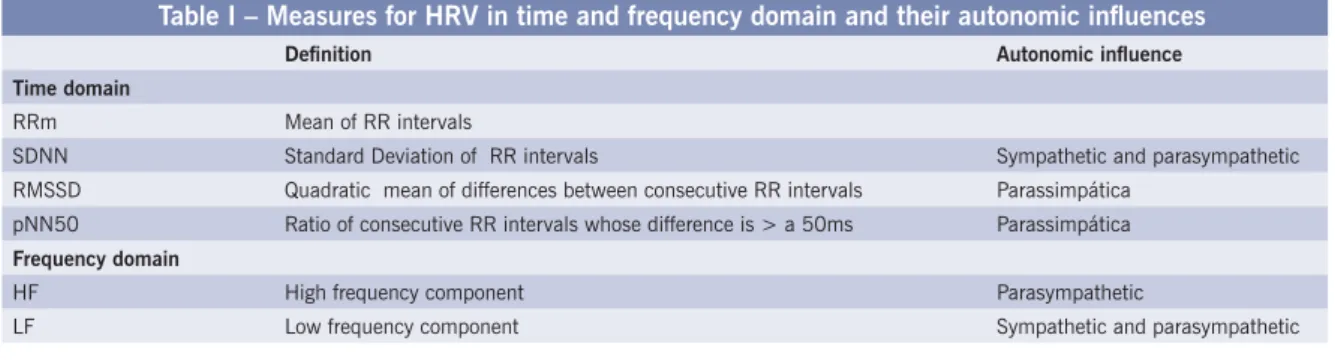
HRV signals were analyzed for time domain and frequency domain at all stages ( Table I). In regard to time domain, variables extracted from normal, consecutive RR intervals series were: a) mean of normal RR intervals (RRm); b) SD of normal RR intervals (SDNN); c) quadratic mean between normal, consecutive RR intervals (RMSSD), and d) ratio of normal, consecutive RR intervals, whose difference is equal to or higher than 50ms as compared to record (pNN50). As for frequency domain, the series of normal, consecutive RR intervals was linearly interpolated for the calculation of power spectral density function (PSDF) through the use of fast Fourier transformation (FFT). Before FFT was calculated, mean of series was subtracted, and series processed by stapling through the use of a Hanning window to avoid artifacts due to the discontinuity of the end points. Spectral estimates have been calculated by elevating the spectrum of amplitude of normal RR intervals series to square values during the time of analysis, in compliance with Predictor IIc protocol. The two spectrum components in the short term series – calculated as area under the curve (AUC) for PSDF - were: a) high frequency (HF), defi ned between 0.15 to 0.40Hz frequencies; and b) low frequency (LF), defi ned between 0.05 - 0.15Hz frequencies8,27.
A normality test was carried out for each variable in each group and at each stage through standard asymmetry and standard curtosis test. Null normality hypothesis was accepteded when the two tests reported values between -2 and +2. Variables where normality hypothesis was rejected were turned into their natural logarithms and reevaluated by normality test.
Intergroups and intrastages comparisons for variables reporting null, accepted normality hypothesis were carried out through the use of Student’s “t” test to compare mean values for samples with equal and unequal variances. For the purpose of comparison of variances, Sinedekor’s “f” test was used, when the null hypothesis for equality among variances was tested. Variables whole null hypothesis
Table I – Measures for HRV in time and frequency domain and their autonomic infl uences
Defi nition Autonomic infl uence
Time domain
RRm Mean of RR intervals
SDNN Standard Deviation of RR intervals Sympathetic and parasympathetic
RMSSD Quadratic mean of differences between consecutive RR intervals Parassimpática
pNN50 Ratio of consecutive RR intervals whose difference is > a 50ms Parassimpática
Frequency domain
HF High frequency component Parasympathetic
for normality was rejected - even after logarithmic transformation – were compared through Mann-Whitney test. For intragroups, intrastage comparison of variables, paired “t” Student test was used with zero difference null hypothesis between the stages. Statistical signifi cance was defi ned by p<0.05 for all tests.
Calculations were carried out using the software
Statgraphics Plus, Version 5.0 (Manugistic Inc, Rockville, MD, EUA).
Study Protocol was submitted to the Ethics Committee at Pedro Ernesto University Hospital at Rio de Janeiro Federal University -Hospital Universitário Pedro Ernesto – UERJ), where it was approved. All individuals presented a written informed consent to be enrolled in the study.
R
ESULTS
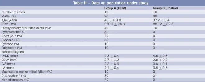
From the twenty (20) individuals enrolled in the study, two (2) – one (1) in each group – did not complete the protocol for autonomic evaluation. Patients’ withdrawal at Stage III – as a result of a dizziness condition - did not allow exam completion. However, those patients were not excluded from the sudy. Their records were incorporated to the analysis for autonomic function in the 3 fi rst stages. Relevant clinical and echocardiographic data are shown in Table II.
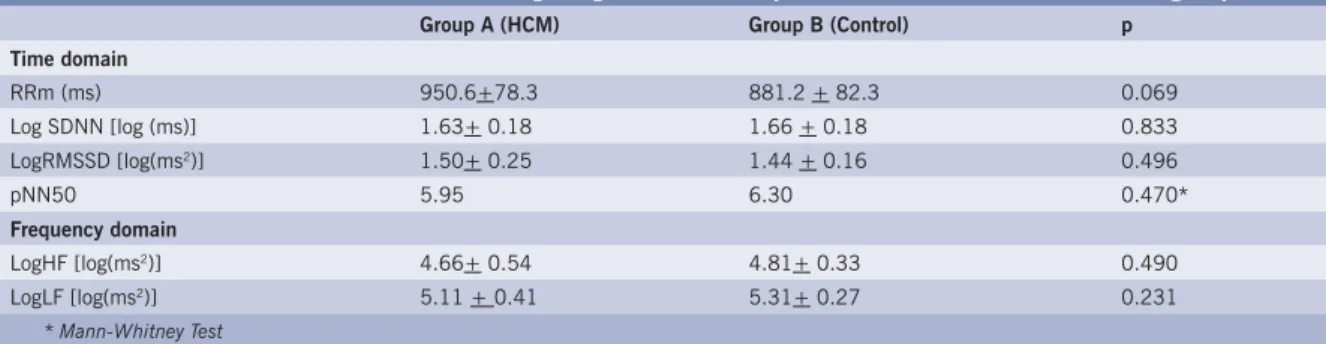
Comparison of intergroup HRV analysis at each stage - Table III shows at rest HRV in the 2 groups. No statistically signifi cant difference was reported for RRm between HCM patients and control group individuals at rest [(mean+ SD) 950.6+78.3 vs 881.2+ 82.3]. No difference was reported, either, for any of the other measures in the time domain and in the frequency domain between the 2 groups.
When the 2 groups were compared for Stages I and II (under control breathing and bend test, respectively), no difference was reported for the measures under study
Table II – Data on population under study
Group A (HCM) Group B (Control)
Number of cases 10 10
Males (%) 90 80
Age (years) 40.3 ± 9.8 37.2 ± 6.4
RRm (ms) 950.6 + 78.3 881.2 + 82.3
Family history of sudden death (%)* 40 10
Symptomatic (%) 80 0
Chest pain (%) 70 0
Dyspnea (%) 60 0
Syncope (%) 10 0
Palpitation (%) 10 0
Echocardiogram
LVDD (mm) 4.3 + 0.4 4.6 + 0.3
SDLV (mm) 2.7 + 1.2 2.8 + 0.2
IVS (mm) 2.2 + 0.6 0.8 + 0.1
LA (mm) 4.1 + 0.4 3.5 + 0.3
Moderate to severe mitral failure (%) 10 0
Obstructive** (%) 30 0
Non-obstructive (%) 70 0
RR m – mean of RR intervals; LVDD- left ventricle diastolic diameter; SDLV- systolic diameter of left ventricle; IVS- interventricular septum thick-ness; LA- left atrium diameter. *Unexpected death of fi rst degree family members under 45 years old28. ** Gradient on left ventricle outfl ow higher than 30mmHg at rest29
– either in the time domain or in the frequency domain (Table IV and V).
However, when analyzing Stage IV (bend test associated to controlled breathing) authors observed that parameters modulated by vagal activity (LogRMSSD and LogHF) reported values signifi cantly higher in the HCM group as compared to control groups, as shown in Table VI.
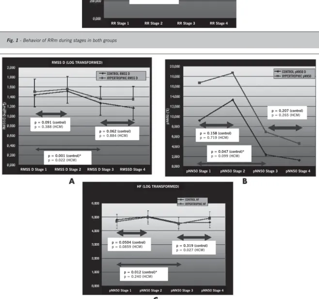
Progressive behavior of autonomic function measures during protocol - while analyzing group behavior in the two groups during stages - both groups reported similar tendency for RRm during stages, showing some increase (although not signifi cant at that point) in mean values during controlled breathing when Stage II was compared to Stage I; signifi cant reduction at tend test (reported when comparing Stage III to Stage I), thus revealing strong infl uence of controlled breathing and of bend maneuvre on vagal and sympathetic activities respectively (fi gure 1).
While evaluating those measures under vagal infl uence (LogRMSSD, pNN50 and LogHF), some differences were reported between groups, since the bend test showed signifi cant decrease in pNN50 and in LogHF in Group B. Group A, in its turn, did not report such signifi cant decrease. RMSSD reported signifi cant, similar decrease in both groups. In the last stage, when controlled breathing was associated to the bend test, signifi cant increase in HCM patients’ LogHF in regard to Stage III, thus reinforcing marked parasympathetic activity in this group of patients (fi gure 2).
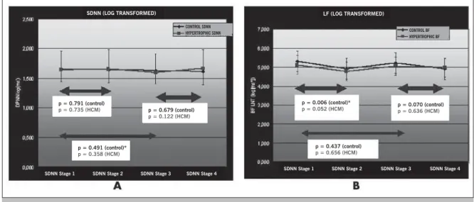
Pa r a m e t e r s u n d e r b o t h s y m p a t h e t i c a n d parasympathetic infl uence (LogDPNN and LogLF) did not report signifi cant changes between stages in either group, except for LogLF in the transition from Stage I to Stage II, when excpected signifi cant decrease was reported by the control group (fi gure 3).
Table III – Measures for HRV during Stage I (at rest) in patients with HCM and in control group
Group A (HCM) Group B (Control) p
Time domain
RRm (ms) 950.6+78.3 881.2 + 82.3 0.069
Log SDNN [log (ms)] 1.63+ 0.18 1.66 + 0.18 0.833
LogRMSSD [log(ms2)] 1.50+ 0.25 1.44 + 0.16 0.496
pNN50 5.95 6.30 0.470*
Frequency domain
LogHF [log(ms2)] 4.66+ 0.54 4.81+ 0.33 0.490
LogLF [log(ms2)] 5.11 + 0.41 5.31+ 0.27 0.231
* Mann-Whitney Test
Table IV – Measures for HRV during Stage II (controlled breathing) in patients with HCM and in control group
Group A (HCM) Group B (Control) p
Time domain
Log SDNN [log(ms)] 1.66 +0.18 1.65+0.10 0.837
LogRMSSD [log(ms2)] 1.55 + 0.24 1.52 + 0.12 0.661
pNN50 9.25 8.80 0.740*
Frequency domain
LogHF [log(ms2)] 4.98 + 0.57 5.04 + 0.35 0.765
LogLF [log(ms2)] 4.78 + 0.45 4.92 + 0.31 0.429
* Mann-Whitney Test
Table V – Measures for HRV during Stage III (bend test) in patients with HCM and in control group
Group A (HCM) Group B (Control) p
Time domain
Log SDNN [log(ms)] 1.60 + 0.16 1.63 + 0.16 0.699
LogRMSSD [log(ms2)] 1.35 + 0.21 1.27 + 0.14 0.302
pNN50 5.70 1.55 0.076*
Frequency domain
LogHF [log(ms2)] 4.51 + 0.46 4.56 + 0.30 0.778
LogLF [log(ms2)] 5.06 + 0.36 5.21 + 0.43 0.410
* Mann-Whitney Test
Table VI – Measures for HRV during Stage IV (bend test associated to controlled breathing) in patients with HCM and in control group
Group A (HCM) Group B (Control) p
Time domain
Log SDNN [log(ms)] 1.66+0.11 1.61 +0.12 0.366
LogRMSSD [log(ms2)]) 1.35+0.14 1.17+ 0.16 0.019
pNN50 4.20 0.20 0.069*
Frequency domain
LogHF [log(ms2)] 4.89+0.22 4.62+0.26 0.032
LogLF [log(ms2)] 4.98+ 0.29 4.91+ 0.45 0.732
* Mann-Whitney Test
in the previous sections in this paper showing the predominance of parasympathetic activity at 4th stage for
HCM patients, the authors evaluated HR (RRm expressed) at rest in Group A for patients on negative chronotropic drugs (betabloquers and calcium antagonists), and those off drugs. No signifi cant difference was found in results obtained (on drugs = 925.8 + 105.6, off drugs = 975.6 + 33.5; p=0.301).
HRV and echocardiographic measures - When comparing within the HCM group those patients reporting
interventricular septum values (IVS) >2 and <2, no difference was found in the analysis of LogHF, LogLF and LogRMSSD at any of the 4 stages.
The same happened when the HCM group was split into 2 subgroups following LA size, and using 4.4cm as the cut point.
D
ISCUSSION
Fig. 1 - Behavior of RRm during stages in both groups
of research papers in the last decade, controversies are still pending. It should be pointed out that literature has not reported – up to this point in time – any experiment pursuing the same objectives in this country.
In the present work authors have observed that: a) the HRV analysis – both in the time domain and in frequency
domain – has brought similar information in regard to autonomic infl uences in patients with HCM and normal individuals while at rest, under controlled breathing, and during bend test; b) measures taken by higher vagal infl uence were higher in patients with HCM at the last stage (where controlled breathing took place with patient
CONTROL RR HYPERTROPHIC RR
p = 0.517 (control)
p = 0.717 (HCM)
p = 0.00029 (control)*
p = 0.00019 (HCM)*
RR Stage 1 RR Stage 2 RR Stage 3 RR Stage 4
Fig. 2 - Behavior of LogRMSSD (A), pNN50 (B) and LogHF (C) during stages in both groups
RMSS D (LOG TRANSFORMED)
CONTROL RMSS D HYPERTROPHIC RMSS D
p = 0.091 (control)
p = 0.388 (HCM)
p = 0.062 (control)
p = 0.884 (HCM)
p = 0.001 (control)*
p = 0.022 (HCM)
RMSS D Stage 1 RMSS D Stage 2 RMSS D Stage 3 RMSSD Stage 4
CONTROL pNN50 D HYPERTROPHIC pNN50
p = 0.158 (control)
p = 0.719 (HCM)
p = 0.047 (control)*
p = 0.099 (HCM)
p = 0.207 (control)
p = 0.265 (HCM)
pNN50 Stage 1 pNN50 Stage 2 pNN50 Stage 3 pNN50 Stage 4
CONTROL HF HYPERTROPHIC HF
p = 0.0504 (control)
p = 0.0859 (HCM) p = 0.319 (control) p = 0.027 (HCM)
p = 0.012 (control)*
p = 0.240 (HCM)
pNN50 Stage 1 pNN50 Stage 2 pNN50 Stage 3 pNN50 Stage 4
bent at 60º); c) along the two stages, measures of vagal activity – such as pNN50 and LogHF – reported less marked increase in the HCM group at bend test; when in transition to the last stage, LogHF showed signifi cant increase, pointing towards a tendency to report higher parasympathetic activity in that group.
Contrarily to what was observed by a number of studies already cited that have analyzed HRV through the use of 24 hours Holter16-19,30-32, no signifi cant compromising of
the autonomic function was observed in HCM patients. In the present paper, the most observed differences were those in the last stage of autonomic evaluation, with parasympathetic activity predominating, while as previously referred to in this paper, that arm of the autonomic response was compromised. However, Fei and collaborators questioned the increase in sympathetic activity reported by patients with HCM in a study published in 1995, when spectral analysis through 24-hour Holter was also used20.
It should also be pointed out that the reliability of measures from the 24-hour recording and corresponding interpretation – particularly spectral components – are questionable, since they may hide information on the autonomic modulation of RR intervals7. The explanation is
simple, since physiologic mechanisms responsible for HR modulation are not stationary in the 24-hour period.
Taking that inco account, the authors have chosen to use a protocol with short-term records, which allowed better control over stimuli in the two groups, which in its turn led to easer interpretation, as done by Limbruno and collaborators in their work21,22. They did not fi nd
any compromising in parasympathetic activity, either; quite the opposite, they confi rmed sympathetic activity reduction at rest and at bend test in obstructive HCM. However, for those patients with non-obstructive HCM – 70% of our patients – Limbruno and collaborators have not found signifi cant difference in HRV spectral analysis
either while comparing with normal individuals. The same was observed by Uemura and collaborators17,22.
Such fi ndings refl ect a contradiction with studies that have addressed the kinetics of catecholamines in HCM. When detecting a reduction of neuronal uptake in nervous terminations, those studies suggest that the disease would be characterized by the increase in sympathetic activity due to the longer presence of norepinephrine in the synaptic gap3-5.
Our hypothesis is that low availability of norepinephrine in sympathetic terminations may induce the opposite, which means to say, to sympathetic activity reduction, particularly in patients with higher level of intraventricular obstruction.
Additionally, the fact that patients with HCM report higher values of LogRMSSD, pNN50 and LogHF at Stage IV (characterized by controlled breathing while at 60º bend) deserves special attention, since none of the research works mentioned earlier has tested that kind of stimulus. If one follows the evolution of those measures across the stages (Illustrations 1, 2 and 3), a tendency towards amortizing decrease for LogRMSSD and pNN50, and the return to higher levels as compared to previous LogHF stage both groups will be observed, thus refl ecting increased parasympathetic activity triggered by controlled breathing, which is opposed to a factor that promotes sympathetic activity – the 60º bend position. However, such response can be seen to be more intense in the HCM groups, which suggests not only the preservation of parasympathetic modulation, but the possibility that exacerbated vagal refl exes may be triggered in response to the stimulus generated by the bend test, as suggested by Gilligan and collaborators in their research work to investigate syncope in HCM33.
In regard to the form of stimulation used in Stage IV in our work, it could be suggested that controlled breathing exacerbated vagal refl exes even more, thus resulting in
Fig. 3 - Behavior of LogSDNN (A) and LogLF (B) during stages in both groups
CONTROL SDNN HYPERTROPHIC SDNN
p = 0.791 (control)
p = 0.735 (HCM) p = 0.679 (control)
p = 0.122 (HCM)
p = 0.491 (control)*
p = 0.358 (HCM)
SDNN Stage 1 SDNN Stage 2 SDNN Stage 3 SDNN Stage 4
SDNN (LOG TRANSFORMED) LF (LOG TRANSFORMED)
CONTROL BF HYPERTROPHIC BF
p = 0.006 (control)*
p = 0.052 (HCM) p = 0.070 (control)p = 0.636 (HCM)
p = 0.437 (control)
p = 0.656 (HCM)
R
EFERENCES
1. Maron BJ. Hypertrophic cardiomyopathy. Lancet. 1997; 350: 127–33.
2. Maron BJ, McKenna WJ, Danielson GK et al. American College of Cardiology/European Society of Cardiology clinical expert consensus document on hypertrophic cardiomyopathy. A report of the American College of Cardiology Foundation Task Force on Clinical Expert Consensus Documents and the European Society of Cardiology Committee for Practice Guidelines. J Am Coll Cardiol. 2003; 42: 1687-713.
3. Goodwin JF. The frontiers of cardiomyopathy. Br Heart J. 1982; 48: 1-18.
4. Brush JE JR, Eisenhofer G, Garty M et al. Cardiac norepinephrine kinetics in hypertrophic cardiomyopathy. Circulation. 1989; 79: 836-44.
5. Schafers M, Dutka D, Rhodes CG et al. Myocardial presynaptic and postsynaptic autonomic dysfunction in hypertrophic cardiomyopathy. Circ Res. 1998; 82: 57-62.
6. Lown B, Verrier RL. Neural activity and ventricular fi brillation. N Engl J Med. 1976; 294: 1165-70.
7. Schwartz PJ, Priori SG. Sympathetic nervous system and cardiac arrhythmias. In: Zipes DP, Jalife J (Eds.) Cardiac Electrophysiology: From Cell to Bedside. Philadelphia, Pa: WB Saunders Co, 1990; 330-43.
8. Task force of the european society of cardiology the north american society of pacing electrophysiology. Heart Rate Variability.Standards of Measurement, Physiological Interpretation, and Clinical Use. Circulation. 1996; 93: 1043-65.
9. Wolf MM, Varigos GA, Hunt D et al. Sinus arrhythmia in acute myocardial infarction. Med J Aust 1978; 2: 52-3.
10. Odemuyiwa O, Malik M, Farrell T et al. Comparison of the predictive characteristics of heart rate variability index and left ventricular ejection
fraction for all-cause mortality, arrhythmic events and sudden death after acute myocardial infarction. Am J Cardiol. 1991; 68: 434-9.
11. Ewing DJ, Campbell IW, Clarke BF. The natural history of diabetic autonomic neuropathy. Q J Med. 1980; 193: 95-108.
12. Smith S. Reduced sinus arrhythmia in diabetic autonomic neuropathy: diagnostic value of an age related normal range. Br Med J 1982; 285: 1599-601.
13. Malpas SC, Maling TJB. Heart rate variability and cardiac autonomic function in diabetes. Diabetes. 1990; 39: 1177-81.
14. Dreifus LS, Agarwal JB, Botvinick EH et al (American College of Cardiology Cardiovascular Technology Assessment Committee). Heart rate variability for risk stratifi cation of life-threatening arrhythmias. J Am Coll Cardiol. 1993; 22: 948-50.
15. Inoue S, Nezuo S, Sawayama T et al. Autonomic function and severity of hypertrophic cardiomyopathy by power spectrum analysis on heart rate variability. Kokyu To Junkan. 1992; 40: 1209-13.(abstract).
16. Ajiki K, Murakawa Y, Yanagisawa-Miwa A et al. Autonomic nervous system activity in idiopathic dilated cardiomyopathy and in hypertrophic cardiomyopathy. Am J Cardiol. 1993; 71: 1316-20.
17. Uemura S, Tomoda Y, Fujimoto S et al. Heart rate variability and ventricular arrhythmia in clinically stable patients with hypertrophic cardiomyopathy. Jpn Circ J. 1997; 61: 819-26.
18. Bonaduce D, Petretta M, Betocchi S et al. Heart rate variability in patients with hypertrophic cardiomyopathy: association with clinical and echocardiographic features. Am Heart J. 1997; 134: 165-72.
19. Doven O, Sayin T, Guldal M et al. Heart rate variability in hypertrophic obstructive cardiomyopathy: association with functional classifi cation and left ventricular outfl ow gradients. Int J Cardiol. 2001; 77: 281-6. a pattern of autonomic response that differed from the
control group.
It should be reminded, at this point, that while analysing RRm in HCM, and comparing those on negative chronotropic drugs and those off those drugs, no difference was found to justify the delayed action of those drugs (even though withheld for a period of 5 half-lives at least) in fi ndings.
From the sample of patients with HCM used for the study, 80% was symptomatic, which does not agree with fi ndings by Bonaduce18 and Counihan30, who observed
higher HRV reduction in those patients. But those works, as already discussed previously, were based on 24-hour electrocardiographic records.
Considering the correlation between HRV and echocardiographic measures such as LA and IVS thickness while analyzing HCM patients, such data were not seen to be independent variables to interfere in autonomic function behavior. Opinions are controversial in that area, too, with some authors in defense of the relevance of such for autonomic modulation22,26 and other
opposing that assumption16. The fact is that even those
who did fi nd the correlation of such data (particularly septal thickness) with autonomic function disorders did not report uniform results. Gillian and collaborators found correlation in parasympathetic activity compromising
while Limbruno and collaborators observed sympathetic activity compromising22,26.
It should also be pointed out that the signifi cant differences found in the variables - that evaluate autonomic function - allow the identifi cation of autonomic modulation patterns typical of HCM. However, due to the small number of samples under study, those conclusions must be validated by larger samples so as to reinforce statistical power for the differences found.
So, we have concluded that: 1) the study showed that autonomic function of patients with HCM evaluated by non-pharmacologic stimulation did not differ from that observed in a control group adjusted for age, gender, and HR at rest, under controlled breathing, and at bend test; 2) signifi cant increase in parasympathetic modulation was observed during controlled breathing when associated to bed test in patients with HCM; 3) no correlation was found between changes in HRV and IVS and LA measures.
20. Fei L, Slade AK, Prasad K et al. Is there increased sympathetic activity in patients with hypertrophic cardiomyopathy? J Am Coll Cardiol. 1995; 26: 472-80.
21. Limbruno U, Strata G, Mengozzi G et al. Spectrum analysis of heart rate variability in obstructive hypertrophic myocardiopathy. Evidence of altered autonomic function. Cardiologia. 1992; 37: 847-52.
22. Limbruno U, Strata G, Zucchi R et al. Altered autonomic cardiac control in hypertrophic cardiomyopathy. Role of outfl ow tract obstruction and myocardial hypertrophy. Eur Heart J. 1998; 19: 146-53.
23. Maron BJ. Hypertrophic Cardiomyopathy: A Systematic Review. JAMA. 2002; 287: 1308-20.
24. Henry WL, Demaria A, Gramiak R et al. Report of the American Society of Echocardiography Committee on nomenclature and standards in two-dimensional echocardiography. Circulation. 1980; 62: 212-5.
25. Schiller NB, Shah PM, Crawford M et al. Recommendations for quantitation of the left ventricle by two-dimensional echocardiography. American Society of Echocardiography Committee on Standards, Subcommittee on Quantitation of Two-Dimensional Echocardiograms. J Am Soc Echocardiogr. 1989; 2: 358-67.
26. Gilligan DM, Chan WL, Sbarouni E et al. Autonomic function in hypertrophic cardiomyopathy. Br Heart J. 1993; 69: 525-9.
27. Barbosa Filho J, Barbosa PRB, Cordovil I. Modulação autonômica do coração na Hipertensão arterial sistêmica. Arq Bras Cardiol. 2002; 78: 181-8.
28. McKenna WJ, Behr ER. Hypertrophic cardiomyopathy: management, risk stratifi cation, and prevention of sudden death. Heart. 2002; 87: 169-76.
29. Fananapazir L, Chang AC, Epstein SE et al. Prognostic determinants in hypertrophic cardiomyopathy. Prospective evaluation of a therapeutic strategy based on clinical, Holter, hemodynamic, and eletrophysiological fi ndings. Circulation. 1992; 86: 730-40.
30. Counihan PJ, Fei L, Bashir Y et al. Assessment of heart rate variability in hypertrophic cardiomyopathy. Association with clinical and prognostic features. Circulation. 1993; 8: 1682-90.
31. Tanabe T. Impaired heart rate variability in patients with symptomatic NYHA class II-III hypertrophic cardiomyopathy. Rinsho Byori. 1998; 46: 1030-6. (abstract).
32. Jimenez AA, Luengo CM, Jimenez AS, et al. Appraisal of the state of the autonomic nervous system in hypertrophic cardiomyopathy by the analysis of heart rate variability. Rev Esp Cardiol. 1998; 51: 286-91.