Late Heart Evaluation of Children with
Rheumatic Mitral Regurgitation Submitted to
Reconstructive Surgery with Implantation of
Gregori’s Ring
Vitor Hugo Soares Machado e Francisco Gregori Júnior
Universidade Estadual de Londrina, Hospital Evangélico de Londrina, Santa Casa de Londrina e Hospital João de Freitas de Arapongas - Londrina, PR - Brazil
Mailing Address: V itor Hugo Soares Machado • Rua Takabumi Murata, 769 – 86055-580 – Londrina, PR - Brazil E-mail: [email protected] Received on 05/10/04 • Accepted on 06/01/05
O
BJECTIVEAssess late cardiological evolution of children with rheumatic mitral regurgitation (RMR) who underwent mitral valve reconstructive surgery with implantation of Gregori’s ring (MVR), from 1987 to 2003.
M
ETHODSA study was conducted to analyze a series of cases of 43 children with RMR who underwent MVR with ages ranged from fi ve to twelve years (mean age 9.7 ± 2.2 years); 25 of the patients were females (58.1%). Patients were evaluated as per the following clinical parameters: functional class of heart failure, heart auscultation, electrocardiogram, chest X-ray and echocardiographic fi ndings.
R
ESULTSForty-three patients underwent preoperative evaluation during the postoperative period, the number of patients evaluated decreased to 31 due to the fact that six patients had died and six others underwent valve replacement surgery. Follow-up was carried out for one hundred percent of the patients. A signifi cant reduction of heart failure functional class was observed. Mitral systolic murmur became lesss intense during the postoperative period. The cardiac area on chest X-ray and the presence of left ventricular overload on electrocardiogram were reduced, as well as the mitral regurgitation level on echocardiographic study. After 188 months, the survival rate was 82%, and the annual mortality rate, 0.38%. Thirty-one (72.6%) patients did not require reoperation and the annual rate of patients who required further surgery was 0.51%.
C
ONCLUSIONMVR is an effective procedure for treating RMR in children, resulting in a signifi cant improvement of functional class, mitral systolic murmur and level of mitral regurgitation, as shown on Doppler echocardiogram.
K
EY WORDSMitral valve reconstructive surgery started to be used in several cardiac surgery centers after the development of the technique by Carpentier et al1, which proved to be superior to valve replacement techniques. In 1987, mitral valve reconstructive surgery with implantation of Gregori’s ring started to be performed.
The mitral valve (left atrioventricular ring) is made up of the mitralannulus, the leafl ets, the chordae tendineae, the papillary muscles and the left ventricle wall underlying the annulus. The main anatomopathological changes affecting the mitral valve are caused by rheumatic disease or myxomatous degeneration. These two diseases may lead to mitral regurgitation, stenosis or double mitral lesion. The dilatation of the mitral valve annulus is the most common cause of mitral regurgitation. The annulus is made up of an anterior portionand a posterior portion. The anterior portion is attached by the right and left fi brous trigones of the heart, whereas the posterior portion is supported by the left ventricular free wall. During the ventricular systole, the annulus contracts like a sphincter taking on akidney-shaped appearance. When the mitral annulus is dilated, the posterior portion pulls away from the anterior portion, increasing the anteroposterior diameter mainly to the right. Leafl ets may be retracted or redundant. In general, leafl et retraction is typical of rheumatic disease sequelae, whereas redundancy usually results from myxomatous degeneration. The posterior leafl et is more susceptible to retraction than the anterior leafl et. Commissural fusion is a common feature when the etiology is rheumatic mitral valve disease. The chordae tendineae may be retracted, elongated or ruptured. The retraction of the primary chordae tendineae may cause them to fuse together or adhere to the papillary muscles. Secondary or tertiary chordae tendineae are also important because, when fi brosed, they may cause localized retractions of the leafl ets.
In the rheumatic disease, papillary muscles are thick and fi brous, while this is not the case with myxomatous degeneration in which the muscles are usually slender.
The left ventricular wall, the papillary muscles, the chordae tendineae, the leafl ets and the annulus all play an important role in the physiology of the left ventricle contraction³-5.
Lesions producedby rheumatic disease can impair
mitral valve function due to the loss of its functional integrity in the following cases: dilatation of the valve annulus, hypoplasia of the posterior leafl et, commissural fusion, chordae shortening or elongation, and rupture of anterior and/or posterior chordae, which consequently leads to mitral regurgitation and/or stenosis.
Gregori’s ring draws the anterior and posterior leafl ets together, more effectively to the right, even correcting the posteromedial commissure widening. This new ring leaves aside the anterior portion, considered useless and even undesirable, especially when it is used in growing children.
This study assessed,by means of clinical and Doppler echocardiography parameters, children with rheumatic
mitral regurgitation who underwent mitral valve reconstructive surgery with implantation of Gregori’s ring during their late clinical evolution.
M
ETHODS
The cases were analyzed by means of a longitudinal study featuring descriptive and analytical components and divided into two phases: the fi rst one is called the preoperative phase, and the second one, the current postoperative phase.
During the preoperative phase, children were submitted to examinations and tests, and were then referred to mitral valve reconstructive surgery with implantation of Gregori’s ring, through the Sistema Único de Saúde – SUS (Unifi ed Health System) in hospitals located in Londrina and Arapongas (state of Paraná). For the postoperative phase, patients were evaluated in private clinics in Londrina.
During the preoperative phase, a total of 43 children, up to twelve years of age with rheumatic mitral regurgitation were submitted to mitral valve reconstructive surgery with implantation of Gregori’s ring in consecutive operations, by the same cardiac surgery service, between October 1987 and July 2002. The mean follow-up was 86.47 +/- 53.27 months, with a minimum of 0 (in-hospital death) and a maximum of 188 months.
For the postoperative phase, a thirty-one patient sample was obtained during 2002 and 2003. Six patients who had to be reoperated for mitral regurgitation correction were excluded, and six other patients died.
Sample size was determined as was convenient and
established according to the number of patients who had undergone surgery at the Serviço de Cirurgia Cardíaca de Londrina (Cardiac Surgery Service in Londrina).
During the preoperative phase, medical charts of the 43 patients were reviewed and the addresses necessary to locate these individuals were obtained. Patients underwent heart auscultation, electrocardiograms, chest X-rays, and Doppler echocardiograms. During the postoperative phase, a total of 31 patients were enrolled in a specifi c protocol and, after the consent form was signed by the person responsible for the child, they were evaluated with clinical examinations, electrocardiograms, chest X-rays and Doppler echocardiograms.
implanted (Fig. 1) in the mitral annulus using U-stitches passed through the outer surface of the annulus. Several reconstructive techniques were employed alone or in combination with others, as shown on Table I.
The Braile-Biomédica (São José do Rio Preto – SP) prosthesis is semicircular and made of stainless steel coated with a thin layer of silicon and Dacron velvet. The anterior portion is open and the lower portion, to the right side, is straightened (Fig. 1). Ring sizes range from 24 to 36, with corresponding ring sizers. The selection of the ideal ring sizeshould be based on the distance between right and left fi brous trigones of the heart. Ring sizes 24, 26, 28, 30, 32, 34, and 36 mm were used.
During the clinical evaluation, patients were analyzed as to the functional class of their heart failure according to the New York Heart Association Criteria Committee10, comparing preoperative and postoperative values.
During the preoperative phase, each patient was auscultated for cardiac murmurs in the hospital by the surgical team who reported on the intensity and type of murmur. During the postoperative phase, patients underwent auscultation performed by the author in the medical offi ce and rated according to an auscultation scale scored from 0 to 4+.
The preoperative Doppler echocardiography transtho-racic study was performed by one single echocardiographist using mechanical transducers ranging from 2.5 to 5.0 MHz; after surgery, it was done by the author using electrical transducers ranging from 2.5 to 3.5 MHz with second harmonic signals. The incidences, measurements and interpretations abided by the recommendations of the American Association of Echocardiography.
Measurements of right and left cavities were expressed in millimeters; the ventricular function, in % (shortening percentage and Teichholz11 method; in the ejection
Table I – Procedures and surgeries associated with mitral annuloplasty by means of Gregori’s ring
Associated procedures* No. %
Chordae shortening as per Gregori’s technique 25 58
Chordae shortening as per Carpentier’s technique 21 49
Chordae transposition 9 21
Chordae shortening as per Fratter’s technique 9 21
Partial rectangular resection of the posterior leafl et 2 4.6
Bilateral Wooler7 technique 1 2.3
Debridement of calcium deposits 1 2.3
Transposition of chordae from the posterior leafl et of the tricuspid valve to the anterior leafl et of the mitral valve 1 2.3
Commissurotomy
Bilateral papillotomy 1 2.3
1 2.3
Associated surgeries No. %
Aortic valve replacement 2 4.6
Aortic valvoplasty 2 4.6
Tricuspid valvuloplasty (De Vega8) 1 2.3
Cox9 surgery 1 2.3
* Isolated or combined procedures
fraction). The mean mitral valve gradient and the pulmonary artery pressure were expressed in millimeters of mercury (mmHg) and the left ventricular mass, in grams (g). The calculation of the mitral valve area was expressed in square centimeters according to the Pressure Half Time (PHT) method. The quantifi cation of the mitral refl ux was obtained by color Doppler mapping, as per Colette12 classifi cation that quantifi es mitral regurgitation based on the extension and magnitude of the regurgitation jet duringleft ventricular systole.
The cardiothoracic index was utilized to estimate heart size on chest X-rays, using a scale of scores from 0 to 4+.
To assess the left ventricle overload on the
electrocardiogram, Sokolow & Lyon13 criteria were used, and for the left atrial overload evaluation, the Morris Jr.14 index was used. Patients were evaluated as to the presence or absence of atrial fi brillation.
n
I II III IV
0 5 10 15 20 25 30 35 40
C. F. NYHA FI n = 43
C.F. NYHA FII n = 31
0
23 (74,2%)
0 03 (9,7%)
03 (7%)
02 (6,4%)
40 (93%)
03 (9,7%)
Results were displayed in tables and fi gures. The Wilcoxon test (paired samples) was used for the comparison between phases with ordinal variables.
The (postoperative) numerical variables with normal
distribution were presented as mean, median, standard deviation, minimum value, maximum value and quartile values. The association between death and gender was calculated using Fisher’s exact test. Survival analysis and the time to valve replacement were performed using the Kaplan & Meier product-limit estimation15. All decisions werebased on a 5% alpha error, and P values * 0.05 were considered signifi cant. All analyses were performed using SPSS 9.1 software for Windows and Statistica 6.0.
The protocol was validated by the Research Ethics Committee of the State University, and each patient was informed before being enrolled in the study.
R
ESULTS
A total of 43 children underwent preoperative evaluations. Eighteen (41.9%) patients were males and 25 (58.1%) were females, with no signifi cant gender difference (p = 0.286). The postoperative evaluation was performed with only 31 participants, as 6 patients died (all females) and 6 (excluded) were submitted to valve replacement (3 females and 3 males). This explains why, at this phase, the study had fi fteen male patients (48.3%) and sixteen female patients (51.7%), with no signifi cant difference between the genders; these patients represented 72% of the cases enrolled at the beginning ofthe study sixteen years before. Patients’ ages during the preoperative phase (n = 43) ranged from fi ve to twelve years (mean age 9.7 ± 2.2 years). During the postoperative phase (n = 31), ages ranged from eleven to twenty-six years (mean age 17.6 ± 4.6).
A signifi cant improvement in the functional class was
observed in the postoperative period as compared to the preoperative period (p < 0,001) (Fig. 2). During the preoperative phase, three patients (7%) were classifi ed as FC III and forty (93%) were classifi ed as FC IV.
In the postoperative period, 23 patients (74.2%) were classifi ed as FC I; three (9.7%) were FC II; two (6.4%) were FC III, and three (9.7%) were classifi ed as FC IV.
A signifi cant decrease (p < 0.001) in the intensity of the mitral systolic murmur (MSM) was seen in the postoperative phase compared to the preoperative phase. During the preoperative period, 16 (37.2%) patients were MSM IV, 19 (44.2%) were MSM III, six (14%) were MSM II and 2 (4.7%) had no report of a murmur. During the postoperative, of the 31 MSM patients evaluated, one (3.2%) was MSM IV, six (19.4%) were MSM III, 12 (38.7%) were MSM II and 12 (38.7%) were MSM I.
There was a signifi cant reduction (p < 0,001) in the cardiac area as shown on the postoperative chest X-ray. In the preoperative phase, three patients (7%) had a normal cardiac area on the X-ray, four (9.3%) had a slight increase, eight (18.6%) had a moderate increase, 25 (58.1%) had a signifi cant increase in the cardiac area, and for 3 patients (7%) no chest X-ray reports were found. Thirty-one patients were evaluated in the postoperative period: 22 (71%) of them had anormal cardiac area on the X-ray, 3 (9.7%) showed a slight increase, fi ve (16.1%) had a moderate increase and 1 (3.2%) had a signifi cant increase in the cardiac area.
A signifi cant reduction (p < 0.001) in left ventricle overload was seen on the electrocardiogram (LVO-ECG). In the preoperative phase, thirty patients (69.8%) had LVO-ECGs exams performed; nine (20.9%) did not have this test and for four patients (9.37%) no electrocardiograms or reports were found. During the postoperative phase, seven patients (22.6%) had LVO-ECGs and 24 (77.4%) did not.
During the preoperative phase, 37 (86%) patients showed sinus cardiac rhythm, 2 (4.7%) had atrial fibrillation and 4 (9.3%) had no ECGs or reports. Postoperatively, 26 patients (83.9%) were in sinus rhythm and 5 (16.1%) were in atrial fi brillation. No signifi cant difference in rhythm was observed between thepre- and postoperative phases (p < 0.083).
Thirty patients (69.8%) had left atrial overload on the electrocardiogram (LAO-ECG) in the preoperative phase, eight (18.6%) did not, and for fi ve patients (11.6%) no electrocardiogram reports were found. During the postoperative period, twenty patients (64.5%) had LAO-ECGs and 11 (35.5%) did not. No signifi cant difference was observed between the pre- and postoperative phases (p < 0.285).
Thirty-one patients with Gregori’s ring underwent Doppler echocardiograms during the postoperative phase. The left ventricular mass had an average of 220.45 ± 94.76 grams, with a minimum value of 80 g and a maximum value of 498 g. As to the left atrium size, minimum and maximum diameters were 29 mm and 84 mm, respectively (mean diameter 49.26 ± 13.20 mm). The left ventricle diastolic diameter measured in millimeters had an average of 51.06 ± 7.07 mm, with value of 40 mm, minimum, and of 69 mm, maximum. The mitral area was estimated using the Pressure Half
Time (PHT) method; minimum area was1.0 cm² and
maximum area was 2.8 cm² (mean area 1.9 ± 0.48 cm²). Echocardiogramsperformed during the preoperative period indicated that two (4.7%) patients had moderate mitral regurgitation, and 41 (95.3%) had signifi cant insuffi ciency (Fig. 3).
During the postoperative phase, fi ve patients (16.1%) showed no mitral regurgitation, fourteen had mild
Ausente Discreto Moderado Importante
0 5 10 15 20 25 30 35 40 45
IM Eco FI n = 43
IM Eco FII n = 31 n
0
14 (45,2 %)
10 (32,3 %)
02 (6,4 %) 05 (16,1 %)
0
41 (95,3%)
02 (4,7%)
Fig. 3 - Distribution of MI (Mitral regurgitation) as shown on echocardiograms in the preoperative (FI) and postoperative (FII) phases (p < 0.001)
insuffi ciency, ten (32.3%) had moderate insuffi ciency, and two (6.4%) had signifi cant mitral regurgitation. A signifi cant decrease in mitral regurgitation was observed on echocardiograms comparing the pre- and postoperative phases (p < 0.001).
The ejection fraction (EF) estimated by the Teichholz7 method indicated a minimum value of 54% and a
maximum valueof 76% (mean: 67.78% ± 6.09%). Peak
pulmonary artery pressures measured in millimeters of mercury (mmHg) ranged from 25 mmHg (minimum) to 78 mmHg (maximum) with a mean value of 41.32 ± 13.03 mmHg. Mitral valve mean gradient measured in mmHg ranged from 3 mmHg (minimum) to 20 mmHg (maximum) with a mean value of 7.19 ± 3.66 mmHg.
Aortic valve maximum gradient measured in mmHg ranged from 4 mmHg (minimum) to 39 mmHg (maximum), with a mean value of 7.94 ± 6.13 mmHg.
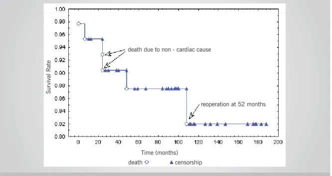
A total of six patients died between 1987 and 2003 (14%), whereas 37 (86%) survived. After 188 months, annual mortality rate was 0.38%.
The median postoperative time to death was 24 months, with corresponding quartiles of nine and sixty-three months (1st and 3rd) and an amplitude between zero and 108 months. Of the six patients who died, one (2.3%) died in-hospital and fi ve (11.6%) died later, two (4.6%) due to non-cardiac causes and three (6.9%) due to cardiac causes. All patients who died were females (6/25), which shows an association between death and gender (p = 0.03 as per Fisher’s exact test). All six patients who died were FC IV.
Figure 4 shows the actuarial curve (Kaplan & Meier15) for the survival analysis.
MI Echo FI
n = 43
MI Echo FII
n = 31
Eight patients (18.6 %) had to undergo reoperation (annual reoperation rate of 0.51%), and 31 patients (72.6%) were free of reoperations at 188 months. The main cause (100%) for reoperation was the recurrence
Fig. 4 - Analysis of postoperative survival. Patient survival probability 188 months after surgery is 82%
of rheumatic activity and incorrect prophylaxis use. In this study, six (14%) patients had to undergo mitral valve replacement (MVR). Patients aged from nine to twelve years underwent the new surgery after 12, 59, 84, 88,
93 and 139 months. The median time for mitralvalve replacement was 86 months. Two patients (33.3%) received metal prostheses and four (66.7%) received bioprostheses. All patients who underwent reoperation had recurrence of rheumatic activity; the reason for valve replacement for two of them (33.3%) was mitral stenosis
and four (66.7%) had mitral regurgitation. All six patients who underwent valve replacement survived.
Figure 5 displays the actuarial curve (Kaplan & Meier15) for patients free of events (valve replacement).
Two children (4.6%) who had initially undergone surgery at the age of fi ve had to be reoperated; both
Fig. 5 – Analysis of time to valve replacement. The probability is that the patient will survive 188 months free of events (valve replacement)
Sur
vival R
ate
death due to non - cardiac cause
reoperation at 52 months
Time (months)
death censorship
Sur
vival R
ate
Time (months)
experienced recurrences of rheumatic activity and had late deaths, six and 52 months after the fi rst surgery.
D
ISCUSSION
Mitral valve reconstructive surgery has increasingly become the method of choice thanks to the concern of surgeons with the preservation of the valvar apparatus, and its importance in maintainingventricular function, which resulted in decreased morbidity and mortality rates. This favorable evolution has alsobeen observed in children with high functional class (FC III and IV). In this study, functional class improved signifi cantly between the pre- and postoperative phases, as shown in Fig. 2, confi rming the results of former studies16-23. Antunes et al20 analyzed patients with rheumatic disease, and of all the patients formerly classifi ed as FC III or IV,
85% became FC I. This improvement observedduring
the postoperative period is due to the preservation of the continuity between the chordae tendineae and the mitral annulus.
The systolic murmur intensity level in the mitral area decreased signifi cantly after surgical correction. These results are similar to those indicated by other studies17 that showed the predominance of a mild murmur during the postoperative phase with an improvement in 93% of the cases, in comparison with the preoperative period. Carvalho et al23 demonstrated that 92.4% of all patients evaluated presented predominantly with just a mild murmur or no murmur at all during the postoperative phase.
The cardiac area on the X-ray and the presence of LVO on ECG showed signifi cant reductions after surgical correction.
A signifi cant decrease was observed in the degree of mitral regurgitation on echocardiogram from the preoperative to the postoperative phase (Fig. 3). The Doppler echocardiographic study was a great advance in assessing not only the indication, but also the results of the surgery. This non-invasive method in most cases replaces the hemodynamic study, especially in children. It is considered a “gold standard” for the evaluation of cardiac valves due to its image technology features such as second harmonic imaging, transesophagic echocardiography, contrast echocardiogram and real-time tridimensional reconstruction. Until the year 2000, all children underwent previous cardiac catheterization during the preoperative phase. From that time on, however, the indication for surgery was made based on the results from the Doppler echocardiogram, as well as on clinical parameters. There are several studies correlating Doppler echocardiographic studies with the results of mitral valvoplasty16,19,22,24-29. According tothe results achieved by Spencer et al24,from a total of 95 patients who were followed, 82 did not have regurgitation, whereas 12 had mild regurgitation. According to Carvalho et al23, 85% of the patients had no regurgitation at all, or a very mild one, with asignifi cant
decrease after treatment. This same incidence was also observed by Gregori Jr. et al22, who used the same ring and the same Carpentier surgical technique.
The presence of a mild or moderate regurgitation is very common during the postoperative phase of mitral valvoplasty, as described by some authors16,19,22-26,29, and the late clinical evolution is always satisfactory. Fix et al28 conducted a pioneer study comparing the late evolution of patients who were classifi ed as having regurgitation intensity of I or II (76 patients). After four years, they observed that mortality, thromboembolism, functional classifi cation, survival (86%) and number of re-hospitalizations were the same. There was, however, a small difference in the rate of reoperations in patients with amild and/or moderate regurgitation (83% x 94%).
The low in-hospital mortality rate (one patient – 2.3%) in this study is within the results reported in medical literature17,18,20,26,30-36. Pomerantzeff et al34 reported a mortality rate of 2.6% in 301 patients who had undergone surgery. Gregori Jr. et al22 noted a 1.9% mortality rate in 105 patients who underwent surgery. In a study conducted with 275 patients, Lessana et al37 registered a 4% mortality rate. In 1983, Carpentier et al32 conducted a study with the largest number of cases worldwide (1,421 patients) and registered a 3.6% in-hospital mortality rate.
Cosgrove et al18 correlated functional class and mortality. FC III patients had a 4.9% rate and those in FC IV, a12.5% rate.
As described in medical literature38, intraoperative mortality is higher in the acute phase of rheumatic fever, and surgery is indicated for patients with severe valve dysfunction for whom clinical treatment is ineffective. But even for these patients, annuloplasty proved to be more effective than valve replacement since it preserves the whole subvalvar apparatus, which should result in better ventricular function after the postoperative period 34.
According to Kumar et al39, the intraoperative mortality rate in children and adolescentsup to 15 years of agewith rheumatic disease undergoing reconstructive surgery was 4.8%. In this study, a total of six patients died (14%), 37 survived (86%), and the annual mortality rate was 0.38%. Five patients (12.6%) had late deaths. One death (2.3%) occurred in-hospital. Eight patients (18.6%) had to be reoperated,with an annual reoperation rate of 0.51%, and 31 patients (72.6%) were free of reoperations during the 188-month follow-up period.
In a study with 551 patients, Carpentier et al31 registered a 7% mortality rate in a ten-year period, with survival rate of 82%.
Reconstructive surgery is technically more diffi cult to perform, with high rates of failure for the group of patients with rheumatic disease (reoperation rate ranges from 4.8% to 27%) 40-43. The rate of reoperations increases inversely with age41,44. Antunes et al20 have also reported that rheumatic disease is the main cause for reoperation. In his experience with 201 patients who had undergone surgery, reoperation incidence was 10.4% during an average period of 9.9 months. The authors of the study also observed that leafl et retraction was the most common cause of valve dysfunction. After 54 months, 78% of patients were free from reoperation. This high rate of success was justifi ed by the fact that the technique preserves the natural valve of the organism (and it is advantageous even though 44.4% of the patients were under 15 years of age).
Compared to valve replacement, mortality and morbidity rates associated with mitral valve reconstructive surgery have decreased, as has been published by other authors18,20,35,45-48. Mortality rate due to isolated mitral valve replacement ranges from 3% to 8% in several medical centers (mean value of 6.4%)49 with 13,936 patients having undergonesurgery due to mitral
regurgitation and/or stenosis. According to the same database (National Cardiac Surgery Database), the mortality rate for isolated reconstructive surgery was 3%, in a total of 4,167 patients.
Adebo & Ross27 observed that, after six years, the survival rate of patients who had undergone annuloplasty was 95%, whereas that of patients who underwent valve replacement
ranged from 66% to 78% with mechanical prostheses,
and 82% to 90% with biological prostheses. Galloway et al45 observed in their practice that 75% of the patients were free of events caused by the conventional procedure, whereas 45% were free of events when submitted to valve replacement, a statistically signifi cantincidence.
There is controversy about the utilization of rings, probably due to the impairment of ventricular function. After analyzing 27 patients who received fl exible and rigid rings, David et al50 confi rmed the good performance
of the fl exible ring. They observed also that both rings caused an important decrease in the left ventricle diastolic diameter (LVDD), whereas the systolic diameter (LVSD) was reduced mainlywith the fl exible ring. After four months, they noted a better LV performance in the series of patients who received the fl exible ring. They alsoreported that the rigid ring hinders fi nal ventricular contraction and recommended its use mostly in cases of myxomatous degeneration. According to these authors, the rigid ring does not alter LV contraction because the patient’s natural ring has been hardened by the disease itself. A better systolic function in the inferobasal portion of the LV was observed in postoperative studies conducted by Duran & Ubago51 in patients with fl exible rings. However, Deloche et al16 showed that there were no differences between the two types of rings, pointing out that the incidence of dehiscence with fl exible and rigid rings was 2.8% and 0.5%, respectively. The fl exible ring interfered less with mitral annulus motion and caused less reduction in the basal contractility of the LV. The rigid ring caused mild aortic subvalvar insuffi ciency due to its compression, reducing the posterior leafl et motion as well, though without any clinical signifi cance. These facts were observed by video-endoscopy in experimental work with porcines52.
The ring used in the surgeries analyzed in the present study is considered arigid, and is fi xed in its posterior portion and open in the anterior one (Fig.1).
C
ONCLUSIONS
Mitral valve reconstructive surgery with implantation of Gregori’s ring is an effective procedure to treat children with rheumatic mitral regurgitation. The procedure signifi cantly improves heart failure functional class, mitral systolic murmur and degree of mitral regurgitation evaluated by Doppler echocardiogram. Moreover, echocardiographic
findings showed that the implantation of an open
prosthesis allowed the development of the mitral annulus during the child’s normal development and growth.
R
EFERENCES
1. Carpentier A, Deloche A, Dauptain J et al. A new reconstructive operation for correction of mitral and tricuspid insuffi ciency. J Thorac Cardiovasc Surg 1971; 61: 1-13.
2. Gregori Jr F. Cirurgia reparadora da valva mitral com novo modelo de anel protético. São Paulo, 1990. Tese (Doutorado em Medicina) - Escola Paulista de Medicina, Universidade Federal de São Paulo.
3. Lillehei CW, Levy MJ, Bonnabeau RC. Mitral valve replacement with preservation of the papillary muscles and the chordae tendineae. J Thorac Cardiovasc Surg 1964; 47: 532-43.
4. David TE, Burns RJ, Bacchus CM, Druck MN. Mitral valve replacement for mitral regurgitation with and without preservation of chordae tendineae. J Thorac Cardiovasc Surg 1984; 88: 718-25.
5. Sarris GE, Cahill PD, Hansen DE, Derby GC, Miller DC. Restoration of left ventricular systolic performance after reattachment of mitral chordae tendineae: the importance of valvular-interaction. J Thorac Cardiovasc Surg 1988; 95: 969-79.
6. Frater RWM, Gabbay S, Shore D, Factor S, Strom J. Reproducible
replacement of elongated or ruptured mitral valve chordae. Ann Thorac Surg1983; 35: 14-19.
7. Wooler GH, Nixon PGE, Grimshaw VA. Experiences with repair of the mitral valve in mitral incompetence. Thorax 1962; 17: 49-57.
8. De Vega NG. La anuloplastia selectiva, regulable y permanente. Una tecnica original para el tratamiento de la insufi ciencia tricuspide. Rev Esp Cardiol 1972; 25: 555-6.
9. Cox JL, Boineau JP, Schuessler RB, Kater KM, Lappasd DG. Five-year experience with the maze procedure for atrial fi brillation. Ann Thorac Surg 1994; 56: 814-24.
10. New York Heart Association Criteria Committee Inc.: Diseases of the Heart and Blood Vessels Nomenclature and Criteria for Diagnosis. 6th ed. Boston: Little, Brown and Co., 1964: 114.
12. Colette V. Pulsed Doppler echocardiographic indices for assessing mitral regurgitation. Br Heart J 1984; 51: 130-8.
13. Sokolow M, Lyon TP. The ventricular complex in left ventricular hypertrophy as obtained by unipolar precordial and limb leads. Am Heart J 1949; 37: 161.
14. Morris Jr JJ, Dunlap WM, Thompson Jr HK, Mc Intosh HD, Estes Jr EH. P wave analysis in the electrocardiographic diagnosis of left ventricular hypertrophy. Circulation 1965; 32 (suppl II): 154.
15. Kaplan EL, Meier, P. Nonparametric estimation from incomplete observations. Journal of the American Statistical Association, 1958; 53: 457-81.
16. Deloche A, Jebara VA, Relland JYM et al. Valve repair with Carpentier techniques: the second decade. J Thorac Cardiovasc Surg 1990; 99: 990-1002.
17. Lessana A, Carbone C, Romano M et al. Mitral valve repair: results and the decision-making process in reconstruction: report of 275 cases. J Thorac Cardiovasc Surg 1990; 99: 622-30.
18. Cosgrove DM, Chavez AM, Lytle BM et al. Results of mitral valve reconstruction Circulation 1986; 74 (3 Pt 2): 182-7.
19. David TE, Armstrong S, Sun Z, Daniel L. Late results of mitral valve repair regurgitation due to degenerative disease. Ann Thorac Surg 1993; 56: 7-14.
20. Antunes MJ, Magalhães MP, Colsen PR et al. Valvoplasty for rheumatic mitral valve disease: a surgical challenge. J Torac Cardiovasc Surg 1987; 94: 44-56.
21. Gregori Jr F, Takeda R, Façanha L et al. Nova técnica reconstrutora na insufi ciência valvar mitral por alongamento das cordas tendíneas da cúspide anterior. Arq Bras Cardiol 1990; 54: 205-9.
22. Gregori Jr F, Silva SS, Hayashi SS, Aquino W, Cordeiro C, Silva LR. Mitral valvuloplasty with a new prosthetic ring: analysis of the fi rst 105 cases. Eur J Cardiothorac Surg 1994; 8: 168-72.
23. Carvalho RG, Giublin PR, Lopes LR, Mulinari L, Loures DRR. Plástica da valva mitral com emprego do anel de Gregori-Braile: análise de 66 pacientes. Rev Bras Cir Cardiovasc 1998; 13(4): 295-316.
24. Spencer FC, Colvin SB, Culiford AT, Isom OW. Experiences with the Carpentier techniques of mitral valve reconstruction in 103 patients (1980-1985). J Thorac Cardiovasc Surg 1985; 90: 341-50.
25. Pomerantzeff PMA, Azevedo JG, Ratti M et al. Plástica da valva mitral em pacientes consecutivos. Como é a evolução tardia?: avaliação clínica e ecocardiográfi ca. Rev Bras Cardiovasc 1991; 6: 63-79.
26. Galler M, Kronzon I, Slater J et al. Long-term follow-up after mitral valve reconstruction: incidence of postoperative left ventricular outfl ow obstruction. Circulation 1986; 74(3 pt 2): 99-103.
27. Adebo OA, Ross JK. Conservative surgery for mitral valve disease: clinical and echocardiographic analysis of results. Thorax 1983; 38: 565-71.
28. Fix J, Isada L, Cosgrove D et al. Do patients with less than “echo-perfect” results from mitral valve repair by intraoperative echocardiography have a different outcome? Circulation 1993; 88 (5 Pt 2): 39-48.
29. Souza Uva M, Grare P, Jebara V et al. Transposition of chordae in mitral valve repair: mid-term results. Circulation 1993; 88 (5 Pt 2): 35-8.
30. Carpentier A, et al. Conservative management of the prolapsed mitral valve. Ann Thorac Surg 1978; 26: 294-302.
31. Carpentier A, Chauvaud S, Fabiani JN et al. Reconstructive of mitral incompetence: ten years appraisal. J Thorac Cardiovasc Surg 1980; 79: 338-48.
32. Carpentier A Cardiac valve surgery: the “French Correction”. J Thorac Cardiovasc Surg 1983; 86: 323-37.
33. Lessana A, Tran Viet T, Ades F et al. Mitral reconstructive operations: a series of 130 cases. J Thorac Cardiovasc Surg 1983; 86: 553-61.
34. Pomerantzeff PMA, Brandão CM, Monterio AC et al. Plástica da valva mitral no InCor HC-FMUSP: resultados tardios de doze anos de evoluções das técnicas. Arq Bras Cardiol 1993; 61 (Supl.II): 175.
35. Orszulak TA, Schaff HV, Danielson GK et al. Mitral regurgitation due to ruptured chordae tendineae: early and late results of valve repair. J Thorac Cardiovasc Surg 1985; 89: 491-8.
36. Scott ML, Stowe CL, Nunnaly LC et al. Mitral valve reconstruction in the elderly population. Ann Thorac Surg 1989; 48: 213-7.
37. Lessana A, Herreman F, Boffety C et al. Hemodynamic and cineangiography study before and after mitral valvuloplasty (Carpentier’s technique). Circulation 1981; 64 (2 Pt 2): 195-202.
38. Pomerantzeff PMA, Snitcowsky R, Trevisan IV, Marcial MB, Verginelli G, Jatene AD. Surgical treatment of acute episodes of rheumatic fever. Cardiol Young 1992; 2: 244-6.
39. Kumar AS, Rao PN, Saxena A. Results of mitral valve reconstruction in children with rheumatic heart disease. Ann Thorac Surg 1995; 60: 1044-7.
40. Chauvaud SM, Deleuze P, Perier PM, Carpentier AF. Failures in reconstructive mitral valve surgery [Abstract]. Circulation 1986; 74 (Suppl 2): 393.
41. Skoularigis J, Sinovich V, Joubert G, Sareli P. Evaluation of long term results of mitral valve repair in 254 young patients with rheumatic mitral regurgitation. Circulation 1994; 90 (Suppl 2): 167-74.
42. Prabhakar G, Kumar N, Gometza B, Galal O, Al-Haless Z, Duran CMG. Triple valve operation in the young rheumatic patient. Ann Thorac Surg 1993; 55: 1492-6.
43. Aharon AS, Laks H, Drinkwater DC et al. Early and late results of mitral valve repair in children. J Thorac Cardiovasc Surg 1994; 107: 1262-71.
44. Duran CMG, Gometza B, De Vol EB. Valve repair in rheumatic mitral valve disease. Circulation 1991; 84 (Suppl 3): 125-32.
45. Galloway AC, Colvin SB, Baumann FG et al. A comparison of mitral valve reconstruction with mitral valve replacement: intermediate – term results. Ann Thorac Surg 1989; 47: 655-62.
46. Angel WW, Oury JII, Shah P. A comparison of replacement and reconstruction in patients with mitral regurgitation. J Thorac Cardiovasc Surg 1987; 93, 665-74.
47. Sand ME, Naftel DC, Blackstone EH, Kirklin JW, Karp RB. A comparison of repair and replacement for mitral valve incompetence. J Thorac Cardiovasc Surg 1987; 94: 208-19.
48. Yacoub M, Halim M, Radley-Smith R et al. Surgical treatment of mitral regurgitation caused by fl oppy valves: repair versus replacement. Circulation 1981; 64 (supl.II): 210-6.
49. Jamieson WRE, Edwards FH, Schwartz M et al. Risk stratifi cation for cardiac valve replacement. National Cardiac Surgery Database. Ann Thorac Surg 1999; 67: 943.
50. David TE, Komeda M, Pollick C, Burns R J. Mitral valve annuloplasty: the effect of the type on left ventricular function. Ann Thorac Surg 1989; 47: 524-8.
51. Duran CG, Ubago JL. Clinical and hemodynamic performance of a totally fl exible prosthetic ring for atrioventricular valve reconstruction. Ann Thorac Surg 1976; 22: 458-6