1 Laboratory of Sports Physical Therapy, Department of Physical Therapy, Universidade Estadual Paulista Júlio de Mesquita Filho (UNESP),
Presidente Prudente, SP, Brazil
2 Stress Physiology Laboratory, Department of Physical Therapy, UNESP, Presidente Prudente, SP, Brazil
Received: 12/19/2012 Revised: 06/13/2013 Accepted: 09/27/2013
The influence of resistance exercise with emphasis on
specific contractions (concentric
vs.
eccentric) on muscle
strength and post-exercise autonomic modulation:
a randomized clinical trial
Mariana O. Gois1, Fernanda A. S. Campoy1, Thâmara Alves1, Roseana P. Ávila1, Luiz C. M. Vanderlei2, Carlos M. Pastre1
ABSTRACT | Background: Compared to eccentric contractions, concentric contractions result in higher cardiovascular stress. However, we do not know how these two types of contractions inluence cardiac autonomic modulation during the post-exercise recovery period. Objective: to compare the effect of resistance training that is performed with concentric vs. eccentric emphasis on muscle strength and on post-exercise recovery which was assessed by examining heart rate variability (HRV), for the knee extensor muscle group in young healthy adults. Method: For this study, 105 men between 18 and 30 years of age were randomized into 4 groups: concentric control (CONCC), eccentric control (ECCC), concentric training (CONCT) and eccentric training (ECCT). The CONCC and ECCC groups underwent one session of resistance exercise (RE) using the knee extensor muscle group (3 sets of 1 repetition at 100% of the maximal repetition [1MR]) and the CONCT and ECCT groups performed 10 training sessions. The HRV was analyzed at baseline and across four recovery periods (T1, T2, T3 and T4). Results: The ECCT group exhibited increased muscle strength at the end of the study. Regarding cardiac autonomic modulation, the CONCC and ECCC groups exhibited increases in overall variability (SDNN and SD2) at T1 compared to baseline, and the ECCT group demonstrated increases in variables relecting vagal modulation and the recovery process (RMSSD, SD1 and HF [ms2]) at T1, T2 and T4 compared to baseline. Conclusions:
Resistance training with emphasis on eccentric contractions promoted strength gain and an increase in cardiac vagal modulation during recovery compared to baseline.
Keywords: cardiovascular recovery; resistance training; autonomic nervous system; muscle strength; physical therapy. Clinical trial number: RBR-75scwh (recorded in Registro Brasileiro de Ensaios Clínicos – REBEC).
HOW TO CITE THIS ARTICLE
Gois MO, Campoy FAS, Alves T, Ávila RP, Vanderlei LCM, Pastre CM. The inluence of resistance exercise with emphasis on speciic contractions (concentric vs. eccentric) on muscle strength and post-exercise autonomic modulation: a randomized clinical trial. Braz J Phys Ther. 2014 Jan-Feb; 18(1):30-37.http://dx.doi.org/10.1590/S1413-35552012005000141
Introduction
Exercises with an emphasis on concentric or eccentric contractions have been studied in relation to outcomes such as increased strength, cardiovascular adaptation and the induction of tissue damage1-4.
Currently, most studies show that eccentric resistance exercise (RE) is more advantageous than concentric RE when considering the consumption of ATP, lactate concentrations and muscle strength output1. On the other hand, concentric RE results in higher heart rate (HR) and blood pressure (BP) values due to increased activation of the motor units, resulting in greater cardiovascular stress when compared to eccentric contractions, which produce
lower motor activation for the equivalent muscle strength output due to the stimulation of the elastic components present in the muscle-tendon units5-8.
This evidence suggests that concentric and eccentric muscle actions influence clinical and functional responses, particularly the behavior of the cardiac autonomic nervous system (ANS). Given
this inluence, outcomes related to post-exercise
process, there is a predominance of sympathetic and a reduction of parasympathetic autonomic modulations that may be associated with increased cardiovascular risk9-12.
Despite the importance of the post-exercise recovery process, our literature review did not identify any studies that evaluated cardiac autonomic modulation after exercise with emphasis on concentric and eccentric contractions. Therefore, observing the relationship between muscle strength gain and the cost of post-exercise recovery seems pertinent. Thus, the objective of this study was to compare the effect of resistance training that is performed with concentric vs. eccentric emphasis on muscle strength and post-exercise recovery, assessed by examining cardiac autonomic modulation, for the knee extensor muscle group in young healthy adults.
Method
Study population and eligibility criteria For the purposes of this study, data from 105 healthy men between 18 and 30 years of age were analyzed
and they were classiied as physically active using
the International Physical Activity Questionnaire (IPAQ)13. The objectives and procedures of the study were discussed with all of the participants, and individuals signed a consent form after agreeing to participate. All procedures were approved by the Research Ethics Committee of Universidade Estadual Paulista Júlio de Mesquita Filho (UNESP), Presidente Prudente Campus, under protocol number 20/2010.
The inclusion criteria were the absence of known cardiovascular, respiratory and/or metabolic diseases, non-alcoholic, non-smoking, no musculotendinous or osteoarticular injury episodes of the lower limbs and/ or spine in the last year and not having participated in a weight-training program for at least six months.
The participants were instructed to not consume any food and/or drinks containing caffeine for 24 hours before each session and to not dramatically alter their daily activities but, if they did so, to inform the researchers. A week before the start of the exercise protocols, testing and familiarization were carried out on the equipment and with the exercises to be performed in each group. Data collection took place in a room with controlled temperature (21 to 23 °C) and humidity (40 to 60%), and the records were collected between 18 and 21 hours.
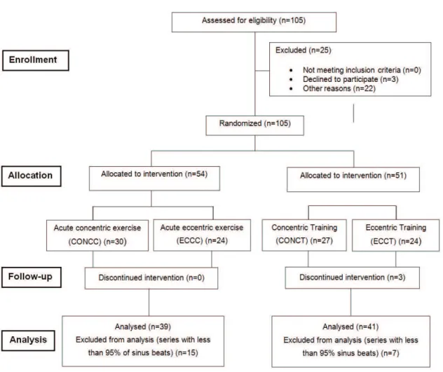
Allocation and groups
The participants were randomized into four groups
according to the lowchart below (Figure 1). The two
control groups underwent only one session of RE. The concentric control group (CONCC) performed RE with a predominance of the concentric phase, and the eccentric control group (ECCC) performed RE with a predominance of the eccentric phase. The two training groups performed RE training, with the concentric training group (CONCT) performing training with a predominance of the concentric phase and the eccentric training group (ECCT) with a predominance of the eccentric phase. For all groups, the knee extensor muscle group was used to perform the exercises.
1MR Test
Before beginning the protocol, one week was allocated to performing the 1MR test to measure the maximum repetition weight for the participant. The test was performed on a weight machine (Ipiranga equipment, Academia Hard line), and the maximum load, in kilograms, that each individual could achieve during knee extension was determined to ascertain training loads and those for the single RE session. The test began by using a weight equal to 50% of the body mass of the individual, and increments of 30% of this value were applied; the perceived strength of the individual was also considered. The test was completed when the volunteer reached his maximum load when executing the knee extension movement without mechanical failure. No more than
ive attempts to establish the maximum load were
permitted. If this occurred, the test was considered invalid, and the volunteer was given the test on another day14.
If attempts at increments of 30% were higher than the volunteer could achieve, the increment was reduced to intermediate values (intervals of 5 kg). If the subject reached values close to his maximum capacity and responded favorably to the slightly increased load, the load was increased using these intermediate values.
secured using Velcro straps on the trunk and thighs to decrease compensatory movements.
Muscle strength
Muscle strength was evaluated using the 1MR test, as described above. The evaluation of muscle strength was performed before and after the end (96 hours) of both exercise protocols.
Protocols
The CONCC and ECCC groups underwent only one RE session, which corresponded to three sets of one repetition at 100% of 1MR. The CONCT and ECCT groups underwent ten sessions of RE training, with gradual incremental adjustments.
The prescription dynamic was based on the classical form of increasing loads, respecting the volume by intensity interdependence proposed by Chiesa16 and adapted for evolution over ten sessions,
performed thrice weekly, with an interval of 48 hours in between them.
Sessions 1 and 2 comprised three sets of eight repetitions at 80% of 1MR, with 3 minutes of rest between each set; sessions 3 and 4 comprised three sets of six repetitions at 85% of 1MR, with 2 minutes of rest between each set; sessions 5 and 6 comprised three sets of four repetitions at 90% of 1MR, with 1 minute and 30 seconds of rest; sessions 7 and 8 comprised three sets of two repetitions at 95% of 1MR, with 40-second rest intervals, and sessions 9 and 10 comprised three sets of one repetition at 100% of 1MR, with 20-second rest intervals.
Description of the exercise
Each group was asked to perform a contraction of the quadriceps muscle at different contraction speeds so that one phase, concentric or eccentric, would predominate. For the CONCC and CONCT groups,
the participants performed knee extensions beginning
at 90° of lexion with the non-dominant lower limb.
The muscle contraction was to occur over 3 seconds, until the full range of motion (ROM) was complete. The return of the limb to its original position occurred over 1 second.
The individuals in the ECCC and ECCT groups performed the knee extensions beginning at 90° of
lexion over 1 second, and the eccentric contraction during lexion occurred over 3 seconds.
Heart rate variability
To capture the RR intervals (iRR), a Polar brand heart rate monitor (model S810i, Polar Electro, Finland) was used. This equipment has been previously validated17. Before the single session of
control groups and inal session, 10th, of training groups, participants remained at rest, breathing spontaneously and in a supine position for 20 minutes, to collect the baseline HRV. After the baseline HRV measurements, participants performed the exercise session and continued to be monitored during post-exercise recovery while breathing spontaneously and in a supine position for 45 minutes.
The data obtained were initially subjected to digital
iltering performed using the software from the Polar
Precision Performance, version 3.0 device. Only
series with more than 95% of the sinus beats were
included in the study. Subsequently, manual iltering
was conducted, characterized by visual inspection of the iRRs and exclusion of abnormal intervals, and series with 256 beats were analyzed by Kubios HRV software18. Five windows were selected for analysis:
baseline (pre-exercise), T1, T2, T3 and T4. The irst
recovery window (T1) was started 2 minutes after
stopping the exercise. To form this irst window, 256
beats were counted. The T2 recovery window began soon after the end of T1, again selecting 256 beats. The same procedure was repeated with windows T3 and T4, which began after the end of the 256 beats of windows T2 and T3, respectively.
Linear methods
The indices analyzed in the time domain were the SDNN, which is the standard deviation of all normal iRR, and the RMSSD, which was calculated by taking the square root of the sum of the squared differences between the iRR on record, divided by the number of iRR at a particular time minus one iRR. Both indices were expressed in milliseconds19.
In the frequency domain, the low frequency (LF [0.04 to 0.15 Hz]) and high frequency (HF [0.15 to 0.4 Hz]) spectral components were analyzed and expressed in ms2 and normalized units (nu). Spectral analysis was performed using Fourier’s fast transform algorithm19.
Nonlinear methods
For the nonlinear methods, the standard deviation of the instantaneous beat-to-beat variability (SD1) and the standard deviation of the long-term continuous iRR (SD2) were analyzed using a Poincaré Plot19.
Data analysis
A Shapiro-Wilk test was used to determine the normality of the data distributions. The strength measurements, anthropometric variables and ages were compared between groups using a Student’s t
testfor parametric data and a Mann-Whitney test for nonparametric data.
For comparisons between contractions (concentric
vs. eccentric), groups (control vs. training) and time (baseline vs. T1, T2, T3 and T4), repeated measures analysis of variance (ANOVA) was used. Parametric analyses were used for the SDNN, RMSSD, SD1, SD2, LF (nu), and HF (nu) variables, and nonparametric analyses were used for the LF (ms2) and HF (ms2) variables, in a two factor scheme, complemented with the SNK (
Student-Newman-Keuls) and Dunn tests, respectively.
A power analysis was performed to calculate the sample size based on a pilot study. For this analysis, the primary variable (muscle strength) was expected to have a gain of 6 kg and a standard deviation of 7. The power analysis thus indicates the need for 21 participants per group to achieve 80% power. All
tests were performed with a signiicance level of 5%.
Results
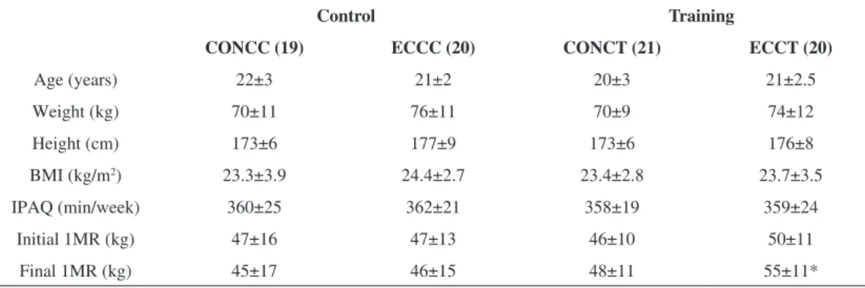
Table 1 presents the participants’ anthropometric variables, age and strength measurements. The 1MR test revealed an increase in muscle strength at the
inal timepoint in relation to the baseline for the
ECCT group.
Tables 2, 3 and 4 show the behavior of the HRV indices during the recovery period in all groups.
No signiicant differences were observed between
compared to baseline, showing better recovery of cardiac autonomic modulation in this group.
Discussion
The results of this study suggest a greater strength gain and better post-exercise recovery for the group that underwent training with emphasis on eccentric contraction. Regarding strength gain, there was
signiicant difference for the ECCT group when
comparing the starting (50±11) and end (55±11)
times. Note that the indings of this study relating to strength gain corroborate those in the scientiic
literature. Despite the short training period (ten sessions), it is noteworthy that the gain in the training group was approximately 5%. Hortobágyi et al.20 observed that, after 12 weeks of eccentric and concentric isokinetic training, eccentric strength increased. According to the authors, this increase in
strength is related to hypertrophy of type II muscle
ibers and greater neural activation.
Aagaard et al.21 also identiied increased quadriceps muscle strength and increased neuromuscular activation after 14 weeks of eccentric training compared to concentric. This type of stimulation appears to promote increased neural drive and greater
eficiency in iber recruitment patterns. The increases
in strength gains after eccentric training compared to concentric training seem to be well described in the literature. However, the speed of contraction may
have also contributed to this inding because greater
strength development can be observed at lower contraction velocities, according to Corvino et al.22.
Importantly, despite the fact that the studies cited had longer training times than those used in the present study, the ten sessions of resistance training
were suficient to show increases in muscle strength
for the exercise group that emphasized eccentric contractions. Thus, this working model is proposed as
Table 1. Participant characteristics.
Control Training
CONCC (19) ECCC (20) CONCT (21) ECCT (20)
Age (years) 22±3 21±2 20±3 21±2.5
Weight (kg) 70±11 76±11 70±9 74±12
Height (cm) 173±6 177±9 173±6 176±8
BMI (kg/m2) 23.3±3.9 24.4±2.7 23.4±2.8 23.7±3.5
IPAQ (min/week) 360±25 362±21 358±19 359±24
Initial 1MR (kg) 47±16 47±13 46±10 50±11
Final 1MR (kg) 45±17 46±15 48±11 55±11*
CONCC: concentric control; ECCC: eccentric control; CONCT: concentric training; ECCT: eccentric training. 1MR: one maximum repetition; BMI: body mass index; min: minutes. *: signiicantly different than the initial 1MR of the ECCT group.
Table 2. Mean and standard deviation values of the SDNN and RMSSD by group and time of analysis.
Baseline T1 T2 T3 T4
SDNN (ms)
CONCC 48±14 87±23* ♦ 53±15 58±18 62±15*
ECCC 49±15 87±26* # 53±17 59±16 60±19
CONCT 56±18 58±19 61±22 56±16 57±15
ECCT 48±14 54±12 61±10* 59±18* 61±14*
RMSSD (ms)
CONCC 40±17 47±18 46±16 48±18 46±16
ECCC 45±20 50±21 53±25 51±24 50±25
CONCT 43±17 51±24* 47±20 44±20 43±18
ECCT 38±15 47±16* 44±16* 43±15 45±13*
a useful method to achieve early strength gains with a lower amount of work, which may be of interest in clinical practice, particularly when designing rehabilitation programs.
When post-exercise recovery processes are the object of study, the behavior of the parasympathetic modulation should be considered. In this regard, for
the analyzed indices that relect vagal modulation,
no difference was observed between the groups (control vs. training). However, when analyzing
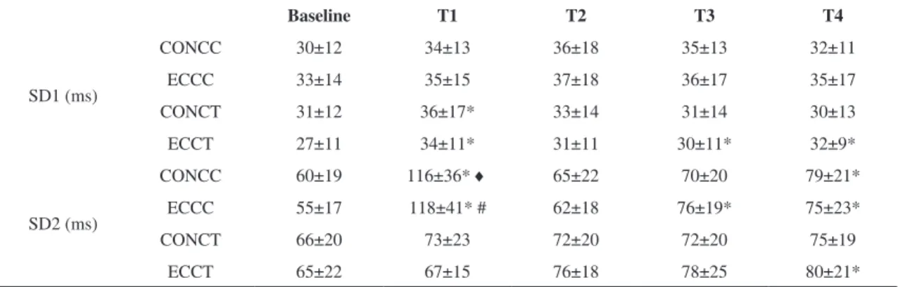
recovery, the control group demonstrated no clear recovery response, while this response was evident in the training groups. The prevalence of a recovery response was highest in the group given eccentric training. Higher RMSSD, SD1 and HF (ms2) values were observed for the ECCT group recovery times compared to baseline.
It is difficult to compare these findings with
the literature because the present study is the irst
to evaluate cardiac autonomic modulation during
Table 3. Mean and standard deviation values of SD1 and SD2 by group and time of analysis.
Baseline T1 T2 T3 T4
SD1 (ms)
CONCC 30±12 34±13 36±18 35±13 32±11
ECCC 33±14 35±15 37±18 36±17 35±17
CONCT 31±12 36±17* 33±14 31±14 30±13
ECCT 27±11 34±11* 31±11 30±11* 32±9*
SD2 (ms)
CONCC 60±19 116±36* ♦ 65±22 70±20 79±21*
ECCC 55±17 118±41* # 62±18 76±19* 75±23*
CONCT 66±20 73±23 72±20 72±20 75±19
ECCT 65±22 67±15 76±18 78±25 80±21*
CONCC: concentric control; ECCC: eccentric control; CONCT: concentric training; ECCT: eccentric training. ♦ p<0.05: versus CONCT; # p<0.05: versus ECCT.
Table 4. Median, minimum, maximum, mean and standard deviation of the spectral components of HF and LF expressed in normalized units and ms2 by group and time of analysis.
Baseline T1 T2 T3 T4
HF (ms2)
CONCC 527 (130;2233) 758 (151;2621) 689 (175;2076) 871 (167;1950) 683(245;2048)
ECCC 617 (38;5011) 691 (150;2951) 787 (1050;2853) 666 (1230;3300) 626 (910;4683)
CONCT 482 (140;2681) 736 (110;3049) 613 (96;2175) 525 (84;2258) 599 (144;2468)
ECCT 425 (50;1514) 685 (1360;2771)* 685 (125;1577)* 549 (138;1906)* 807 (105;2299)*
LF (ms2)
CONCC 433 (209;2550) 908 (180;4747) 760 (183;1963) 756 (197;2117) 881 (4010;4348)
ECCC 813 (60;3546) 824 (1560;2494) 511 (196;2936) 969 (2050;5388) 1061 (109;3181)
CONCT 756 (18;2252) 816 (1080;2373) 1035 (120;4221) 789 (217;4423) 920 (110;5625)
ECCT 577 (2360;1464) 762 (258;2038) 913 (238;3384) 901 (1960;2431) 1052 (1950;3068)
LF (nu)
CONCC 53±14 51±8 52±13 55±15 59±16
ECCC 56±18 53±11 49±14* 56±14 62±14
CONCT 59±14 56±14 62±12 62±15 62±17
ECCT 54±14 51±12 56±14 60±12* 57±11
HF (nu)
CONCC 46±14 47±11 47±14 47±13 40±16
ECCC 44±19 47±11 50±15 45±14 39±17
CONCT 39±11 45±15 42±13 38±14 38±17
ECCT 42±11 47±14 43±11 41±15 44±18
the recovery period after concentric and eccentric actions. The literature review only revealed studies that examine the effect of training on the behavior of HRV, i.e., the adaptation process2,3,and not its behavior as a marker of recovery. This was particularly true for studies that considered types of exercises with different dynamics.
The indices reflecting overall variability19,23 (SDNN and SD2) show an increase in the first window of recovery (T1) for the control groups. By observing the indices as a whole, it seems that such behavior is due to an interaction of sympathetic and parasympathetic components, both of which were increased at T1 for the CONCC and ECCC groups. Some authors9-11 have observed a predominance of sympathetic modulation when analyzing HRV indices in the frequency domain after acute RE stimulation. Heffernan et al.9 observed a decrease in the HF index (nu) and an increase in LF (nu) after RE and endurance exercises, but greater reductions in total power were observed only after RE. Rezk et al.10 and Andrade Lima et al.11 also observed a predominance of sympathetic modulation and a reduced parasympathetic modulation after global upper limb (MMSS) and trunk RE, respectively, performed at different intensities.
It should be noted that the aforementioned studies do not evaluate the two types of contractions in isolation, which makes further comparison impossible. They also differ from the present study in relation to the number of groups of muscles involved, exercise intensity and volume of work, factors that
directly inluence the behavior of cardiac autonomic
modulation after exercise.
In summary, when observing cardiac autonomic behavior during the post-exercise recovery process, groups that underwent only one exercise session showed increased overall variability in the first window of recovery, and the groups that underwent training, especially the ECCT group, showed better values on indices that reflect vagal modulation (RMSSD, SD1 and HF) in the recovery windows when compared to baseline. It is therefore suggested that eccentric training leads to positive adaptation in cardiac vagal control and offers better recovery.
One limitation of this study that should be noted is the posture of the participants when the heartbeats were recorded. At both the baseline and recovery times, participants were in a supine position when the heartbeats were recorded, which did not permit the analysis of immediate cardiac autonomic modulation
after exercise to evaluate vagal reentry because the exercise was performed in a seated position. The
indings of this study suggest that the analysis of
post-exercise recovery periods based on HRV indices
should be considered, especially those that relect
parasympathetic activity. Although there are no differences between contraction types, an exploration of different stresses or pathological conditions may
provide interesting indings and therefore reveal risks
or safety issues for conducting workouts of any type.
Conclusion
In conclusion, the indings of this study suggest
that resistance training performed with an emphasis on eccentric contractions promoted strength gains and increases in cardiac vagal modulation during the recovery process compared to the baseline condition and that there are no differences in HRV indices between contraction types across the times analyzed.
Acknowledgements
The authors would like to thank the Foundation for Research Support of the State of São Paulo (Fundação de Amparo à Pesquisa do Estado de São Paulo - FAPESP: 2010/09687-0), National Council for Scientific and Technological Development
(Conselho Nacional de Desenvolvimento Cientíico
e Tecnológico – CNPq: 476109/2010-8) for their
inancial support.
References
1. Roig M, Shadgan B, Reid WD. Eccentric exercise in patients with chronic health conditions: a systematic review. Physiother Can. 2008;60:146-160. http://dx.doi. org/10.3138/physio.60.2.146
2. Cooke WH, Carter JR. Strength training does not
affect vagal-cardiac control or cardiovagal barorelex
sensitivity in young healthy subjects. Eur J Appl Physiol. 2003;93:719-25. http://dx.doi.org/10.1007/ s00421-004-1243-x
3. Melo RC, Quitério RJ, Takahashi ACM, Silva E, Martins LEB, Catai AM. High eccentric strength training reduces heart rate variability in healthy older men. Br J Sports Med. 2008;42:59-63. http://dx.doi.org/10.1136/ bjsm.2007.035246
4. Seger JY, Thorstensson A. Effects of eccentric versus concentric training on thigh muscle strength and EMG. Int J Sports Med. 2005;26:45-52. http://dx.doi. org/10.1055/s-2004-817892
and older. J Gerontol A Biol Sci Med Sci. 2000;55(4):177-82. http://dx.doi.org/10.1093/gerona/55.4.B177
6. Thompson E, Versteegh TH, Overend TJ, Birmingham TB, Vandervoort AA. Cardiovascular responses to submaximal concentric and eccentric isokinetic exercise in older men. J Aging Physl Act. 1999;7:20-31.
7. Okamoto TB, Masuhara M, Ikuta K. Cardiovascular responses induced during high-intensity eccentric and concentric isokinetic muscle contraction in healthy young adults. Clin Physiol Funct Imaging. 2006;26:39-44. http:// dx.doi.org/10.1111/j.1475-097X.2005.00651.x
8. Vallejo AF, Schroeder ET, Zheng L, Jensky NE, Sattler FR. Cardiopulmonary responses to eccentric and concentric resistance exercise in older adults. Age Ageing. 2006;35:291-7. http://dx.doi.org/10.1093/ageing/afj082 9. Heffernan KS, Kelly EE, Coliier SR, Fernhall B. Cardiac
autonomic modulation during recovery from acute endurance and resistance exercise. Eur J Cardiovasc Prev Rehabil. 2006;13(1):80-6. PMid:16449868.
10. Rezk CC, Marrache RCB, Tinucci T. Post-resistance exercise hypotension, hemodynamics, and heart rate
variability: inluence of exercise intensity. Eur J Appl
Physiol. 2006; 98:105-12. http://dx.doi.org/10.1007/ s00421-006-0257-y
11. Andrade Lima AHR, Forjaz CLM, Silva GQM, Menêses AL, Rofrigues Silva AJM, Ritti-Dias RM. Efeito agudo da intensidade do exercício de força na modulação autonômica cardíaca pós-exercício. Arq Bras Cardiol. 2011;96(6):498-503. http://dx.doi.org/10.1590/ S0066-782X2011005000043
12. Heffernan KS, Sosnoff JJ, Jae SY, Gates GJ, Fernhall B. Acute resistance exercise reduces heart rate complexity and increases QTc interval. Int J Sports Med. 2008;29(4):289-93. http://dx.doi.org/10.1055/s-2007-965363
13. Pardini R, Matsudo S, Araújo T, Matsudo V, Andrade E, Braggion G, et al. Validação do questionário internacional de nível de atividade física (IPAQ)- versão 6: estudo piloto em adultos jovens brasileiros. Rev Bras Ciên Mov. 2001;9(3):45-51.
14. Brown LE, Weir JP. Asep procedures recommendation I: accurate assessment of muscular strength and power. J Exerc Physiol Online. 2001;4(3):1-21.
15. Gleeson N, Eston R, Marginson V. Effects of prior concentric training on eccentric exercise induced muscle damage. Br J Sports Med. 2003;37:119-25. http://dx.doi. org/10.1136/bjsm.37.2.119
16. Chiesa LC. La Musculación Racional: Bases para um entrenamiento organizado. Barcelona: Editorial Paidotribo; 2007.
17. Vanderlei LCM, Silva RA, Pastre CM, Azevedo FM, Godoy FM. Comparison of the Polar S810i monitor and the ECG for the analysis of heart rate variability in the time and frequency domains. Braz J Med Biol Res. 2008;41(10):854-9. http://dx.doi.org/10.1590/ S0100-879X2008005000039
18. Niskanen JP, Tarvainen MP, Ranta-Aho PO, Karjalainen PA. Software for advanced HRV analysis. Comput Methods Programs Biomed. 2004;76(1):73-8. http:// dx.doi.org/10.1016/j.cmpb.2004.03.004
19. Vanderlei LCM, Pastre CM, Hoshi RA, Carvalho TD, Godoy MF. Noções básicas de variabilidade de frequência cardíaca e sua aplicabilidade clínica. Rev Bras Cir Cardiovasc. 2009;24(2):205-17. http://dx.doi.org/10.1590/ S0102-76382009000200018
20. Hortobágyi T, Hill JP, Houmard JA, Fraser DD, Lambert NJ, Israel RG. Adaptative responses to muscle lengthening and shortening in humans. J Appl Physiol. 1996;80(3):765-72. PMid:8964735.
21. Aagaard P, Simonsen EB, Andersen JL, Magnusson SP, Halkjaer-Kristensen J, Dyhre-Poulsen P. Neural inhibition during maximal eccentric and concentric quadriceps contraction: effects of resistance training. J Appl Physiol. 2000;89:2249-57. PMid:11090575. http:// dx.doi.org/10.1097/00005768-199805001-01178 22. Corvino RB, Caputo F, Oliveira AC, Greco CC, Denadai
BS. Taxa de desenvolvimento de força em diferentes velocidades de contrações musculares. Rev Bras Med Esporte. 2009;15(6):428-31. http://dx.doi.org/10.1590/ S1517-86922009000700005
23. Task Force of the European Society of Cardiology of the North American Society of Pacing Electrophysiology. Heart rate variability standards of measurement, physiological interpretation, and clinical use. Circulation. 1996;93(5):1043-65. http://dx.doi.org/10.1161/01. CIR.93.5.1043
Correspondence Carlos Marcelo Pastre Univ Estadual Paulista (UNESP) Departamento de Fisioterapia