Minimally Invasive Coronary Angiography Using a
Multidetector CT
André Ricardo Vale Rodrigues, Maurício Rezende Barbosa, Marcelo Sá Vieira de Brito,
Luciana Costa Silva, Fernando Santana Machado
ECOAR Medicina Diagnóstica - Belo Horizonte, MG - Brazil
Mailing Address: André Ricardo Vale Rodrigues • Rua Buenos Aires, 411/1001 – 30315-110 - Belo Horizonte, MG - Brazil E-mail: [email protected] Received on 01/20/05 • Accepted on 06/14/05
Since the beginning of the 19th century, coronary
thrombosis has been a known cause of death. In the 20th century, this knowledge led to revolutionary
treatment and prevention methods for acute myocardial infarction (AMI)1. The selective cineangiocardiography
was introduced in 1958, and up to a few years ago was the only test available for coronary anatomical studies, in conjunction with autopsies. The non-invasive tests available were focused on analysis of the coronary function using physical or pharmacologic stress in an attempt to induce myocardial ischemia in order to detect it2. The advent of multidetector CTs, work stations with
high processing capacity and the acquisition of images with isotropic resolution, made it possible to perform non-invasive coronary anatomical studies in vivo that are accurate and reproducible3.
Heart failure due to atherosclerosis presents two distinct physiopathological mechanisms, culminating in different clinical entities. In the fi rst mechanism, the slow and gradual progression of a stable and angiographically signifi cant plaque leads to luminal stenosis, reducing the distal coronary fl ow and causing angina pectoris4,5. In the
second mechanism, the abrupt rupture of an unstable plaque can induce thrombosis almost instantly, causing a coronary occlusion and the development of the so called acute coronary syndromes: unstable angina and acute myocardial infarction (AMI)6,7. In this second mechanism,
the atherosclerotic plaque is often small with a high lipid content covered by a tenuous fi brous cap and thus the tendency to rupture8,9. These are often undetected by
conventional invasive angiographies and do not produce any symptoms until they rupture. Since they do not cause a signifi cant luminal obstruction, they may also be undetected by conventional non-invasive methods.
TECHNICAL CONSIDERATIONS
Coronary arteries are small vessels (usually with lumen less than 4mm) that originate at the ascending aorta and are distributed throughout the myocardium,
creating a complex three dimensional structure. Since they are in close contact with the cardiac muscle they present intense motility. These characteristics make the minimally invasive coronary angiography a superlative endeavor, requiring imaging equipment with high spatial and temporal resolution10.
In 1998, tomographies that were able to acquire images simultaneously by means of four rows of detectors and a minimum rotation time of 500 msec were introduced. Since then, the strategy adopted to raise the imaging capacity of the tomographies has been to increase the number of slices that can be acquired simultaneously. This in turn has led to the development of machines with more and more rows of detectors.
The key to acquire good quality images resides in the capacity to make tomographic “slices” during the cardiac cycle phase when there is minimal coronary movement: the telediastole. To achieve this, modern multislice tomographies use sophisticated algorithms that are able to produce a “virtual diastole cardiac arrest.” The systolic phase is classically described as the initial third of the cardiac cycle. As the heart rate increases, the systole time, defi ned on the EKG as the R-T interval, tends to suffer minimal variations. However the diastole, defi ned as the T-R interval, suffers great fl uctuations. Most laboratories use betablockers to maintain low heart rates, enabling sharper images and greater diagnostic precision.
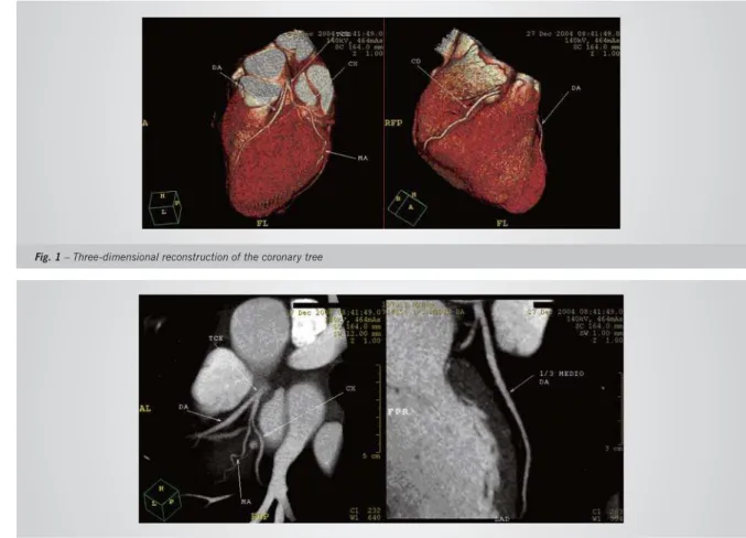
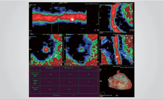
reconstruction, formatting and interpretation. There is considerable variation in image manipulation methods because of the different software packages offered by the various manufacturers. The most common methods include MPR (multiplanar reformation), oblique MPR, curved MPR, MIP (maximum-intensity projection), shaded-surface display and three-dimensional reconstruction (3-D). Considering the large number of settings and projection angles it is important that the images are manipulated so as to visualize the entire vessel volume, taking advantage of the features of each technique10,11 (fi g.1, 2 and 3).
Even though the 3-D images provide a good perception of the coronary tree’s spatial distribution anatomy, it is the
C
LINICALAPPLICABILITYThe role of a coronary angiotomography is not yet completely defi ned, however, its diagnostic capability has turned it into a method that is more and more frequently indicated in daily clinical practice12,13.
The rapid technological evolution of this new technique makes it diffi cult to safely determine what the real importance of the test will be in altering the history of coronary disease in its widest sense. Likewise, it is important to note that a coronary angiotomography involves new methods and that great improvements are still required and hoped for. Nevertheless, documented
Fig. 1 – Three-dimensional reconstruction of the coronary tree
data have already specifi ed some of the main indications as follows.
In native coronaries, some authors have documented that the angio-CT is able to not only accurately detect lesions but also to classify the coronary plaque in accordance with its density and calcium content14.
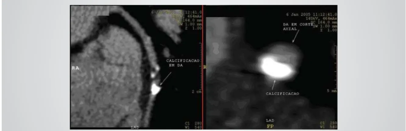
Schroeder et al15 compared plaque density recorded by
the angio-CT with post-mortem histological fi ndings, determining that high lipid plaques have an average density of 42 ± 22 Hounsfi eld units (HU), intermediate plaques of 70 ± 21 HU, and calcifi ed plaques of 715 ± 328 HU (fi g.4). Unfortunately a precise estimate of the histological characteristics of coronary atherosclerosis plaques remains controversial. Viles-Gonzales et al16 did
not fi nd a dependable relation between the radiological density of the lesion recorded by the angio-CT and the histological fi ndings. Once again, the question is raised regarding the methods used to conduct the test and the results obtained.
The prospects for the angio-CT technique in the case of acute coronary syndromes are promising. Patients with unstable angina present coronary atherosclerosis plaques that are consistently less dense than those found in patients with stable angina17,18. Dorgelo et al19, studying
patients diagnosed with acute coronary syndrome, that did not present an elevated ST segment, using CTs with sixteen rows of detectors, obtained an excellent correlation in comparison to a conventional angiography (sensitivity 94%, specifi city 96%). In these cases , 45% of the patients were not submitted to a percutaneous intervention as there was no signifi cant obstruction (27%)
or surgical revascularization was required (18%). Mollet et al20 prospectively studied the entire coronary
tree of 128 patients with stable angina in order to detect lesions that would potentially require intervention, fi nding an excellent correlation with an invasive angiography (sensitivity 92%, specifi city 95%, positive predictive value 79%, and negative predictive value 98%).
Various authors21-28 have studied patients who had
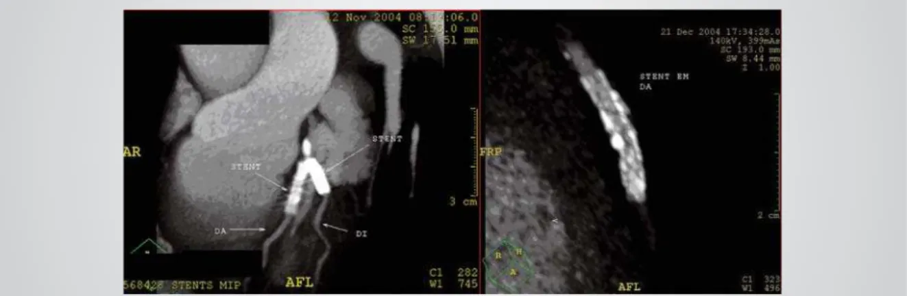
undergone surgical revascularization using multislice CTs. The angio-CT technique proved to be precise for the evaluation of various types of anastomosis and coronary artery bypass grafts (fi g.5), in various locations, in patients that were asymptomatic or had angina symptoms. Although the reports of these various authors indicate an optimistic future as regards non-invasive anatomical coronary artery bypass graft studies, it is important to note that considering technically inferior tests, there are substantial artifact sources for interpretation errors. As mentioned earlier, mammary anastomosis requires a longer breath hold, which is a diffi cult task for patients who are demented, elderly or pneumopathic.
The evaluation of stents using multislice CT methods is still a challenge since the increased density of their metallic structure produces artifacts that greatly reduce the visibility of the intraluminal contrast (fi g.6). Nevertheless, in some cases, the contrast through the prosthesis could be adequately studied as well as its patency along with the degree of distal artery opacifi cation29-33.
Other applications include the follow-up of coronary artery remodelling34,35, detection and graduation of
coronary artery aneurisms in individuals with or without
Kawasaki disease as well as precise detection and characterization of anomalous coronary arteries36-39.
C
ALCIUM ANDHEART DISEASEThe association between the presence of calcium in the coronaries and atherosclerosis plaques is well established and has been known for almost a century40.
In rare cases, patients can present Monckeberg calcifi -cation in the media layer of the coronary artery and not have atherosclerosis41,42. Individuals with
calcium-phosphate metabolism alterations can also present accelerated artery calcifi cation43,44. Nevertheless, for
clinical purposes, coronary calcifi cation represents the existence of atherosclerosis45.
Data obtained from an electron beam tomography (EBT) allow the formulation of a score that describes the calcifi cation intensity as: low (1-10), moderate (11-100), moderately high (101-399) and high (> 400). The incidence of complications is directly related to the quantifi cation supplied by the score and the absence of
tomographically detectable coronary calcifi cation will have an elevated negative predictive value, excluding the possibility of signifi cant atherosclerosis which, for example, is very useful information for a population with atypical precordialgia45. Meneghelo et al46 described the
distribution of EBT determined calcium scores for 2,253 Brazilian men between the ages of 22 and 88 years, fi nding scores higher than zero in 48.8% of the cases, in a pattern that did not follow the Gauss curve.
Recent studies have shown that a multislice CT is at least comparable to a EBT47,48. The number of EBTs
available in Brazil is extremely limited due to the high cost and low versatility. Since most of the prognostic data were obtained based on prospective studies using an EBT, comparison between the methods remains a controversial issue.
To date, there are no studies available regarding the predictive value of coronary calcifi cation levels based on rigorous prospective studies that have been concluded. Therefore, the real impact of the calcium score still needs to be demonstrated in large population cohorts.
D
IAGNOSTICACCURACYIn dealing with studies of angio-CT diagnostic accuracy it is extremely important to emphasize the diffi culty of comparison and interpretation since distinct techniques are used. A detailed analysis of the studies reveals diversity in the following variables:
• Algorithms for reconstruction and reconstructed diac cycle phases;
• The patient studied;
• Type, volume and fl ow of the contrast agent injected; • CT manufacturer;
• Software used for interpretation.
The non-invasive test for coronary visualization requires high temporal and spatial resolution equipment. Since the initial reports in the year 200049,50, various authors
have contributed to the analysis of coronary stenosis
Fig. 4 – Mixed plaque on the mid third of the anterior descending artery. At the left, observe the longitudinal slice using the curved MIP and at the right the cross section
Fig.6 – Stents. At the left, observe the presence of two stents whose metal structure affects luminal observation. At the right, the contrast within the stent can be seen
detection accuracy demonstrating that CTs equipped with sixteen rows of detectors, even in the preliminary studies, are superior to the predecessors with four rows of detectors (tab. 1).
Surgical grafts (saphenous vein, mammary and radial arteries) have less movement than native coronaries during the cardiac cycle and is the technique indicated for revascularized patient follow-up (tab. 2).
Along with the development of more modern multislice CTs that include technology with 32, 40 and 64 detectors60, it is hoped that the image quality will be
sharper and that diagnostic accuracy will improve.
L
IMITATIONSAs previously mentioned, coronary calcifi cation has been considered as a sign of atherosclerosis. However, luminal evaluation of segments that are severely calcifi ed are affected by the production of arterial light artifacts,
similar to those seen in patients with stents.
In order to acquire good quality images, it is desirable to have a cardiac rhythm with a regular R-R interval and a lower heart rate61,62. Most laboratories have been
using betablockers to obtain heart rates lower than 65 BPM63. Of course, the presence of cardiac arrhythmias,
especially tachyarrhythmias with a variable R-R interval, such as in the case of atrial fi brillation, often makes it impracticable to conduct the test. Ectopic beats, even isolated, could affect adequate visualization of the coronary segments64 (fi g.7).
The radiation dose to perform the coronary angioto-mography is considered high and superior to the dose used in electron beam tomographies and conventional angiographies65 (tab. 3).
Similar to the classic angiography, the test requires an venous injection of iodinated contrast agent which could cause nephrotoxic and allergic reactions.
Table 1 – Coronary angio-CT accuracy using tomographs with four and sixteen rows of detectors: preliminary studies with sixteen rows of detectors
Author n Detectors Sensitivity Specifi city No evaluation
Nieman et al51 31 4 81% 97% 27%
Achenbach et al52 64 4 91% 84% 32%
Knez et al53 42 4 78% 98% 6%
Herzog et al54 42 4 72% 92%
-Kopp et al55 102 4 86-93% 96-97% 18%
Becker et al56 28 4 78% 71% 11%
Nieman et al57 53 4 82% 93% 30%
Nieman et al3 59 16 95% 86% 0%
Ropers et al58 77 16 92% 93% 12%
Table 2 – Angio-CT accuracy in the evaluation of by-passes and coronary anastomosis
Author n patients n anastom. Sensitivity Specifi city
Burgstahler et al21 10 21 86% 100%
Ko et al23 39 115 93.3 % 99 %
Tello et al22 26 75 97% 95%
Schlosser et al26 48 131 96% 95%
arrhythmias during the image acquisition, difficulty for the patient to hold their breath, extensive coronary
Fig.7 – Image artifacts. Image at the left showing “steps” produced by respiration during the aquisition phase. At the right, observe the artifact in the third proximal of the circumfl ex artery due to ventricular ectopics
Table 3 – Radiation dose during an angio-CT test
Multislice CT
Dose
Organ (mSv) EBT Siemens®
Schroder et al65,66
Achenbach
et al50,52 Invasive angiography
Bone marrow 2.0 14.1 11.0 10.1 2.5
Lungs 3.9 37.6 27.9 22.7 8.1
Thyroid 0.4 2.7 3.4 1.4 0.5
Esophagus 3.1 27.1 16.9 17.2 4.2
Liver 1.8 13.5 9.8 9.9 2.9
Stomach 2.6 18.2 9.8 9.8 1.6
Colon 1.0 1.0 0.7 0.9 0.7
Bladder 0.3 0.4 0.3 0.3 0.3
Breasts 5.9 44.0 28.7 25.6 6.9
Ovaries 1.5 0.6 1.4 0.9 1.1
Testicles 0.4 0.7 0.4 0.2 0.5
Actual dose (mSv) 0.5 - 2.0 10.9 - 13.0 7.6 - 9.2 6.7 - 8.1 2.1 - 2.5
for coronary risk evaluation, however, further investigation in this area is required.
R
EFERENCES1. Sarmento-Leite R, Krepsky AM, Gottsschall CAM. Infarto agudo do miocárdio: um século de história. Arq Bras Cardiol 2001; 77: 592-601.
2. Rittman EL. Cardiac computed tomography imaging: a history and some future possibilities. Cardiol Clin 2003; 21:491-513.
3. Nieman K, Cademartiri F, Lemos PA, Raaijmakers R, Pattynama PM, Feyter PJ. Reliable noninvasive coronary angiography with fast submillimeter multislice spiral computed tomography. Circulation 2002; 106:2036-8.
4. Hangartner J, Charleston A, Davies Ml, Thomas AC. Morphological
characteristics of a clinically signifi cant coronary artery stenosis in stable angina. Br Heart J 1986; 56:501-8.
5. Roberts W. The coronary arteries and left ventricle in clinically isolated angina pectoris. Circulation 1976; 54:388-90.
6. Mann J, Davies N. Vulnerable plaque relation of characteristics to degree of stenosis in human coronary arteries. Circulation 1996; 94:928-31
8. Ross R. The pathogenesis of atherosclerosis a perspective for the 1990s. Nature 1993; 362:801-9.
9. Davies MJ. The composition of coronary artery plaques. N Engl J Med 1997; 336: 1312-4.
10. Van Ooijen PM, Ho KY, Dorgelo J, Oudkerk M. Coronary artery imaging with multidetector CT: visualization issues. Radiographics 2003; 23:16.
11. Schroder S, Kopp AF, Ohnesorge B. Virtual coronary angioscopy using multislice computed tomography. Heart 2002; 87:195-7.
12. Vignaux O, Paul JF, Duboc D. Multislice CT and MRI of coronary artery disease: current and future role. J Radiol 004; 85:1786-95.
13. Achenbach S, Daniel WG. Noninvasive coronary angiography – an aceptable alternative? N Eng J Med 2002; 345:1909-10.
14. Achenbach S, Moselewski F, Ropers D, Ferencik M, Hoffmann U, MacNeil B et al. Detection of calcifi ed and noncalcifi ed coronary atherosclerotic plaque by contrast-enhanced submillimeter multidetector spiral computed tomography: a segment-based comparison with intravascular ultrasound. Circulation 2004; 109:14-7.
15. Schroeder S, Kuettner A, Leitritz M , Janzen J, Kopp AF, Herdeg C et al. Reliability of differentiating human coronary plaque morphology using contrast-enhanced multislice spiral computed tomography: a comparison with histology. J Comput Assist Tomogr 2004; 28:449-54.
16. Viles-Gonzalez JF, Michael Poon, Javier Sanz J, Rius T, Nikalaou K, Fayad ZA et al. In Vivo 16-slice, multidetector-row computed tomography for the assessment of experimental atherosclerosis: comparison with magnetic resonance imaging and histopathology . Circulation 2004; 110:1467-72;
17. Inoue F, Sato Y, Matsumoto N, Tani S, Uchiyama T. Evaluation of plaque texture by means of multislice computed tomography in patients with acute coronary syndrome and stable angina. Circ J 2004; 68:840-4.
18. Caussin C, Othanessian A, Lancelin B, Rachal S, Hennequin R, Dambrin G et al. Coronary plaque burden detected by multislice computed tomography after acute myocardial infarction with near normal coronary arteries by angiography. Am J Cardiol 2003; 92:849-52.
19. Dorgelo J, Willems TP, Geluk CA , van Ooijen PM, Zijlstra F, Oudkerk M. Multidetector computed tomography-guided treatment strategy in patients with non-ST elevation acute coronary syndromes: a pilot study. Eur Radiol 2004;15:708-13.
20. Mollet NR, Cademartiri F, Nieman K, Saia F, Lemos PA, McFadden EP et al. Multislice computed coronary angiography in patients with stable angina pectoris. J Am Coll Cardiol 2004; 43:2265-70.
21. Burgstahler C, Kuettner A, Kopp AF, Herdez C, Martensen J, Claussen CD et al. Non invasive evaluation of coronary artery bypass grafts using multi-slice computed tomography; initial clinical experience. Int J Cardiol 2003; 90:275-80.
22. Tello R, Hartnell GG, Costello P, Ecker CP. Coronary artery bypass graft fl ow: qualitative evaluation with cine single-detector row CT and comparison with fi ndings at angiography. Radiology 2002; 224::913-8.
23. Ko YG, Choi DH, Jang YS, Chung NS, Shim WH, Cho SY, Yoo KJ. Assessment of coronary artery bypass graft patency by multislice computed tomography. Yonsei Med J 2003; 44:438-44.
24. Kham MF, Herzog C, Landerberger K , Maataouri A, Martens S, Akermann H et al. Visualisation of non-invasive coronary bypass grafts: 4-row vs. 16-row multidetector computed tomography. Eur Radiol 2005;15:118-26.
25. Yoo KJ, Choi D, Choi BW, Lim SH, Chang BC. The comparison of the graft patency after coronary artery bypass grafting using coronary angiography and multi-slice computed tomography. Eur J Cardiothorac Surg 2003; 24:86-91.
26. Schlosser T, Konorza T, Hunold P, Kuhl H, Schmermund A, Barkhausen J. Noninvasive visualization of coronary artery bypass grafts using
16-detector row computed tomography. J Am Coll Cardiol 2004; 44:1224-9.
27. Willmann JK, Weishaupt D, Kobza R , Verdun FR, Seifert B, Marincek B et al Coronary artery bypass grafts: ECG-gated multi-detector row CT angiography – infl uence of image reconstruction interval on graft visibility. Radiology 2004;232:568-77.
28. Dewey M, Lembcke A, Enzweiller C, Hamm B, Rogalla P. Isotropic half-millimeter angiography of coronary artery bypass with 16 slice computed tomography. Ann Thorac Surg 2004; 77:800-4.
29. Kruger S, Mahnken AH, Sinha AM, Borghans A, Dedden K, Hoffmann R et al. Multislice spiral computed tomography for the detection of coronary stent restenosis and patency. Int J Cardiol 2003; 89:167-72.
30. Schuijf JD, Bax JJ, Jukema JW, Lamb HJ, Warda HM, Vliegen HW et al. Feasibility of assessment of coronary stent patency using 16-slice computed tomography. Am J Cardiol 2004; 94:427-30
31. Hong C, Chrysant GS, Woodart PK, Bae KT. Coronary artery stent patency assessment with in-stent contrast enhancement measured at multi-detector row CT angiography: initial experience. Radiology 2004; 233:286-91.
32. Nieman K, Cademartiri F, Raaijmakers R, Pattynama P, de Feyter P. Noninvasive angiographic evaluation of coronary stents with multi-slica spiral CT. Herz 2003; 28:136-42.
33. Ligabue G, Rossi R, Ratti C, Ligabue G, Romagnoli R, Modena MG. Noninvasive evaluation of coronary artery stents patency after PTCA. Radiol Med 2004; 108:128-37.
34. Schoenhegen P, Tuzcu EM, Stillman AE, Moliterno DJ, Halliburton SS, Kuzmiak AS et al. Non-invasive assessment of plaque morphology and remodeling in mildly stenotic coronary segments: comparison of 16-slice computed tomography and intravascular ultra-sound. Coron Artery Dis 2003; 14:459-62.
35. Imazeke T, Sato Y, Inoue F, Anazawa T, Tani S, Matsumoto N et al. Evaluation of coronary artery remodeling in patients with acute coronary syndrome and stable angina by multislice computed tomography. Circ J 2004; 68:1045-50.
36. Sohn S, Kim HS, Lee SW. Multidetetctor row computed tomography for follow-up of patients with coronary artery aneurysms due to Kawasaki disease. Pediatr Cardiol 2004; 25:35-9.
37. Sato Y, Kato M, Inoue F, Fukui T, Imazeki T, Mitsui M et al. Detection of coronary artery aneurysms, stenoses and occlusions by multislice spiral computed tomography in adolescents with Kawasaki disease. Circ J 2003, 67:427-30.
38. Shi H, Aschoff AJ, Brambs HJ, Hoffmann MH. Multislice CT imaging of anomalous coronary arteries. Eur Radiol 2004;14;2172-81.
39. Deibler AR, Kuzo RS, Vohringer M, Page EE,Safoord RE, Patron JN et al. Imaging of congenital coronary anomalies with multislice computed tomography. Mayo Clin Proc 2004; 79:1017-23.
40. Faber A. Die Arteriosklerose, ihre pathologische Anatomie, ihre Pathogenese und Aetiologie. G Fischer 1912.
41. Mönckenberg JG. Mediaverkalkung und atherosklerose. Virchows Arch Pathl Anat 1914; 216:408-16.
42. Lachman AS, Spray TL, Kerwin DM, Shugoll GI, Roberts WC. Medial calcifi cation of Monkenberg. Am J Med 1977; 63:615-22.
43. Oh J, Wunsch R, Turzer M, Bahner M, Raggi P, Querfeld U et al. Advanced coronary and carotid arteriopathy in young adults with childhood-onset chronic renal failure. Circulation 2002; 106:100-5.
44. Raggi P, Boulay A, Chasan-Taber S, Amin N, Dillon M, Burke SK et al. Cardiac calcifi cation in adult hemodialysis patients. A link between end-stage renal disease and cardiovascular disease? J Am Coll Cardiol 2002; 39:695-701.
49. Ohnesorge B, Flohr T, Becker C, Kopp AF, Schoepf UJ, Baum U et al. Cardiac imaging by means of electrocardiographically gated multisection spiral CT: initial experience. Radiology 2000; 217:564-71.
50. Achenbach S, Ulzheimer S, Baum U, Kachebriess M, Ropers D, Giesler T et al. Noninvasive coronary angiography by retrospectively ECG-gated multislice spiral CT. Circulation 2000; 102:2823-8.
51. Nieman K, Oudkerk M, Rensing BJ, van Ooijen P, Munns A, van Geuns RJ et al. Coronary angiography with multi-slice computed tomography. Lancet 2001; 357:599-603.
52. Achenbach S, Giesler T, Ropers D, Ulzheimer S, Derlien H, Schulte C et al. Detection of coronary artery stenoses by contrast- enhanced, retrospectively ECG-gated, multi-slice spiral CT. Circulation 2001; 103:2535-8.
53. Knez A, Becker CR, Leber A, Ohnesorge B, Becker A, White C et al. Usefulness os multislice spiral computed tomography angiography for determination of coronary artery stenoses. Am J Cardiol 2001; 88:1191-4.
54. Herzog C, Abolmaali N, Balzer JO, Baunach AS, Ackermann H, Dogan S. Heart-rate adapted image reconstruction in multidetector row cardiac CT: infl uence of physiological and technical pré-requisite on image quality. Eur Radiol 2002; 12:1670-8.
55. Kopp AF, Schroder S, Kuettner A, Baumbach A, Georg C, Kuzzo R et al. Non-invasive coronary angiography with high resolution multidetector-row computed tomography. Results in 102 patients. Eur Heart J 2002; 23:1714-25.
56. Becker CR, Knez A, Leber A, Treede H, Ohnesorge B, Schoeph UJ et al. Detection of coronary artery stenoses with multislice helical CT angiography. J Comp Assist Tomogr 2002; 26:250-5.
61. Mennicke M, Giesler T, Ropers D, Baum U, Ulzheimer S, Wenkel E et al. Infl uence of heart rate in image quality and detection of coronary stenoses with multislice spiral CT. Biomed Tech 2002; 47(Suppl 1): 782-5.
62. Schroder S, Kopp AF, Kuettner A, Burgstahler S, Herdeg C. Infl uence of heart rate on vessel visibility in noninvasive coronary angiography using new multislice computed tomography: experience in 94 patients. Clin Imaging 2002; 26:106-11.
63. Nieman K, Rensing BJ, van Geuns RJ, Vos J, Pattynama PM, Krestin GP et al. Non-invasive coronary angiography with multislice spiral computed tomography: impact of heart rate. Heart 2002; 88:470-4.
64. Choi HS, Choi BW, Choe KO, Choi D, Yoo KJ, Kim MI et al. Pitfalls, artifacts, and remedies in multi-detector row CT coronary angiography. Radiographics 2004; 24:787-900.
65. Schroeder S, Kopp AF, Baumbach A, Kuettner A, Herdeg C, Rosenberger A et al. Noninvasive detection of coronary lesions by multislice computed tomography: results of the new age pilot trial. Catheter Cardiovasc Interv 2001; 53:352-8.
66. Schroeder S, Kopp AF, Baumbach A, Kuettner A, Georg C, Ohnesorge B et al. Non-invasive characterisation of coronary lesion morphology by multi-slice computed tomography: a promising new technology for risk stratifi cation of patients with coronary artery disease. Heart 2001; 85:576-8.
67. Achenbach S. Clinical use of multi-slice CT coronary angiography. Herz 2003; 28:119-25.