Case Report
1 1 3 Arq Bras Oftalmol. 2016;79(2):113-5 http://dx.doi.org/10.5935/0004-2749.20160033
INTRODUCTION
Pseudophakic cystoid macular edema (PCME), or Irvine-Gass syn-drome, is a common complication following cataract surgery(1). It was first described in 1953 by Irvine, and in 1966, Gass and Norton pu-blished an angiographic study of its characteristics(1,2). The incidence of clinical (symptomatic) PCME has been greatly reduced because of its advances in surgical techniques (approximately 0.1%-2.35%), and most cases of PCME have spontaneous resolution(2). Majority of patients remain asymptomatic without active inflammation on fundus examination and optical coherence tomography (OCT)(3). However, subclinical PCME is detected in almost 30% of patients with postsur-gical angiography and a further 11%-41% patients with OCT(4).
The pathogenesis of PCME remains obscure. Most investigators agree that inflammation is the main etiologic factor in the develop-ment of PCME(5). Inflammatory mediators disrupt the blood-aqueous barrier (BAB) and blood-retinal barrier (BRB), leading to an amplifica-tion of retinal permeability and the development of PCME(2,5).
No recent significant studies have been conducted to establish the best therapeutic options for treating this disorder(4). Non-steroidal anti-inflammatory drugs (NSAIDs) and corticosteroids represent the most common first-line treatments(4,6). Antiangiogenic therapy, sub
cu-taneous injections of interferon α2a, hyperbaric therapy, and vitrecto-my are also therapeutic options(4,5). The dexamethasone implant (DEX implant; Ozurdex; Allergan, USA) is a 0.7 mg biodegradable intravitreal implant that delivers dexamethasone into the vitreous and retina. The drug acts on all inflammatory mediators and has been approved by the US Food and Drug Administration (FDA) for the treat ment of macular edema (ME) secondary to retinal vein occlusion (RVO) and for non-infectious posterior uveitis(4). The DEX implant has also been indicated for the treatment of diabetic ME in patients who are pseu-dophakic or are phakic and scheduled for cataract surgery(4).
In this study, we describe a case series of six patients who were previously diagnosed with PCME and treated with DEX implants.
CASE REPORT
This is a retrospective small case series of six patients who were diagnosed with PCME and treated with DEX implants at the Centro Brasileiro de Cirurgia de Olhos (Goiânia, Goiás, Brazil) and Centro Bra sileiro da Visão (Brasília, Distrito Federal, Brazil) between 2013 and 2015. Spectral domain OCT (SD-OCT) was performed with a Spectralis instrument (Heidelberg Engineering, Heidelberg, Germany) during
Dexamethasone 0.7 mg implants in the management of pseudophakic
cystoid macular edema
Implante de 0,7 mg de dexametasona no tratamento do edema macular cistóide do pseudofácico
José Maurício Bottode Barros Garcia1,2, david Leonardo cruvineL isaac1,2, Marcos Pereirade ÁviLa1,2
ABSTRACT
Pseudophakic cystoid macular edema (PCME) is a common complication follo-wing cataract surgery. Although majority of patients with PCME remain asympto-matic, it remains an important cause of vision loss after cataract surgery. The pathogenesis of PCME remains unclear, but most authors agree that inflammation plays a major role in its development. There is no standard algorithm for treatment procedures for PCME. A biodegradable 0.7 mg dexamethasone intravitreal implant can be used to deliver medication into the posterior segment of eyes. This drug acts on all inflammatory mediators and has been approved for the treatment of macular abnormalities secondary to retinal vein occlusion and for non-infectious posterior uveitis. In this case series, we report six patients who presented with PCME and were treated with a 0.7 mg dexamethasone intravitreal implant. Fa-vorable anatomical outcomes were demonstrated by spectral domain-optical coherence tomography images.
Keywords: Macular edema/etiology; Macular edema dexamethasone implants/ drug therapy; Dexamethasone/administration & dosage; Visual acuity/physiology
RESUMO
O edema macular cistóide do pseudofácico (PCME) é uma frequente complicação no acompanhamento pós-operatório da cirurgia de catarata. Embora a maioria dos pacientes apresente-se sem sintomas, PCME ainda permanece como importante causa de baixa visão após facectomia. Sua patogênese ainda permanece obscura, porém, autores sugerem que fatores que promovem maior inflamação possuem papel fundamental em sua origem. Não há um algoritmo padrão no manejo do PCME. O implante biodegradável de dexametasona 0,7 mg surgiu como possível arma terapêutica, após aplicação intra-vítrea. Essa droga consegue agir sobre me-diadores inflamatórios, além de já ter sido aprovada no tratamento do edema de macula secundário à oclusões venosas da retina, e uveítes posteriores de origem não infecciosa. Na seguinte série de casos, relatamos a evolução de 6 pacientes com PCME, submetidos a terapia com implante de dexametasona 0,7 mg. A melhora anatômica foi documentada com imagens de SD OCT.
Descritores: Edema macular/etiologia; Edema macular/quimioterapia; Dexameta-sona/administração & dosagem; Acuidade visual/fisiologia
Submitted for publication: May 11, 2015 Accepted for publication: July 21, 2015
1 Ophthalmology Department, Centro de Referência em Oftalmologia, Goiânia, GO, Brazil. 2 Ophthalmology Department, Universidade Federal de Goiás, Goiânia, GO, Brazil.
Funding: No specific financial support was available for this study.
Disclosure of potential conflicts of interest: None of the authors have any potential conflict of interest to disclose.
Dexamethasone 0.7 mg implants in the management of pseudophakic cystoid macular edema
1 1 4 Arq Bras Oftalmol. 2016;79(2):113-5
pre- and post-procedure visits to evaluate macular status (Table 1). The first-li ne approach in all patients was a topical combination of NSAIDs and corticosteroids for a course of 3 months, what did not result in the clinical resolution of the described cases. Mild glaucoma had been previously diagnosed in two of the patients (Figures 1, 2, and 3). The patients ranged in age from 64 to 74 years (median: 69.75). Three of the patients were men (50.0%). The follow-up period after the procedure ranged from 48 to 201 days. The initial central retinal thickness (CRT) on SD-OCT ranged from 406 to 773 µm (median 542.5), and the final CRT ranged from 218 to 288 µm (median: 219.5). No patient presented significant changes in intraocular pressure du-ring the follow-up period.
DISCUSSION
The pathogenesis of PCME remains unclear, but it is generally agreed that inflammation plays a major role in its development(2,4,5,7). Breakdown of the BAB and BRB may be associated with diabetes and glaucoma(2). Patients with diabetes, particularly those with diabetic retinopathy, are at an increased risk of developing PCME(8). In gene-ral, cataract surgery results in intraocular inflammation produced by inflammatory mediators that cause increased expression of vascular endothelial growth factor (VEGF), which potently induces increased permeability of the perifoveal capillary net and breakdown of the
BRB(5). However, to date, no studies have shown increased ocular VEGF levels in patients with PCME in the absence of ischemic ocular disease(2).
The DEX implant has been approved by the US FDA for the treat-ment of ME secondary to RVO, for non-infectious posterior uveitis and for the treatment of diabetic ME in patients who are pseudophakic or are phakic and scheduled for cataract surgery(4).
Dexamethasone has a six-fold more potent anti-inflammatory pro file than triamcinolone acetonide(8). A phase II study on the dexa-methasone drug delivery system conducted a subgroup analysis that included patients with PCME and patients with uveitis(4,9). A visual gain of at least 15 ETDRS letters was reported at the 90-day follow-up in 53.8% of the patients who were treated with 0.7 mg intravitreal dexa-methasone implants(9). In the EPISODIC study, cases of ocular hyper-tension were reported but were controlled with pressure-lowering me dications. No filtering surgery was required(4). In our group of pa tients, including two with previously diagnosed glaucoma, there was no need to change their current therapies.
In conclusion, when considering the relationship between in-flammation and the development of PCME, it is reasonable to hypo-thesize that DEX implants may offer a promising treatment option for patients who develop PCME following cataract surgery. Controlled studies with larger sample sizes should be performed to confirm these preliminary findings.
Table 1. Patient demographics and treatment history
Patient
Age
(years) Gender Eye
Follow-up after Ozurdex® (days)
Initial SD-OCT (CRT)
Final SD-OCT (CRT)
Initial BCVA (Snellen)
Final BCVA (Snellen)
1 64 M OS 091 496 µm 252 µm 20/200 20/30
2 68 F OD 064 529 µm 227 µm 20/200 20/50
3 69 M OD 195 406 µm 218 µm 20/80 20/30
4 74 F OS 080 556 µm 216 µm 20/60 20/30
5 70 M OS 048 773 µm 221 µm 20/50 20/30
6 74 F OS 201 556 µm 288 µm 20/60 20/30
SD-OCT= spectral domain optical coherence tomography; BCVA= best-corrected visual acuity; CRT= central retinal thickness.
Garcia JMBB, et al.
1 1 5
Arq Bras Oftalmol. 2016;79(2):113-5
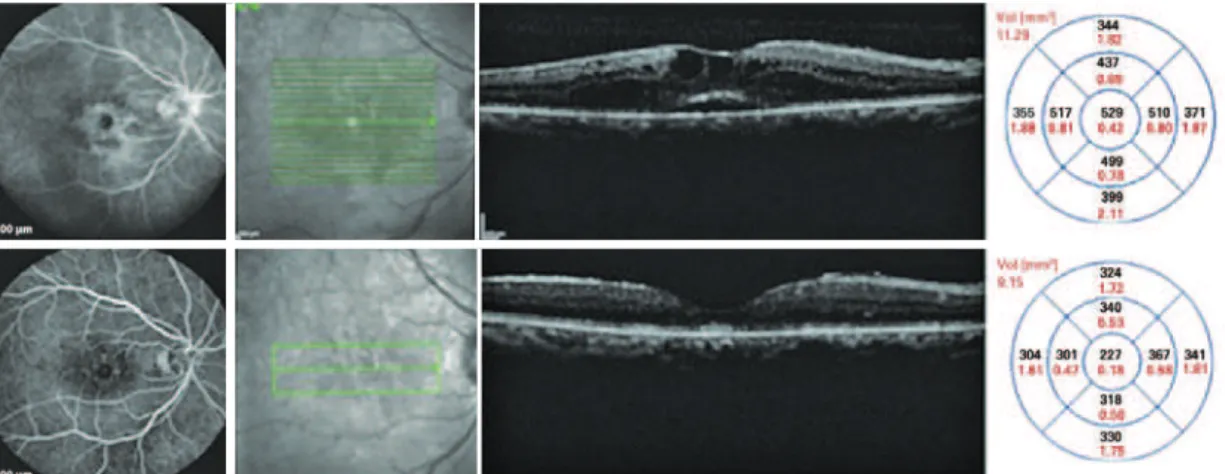
Figure 2. The patient underwent cataract surgery 10 years ago in the OD. Top: FA and SD-OCT before DEX implantation. Bottom: Six months after DEX implantation, late frame FA and SD-OCT showed a reduction of the central retinal thickness of 302 µm. SD-OCT= spectral domain optical cohe-rence tomography; DEX= dexamethasone; FA= luorescein angio gram.
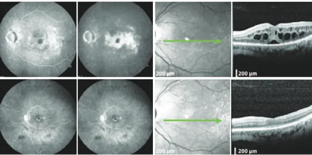
Figure 3. Patient with mild glaucoma under timolol 0.5% BID. A) Infrared photograph. B) Petaloid aspect in central macula displayed in FA of OD. C) SD-OCT de-monstrating ME before DEX implantation. D) SD-OCT 195 days after the procedure, with no signs of ME. SD-OCT= spectral domain optical coherence tomography; ME= macular edema; DEX= dexamethasone; FA= luorescein angiogram; BID= twice daily.
A
B
C
D
E
F
REFERENCES
1. Irvine SR. A newly defined vitreous syndrome following cataract surgery. Am J Ophthal-mol. 1953;36(5):599-619.
2. Guo S, Patel S, Baumrind B, Johnson K, Levinsohn D, Marcus E, et al. Management of pseudophakic cystoid macular edema. Surv Ophthalmol. 2015;60(2):123-37. 3. Kiernan DF HS. Controversies in the management of Irvine-Gass syndrome.
Ophthal-mic Surg Lasers Imaging Retina. 2013;44(6):522-7.
4. Bellocq D KJ, Burillon C, Voirin N, Dot C, Souied E, Conrath J, et al. Effectiveness and sa fety of dexamethasone implants for post-surgical macular oedema including Irvine-Gass syndrome: the EPISODIC study. Br J Ophthalmol. 2015;99(7):979-83. 5. Arevalo JF, Maia M, Garcia-Amaris RA, Roca JA, Sanchez JG, Berrocal MH, et al. Intravitreal
bevacizumab for refractory pseudophakic cystoid macular edema: the Pan-American Collaborative Retina Study Group results. Ophthalmology. 2009;116(8):1481-7.
6. Dutra Medeiros M NR, Garcia-Arumi J, Mateo C, Corcostegui B. Dexamethasone in-travitreal implant for treatment of patients with recalcitrant macular edema resulting from Irvine-Gass syndrome. Invest Ophthalmol Vis Sci. 2013;54(5):3320-4. 7. Brynskov TLC, Halborg J, Kemp H, Sorensen TL. Longstanding refractory pseudophakic
cystoid macular edema resolved using intravitreal 0.7 mg dexamethasone implants. Clin Ophthalmol. 2013;7:1171-4.
8. Khurana RN PJ, Porco TC, Wieland MR. Dexamethasone intravitreal implant for pseu-dophakic cystoid macular edema in patients with diabetes. Ophthalmic Surg, Lasers Imaging Retina. 2015;46(1):56-61.