Case Report
111 Arq Bras Oftalmol. 2016;79(2):111-2 http://dx.doi.org/10.5935/0004-2749.20160032
INTRODUCTION
Posterior scleritis is an ocular inflammatory disorder that is pre-dominantly idiopathic, autoimmune, or rarely, infective(1,2). Anterior
and posterior scleritis due to Mycobacterium tuberculosis are rare but have been reported in the literature(3); however, to our knowledge,
isolated posterior scleritis associated with tuberculosis (TB) has only been described twice(1,4).
We report a patient with posterior scleritis associated with latent TB without associated uveitis, anterior scleritis, keratitis, or any other previous ocular disease history.
CASE REPORT
A previously healthy 43-year-old woman presented with com-plaints of painful reduced visual acuity and periorbital edema in her left eye of 4 days’ duration. Ophthalmological examination showed best corrected visual acuity (BCVA) in the right and left eyes of 6/6 and 6/60, respectively, and left eye proptosis, with no conjunctival or ciliary hyperemia and no anterior chamber reaction. Fundoscopy was unremarkable in the right eye, while the left eye showed choroidal folds, optic disc edema, and exudative inferior retinal detachment.
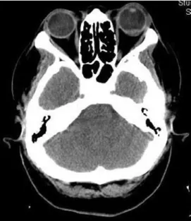
Orbital computerized tomography (CT) scan and magnetic re-sonance imaging revealed a left eye choroidal and transscleral mass
spreading to the Tenon’s space, along with thickening of the optic papilla (Figure 1).
Macular optical coherence tomography (OCT) showed edema with neurosensory retinal detachment in the left eye. B-scan ultra-sonography (Figure 2) revealed sclerochoroidal thickening and the presence of sub-Tenon’s space fluid (T-sign).
On fluorescein angiography, we found disc staining and multiple early pinpoint retinal pigment epithelial leaks (Figure 3).
The patient was treated for posterior scleritis with oral predniso-lone 1 mg/kg body weight once daily. After 2 weeks of treatment, an improvement in pain was noted by the patient, along with BCVA of 6/60 in the left eye with improvement of the macular OCT edema; ho-wever, on fundoscopy, worsening of the optic disc edema was found. The orbital CT scan and B-scan ultrasonography revealed identical results. Etiological investigation showed negative anti-HIV antibody test, C-reactive protein level of 0.3 mg/dL, elevated sedimentation rate (59 mm), negative rapid plasma reagin and treponema pallidum hemagglutination test, normal angiotensin-converting enzyme (25 UI/L), negative cytoplasmic and perinuclear anti-neutrophil cytoplasmic antibodies, negative Borrelia burgdorferi and Toxoplasma gondii IgG and IgM, and negative rheumatoid factor and antinuclear antibodies. The chest radiograph was normal and the patient denied any respiratory symptoms or recent travel in TB-endemic regions. The
Isolated posterior scleritis associated with tuberculosis
Esclerite posterior isolada associada à tuberculose
AnA FilipA MirAndA1, João CArdoso1, nAdine MArques1, sAndrA BArros1, pAulA Telles1, nuno CAMpos1
Submitted for publication: March 23, 2015 Accepted for publication: May 2, 2015
1 Department of Ophthalmology, Hospital Garcia de Orta, Almada, Portugal.
Funding: No specific financial support was available for this study.
Disclosure of potential conflicts of interest: None of the authors have any potential conflict of interest to disclose.
Corresponding author: Ana Filipa Miranda. Av. Torrado da Silva, 2.801-951 - Almada - Portugal - E-mail: [email protected]
ABSTRACT
Ocular tuberculosis (TB) is considered to be rare, although its incidence has varied widely over time and in different populations. Latent TB is diagnosed when a person is infected with Mycobacterium tuberculosis but does not have active TB. During the last decade, interferon-gamma release assay tests have been developed that allow identification of patients with latent TB infection with better specificity than the tuberculin skin test and can differentiate between infection and prior vaccina-tion. Although rare, tuberculous scleritis should be considered in the differential diagnosis of posterior scleritis. Here we describe a patient with posterior scleritis and severe visual loss associated with latent TB without uveitis, anterior scleritis, keratitis, or any other previous ocular disease history. The patient responded well to a combined treatment of antitubercular therapy and oral corticosteroids.
Keywords: Scleritis/diagnosis; Tuberculosis, ocular/diagnosis; Eye infections,
bac-terial; Mycobacterium tuberculosis
RESUMO
A tuberculose (TB) ocular foi considerada rara, embora a sua incidência tenha variado significativamente ao longo do tempo e nas diferentes populações. A TB latente é diagnosticada quando alguém é infetado com Mycobacterium tuberculosissem possuir doença ativa. Durante a última década, testes tendo por baseinterferon gamma release assay foram desenvolvidos, permitindo a identificação de pacientes com infeção por tuberculose latente com maior especificidade que o teste tuberculínico e diferenciar infeção e vacinação prévia. Embora rara, a esclerite tuberculosa deve ser tida em consideração no diagnóstico diferencial de esclerite posterior. Reportamos um paciente com esclerite posterior e baixa grave de acuidade visual associada a TB latente, sem uveíte, esclerite anterior, ceratite ou história de doença ocular prévia. O paciente respondeu favoravelmente a um tratamento combinado de fármacos antituberculose e corticoides orais.
Isolated posterior scleritis associated with tuberculosis
112 Arq Bras Oftalmol. 2016;79(2):111-2
patient had a positive result for the purified protein derivative (PPD) skin test (17 mm induration under corticotherapy 80 mg/day) and interferon-gamma release assay (IGRA), which is a more specific test. Antitubercular chemotherapy was started in combination with the oral corticosteroids, and was a combination of rifampicin 10 mg/kg, isoniazid 5 mg/kg, and pyrazinamide 25 mg/kg. The oral prednisolo-ne treatment was given for 10 weeks, and the antitubercular therapy for 9 months. After 10 weeks of treatment, the patient recovered a visual acuity of 6/6 in the left eye, the optic disc edema and choroidal folds disappeared, and the orbital CT scan (Figure 4) and B-scan ultra-sonography were normal. The patient has since remained asympto-matic for a period of 18 months.
DISCUSSION
This case report is an example of a patient with latent TB and seve-re posterior scleritis, with no signs of anterior segment involvement. Some authors have suggested that infection due to TB should be considered as a possible cause of scleritis if the investigation reveals a positive PPD skin test, particularly if the scleral inflammation does not respond adequately to standard corticosteroid treatment(5).
Given the worsening of the optic disc edema, no im provement of the BCVA, orbital CT scan, and B-scan ultrasonography with oral corticosteroids, and the positive results from the PPD skin and IGRA tests, we assumed that TB was the causative agent of the posterior scleritis, and started antitubercular therapy. An intraocular sample to confirm the diagnosis ofMycobacterium tuberculosis infection was not performed as the patient responded well to the combined treatment of oral corticosteroids and antitubercular therapy. In our investigation, we could not establish the presence of any connec-tive tissue disorder in our patient, and she has had no recurrences in the 18 months of follow-up, which suggests a non-connective tissue disorder as an etiology. Some authors consider a therapeu-tic trial to be justified in patients with severe sight-threatening intraocular inflammation and latent TB infection(6). Furthermore,
the addition of antitubercular therapy to corticosteroids in uveitis patients with latent/manifested TB has been shown to reduce the recurrences of uveitis(7).
The difficulty in optic neuropathy screening in this patient led our infectious diseases specialists to not use ethambutol as part of the antitubercular regimen.
In conclusion, posterior scleritis associated with TB can present with no signs of anterior segment involvement, and responds well to a combination treatment of antitubercular therapy and oral corti-costeroids. Oral corticosteroids, given without antitubercular drugs, are likely to worsen the disease in these patients. The establishment of the diagnosis is very important for well-directed therapy, visual re covery, and no recurrences.
REFERENCES
1. Chen FK, White A, Harney BA. Systemic tuberculosis presenting with bilateral visual loss. Br J Ophthalmol. 2010;94(12):1686-7.
2. Gupta A, Gupta V, Pandav SS, Gupta A. Posterior scleritis associated with systemic tuberculosis. Indian J Ophthalmol. 2003;51(4):347-9.
3. Thompson MJ, Albert DM. Ocular Tuberculosis. Arch Ophthalmol. 2005;123(6):844-9. 4. Velasco e Cruz AA, Chahud F, Feldman R, Akaishi PM. Posterior scleral tuberculoma:
case report. Arq Bras Oftalmol. 2011;74(1):53-4.
5. Keino H, Watanabe T, Taki W, Nakashima C, Okada AA. Clinical features and visual outco-mes of Japanese patients with scleritis. Br J Ophthalmol. 2010;94(11):1459-63. 6. La Distia Nora R, van Velthoven ME, Ten Dam-van Loon NH, Misotten T, Bakker M, van
Hagen MP, Rothova A. Clinical manifestations of patients with intraocular inflamma-tion and positive QuantiFERON-TB gold in-tube test in a country nonendemic for tu berculosis. Am J Ophthalmol. 2014;157(4):754-61.
7. Bansal R, Gupta A, Gupta V, Dogra MR, Bambery P, Arora SK. Role of anti-tubercular the-rapy in uveitis with latent/manifest tuberculosis. Am J Ophthalmol. 2008;146(5):772-9.
Figure 1. CT scan of the patient showing a transscleral mass on
the left eye.
Figure 2. B-scan ultrasonography showing T-sign on the left
eye of the patient.
Figure 3. Fluorescein angiography with multiple early pinpoint
retinal pig ment epithelial leaks.
Figure 4. Normal orbital CT scan of the patient 10 weeks after