Trichoderma
: A Bioinformatic Approach
Saila Viridiana Ca´zares-Garcı´a1, Ma. Soledad Va´zquez-Garciduen˜as2, Gerardo Va´zquez-Marrufo1* 1Centro Multidisciplinario de Estudios en Biotecnologı´a, Facultad de Medicina Veterinaria y Zootecnia, Universidad Michoacana de San Nicola´s de Hidalgo, Morelia, Michoaca´n, Mexico,2Divisio´n de Estudios de Posgrado, Facultad de Ciencias Me´dicas y Biolo´gicas ‘‘Dr. Ignacio Cha´vez’’, Universidad Michoacana de San Nicola´s de Hidalgo, Morelia, Michoaca´n, Mexico
Abstract
The genusTrichodermaincludes species of great biotechnological value, both for their mycoparasitic activities and for their ability to produce extracellular hydrolytic enzymes. Although activity of extracellular laccase has previously been reported in
Trichodermaspp., the possible number of isoenzymes is still unknown, as are the structural and functional characteristics of both the genes and the putative proteins. In this study, the system of laccasessensu strictoin theTrichodermaspecies, the genomes of which are publicly available, were analyzed using bioinformatic tools. The intron/exon structure of the genes and the identification of specific motifs in the sequence of amino acids of the proteins generatedin silicoallow for clear differentiation between extracellular and intracellular enzymes. Phylogenetic analysis suggests that the common ancestor of the genus possessed a functional gene for each one of these enzymes, which is a characteristic preserved inT. atrovirideand
T. virens. This analysis also reveals that T. harzianum and T. reesei only retained the intracellular activity, whereas T. asperellumadded an extracellular isoenzyme acquired through horizontal gene transfer during the mycoparasitic process. The evolutionary analysis shows that in general, extracellular laccases are subjected to purifying selection, and intracellular laccases show neutral evolution. The data provided by the present study will enable the generation of experimental approximations to better understand the physiological role of laccases in the genusTrichoderma and to increase their biotechnological potential.
Citation:Ca´zares-Garcı´a SV, Va´zquez-Garciduen˜as MS, Va´zquez-Marrufo G (2013) Structural and Phylogenetic Analysis of Laccases from Trichoderma: A Bioinformatic Approach. PLoS ONE 8(1): e55295. doi:10.1371/journal.pone.0055295
Editor:Alfredo Herrera-Estrella, Cinvestav, Mexico
ReceivedSeptember 27, 2012;AcceptedDecember 20, 2012;PublishedJanuary 31, 2013
Copyright:ß2013 Ca´zares-Garcı´a et al. This is an open-access article distributed under the terms of the Creative Commons Attribution License, which permits unrestricted use, distribution, and reproduction in any medium, provided the original author and source are credited.
Funding:The authors are grateful for the support of the Coordinacio´n de la Investigacio´n Cientı´fica-Universidad Michoacana de San Nicola´s de Hidalgo (CIC-UMSNH), Research Support Program 2012. SVCG acknowledges a scholarship from CONACYT-Me´xico for doctoral studies. The funders had no role in study design, data collection and analysis, decision to publish, or preparation of the manuscript.
Competing Interests:The authors have declared that no competing interests exist.
* E-mail: [email protected]
Introduction
Laccases (benzenediol:oxygen oxidoreductase, EC 1.10.3.2) are metalloenzymes that belong to the multicopper oxidase (MCO) family. These enzymes catalyze the oxidation of various aromatic substrates with the concomitant reduction of molecular oxygen to water. This redox process is mediated by two centers that contain four atoms of copper in their +2 oxidation state. These copper atoms are classified as T1 (blue copper), T2 or T3 according to their spectroscopic characteristics [1]. Laccases are generally monomeric glycoproteins with molecular weights that range from 60 to 70 kDa, and up to 30% of their molecular weight is made up of carbohydrates [2]. These enzymes are widely distributed in nature, and the physiological functions that they perform depend both on their origin and on their biochemical and structural properties. In fungi, laccase activities have been related to the degradation of lignocellulose material, the production of pigments, sporulation, processes of morphogenesis, phenomena of patho-genesis toward plants and animals [3], the oxidation of antibiotics produced by microorganisms that are antagonists of plant pathogens and antimicrobial components of plants, such as flavonoids or phytoalexins [4]. This great functional versatility is partly due to the fact that laccases possess low substrate specificity and exhibit a broad range of redox potentials [2]. Because of this
flexibility, these enzymes are able to act on ortho- and para-diphenols, methoxy-substituted phenols, aromatic diamines and benzenothiols. Furthermore, these enzymes can oxidate organic and inorganic metallic compounds. In addition, the gamut of substrates for laccases can extend to non-phenolic compounds through the inclusion of redox mediators, with which they are able to oxidize large polymers, such as lignin, cellulose or starch [5]. This peculiarity has been exploited by various biotechnological processes, including biopulping, bioremediation, the breakdown of colorants, the enzymatic conversion of chemical intermediates and the synthesis of pharmaceutical products, among others [6].
number of laccase genes and their corresponding proteins in both basidiomycete [11,14,15] and ascomycete fungi [8,12].
Species of the genus Trichoderma are characterized by rapid growth, the ability to assimilate a large variety of lignocellulose substrates and resistance to toxic chemical products [16]. Several species in the genus, particularlyT. reesei/Hypocrea jecorina, are good producers of extracellular enzymes that degrade plant cell walls, such as cellulases and hemicellulases, for which reason they have been used in the production of recombinant proteins at industrial levels. Other species, such as T. harzianum/H. lixii, T. virens/H. virensand T. atroviride/H. atroviridis,are used as biological control agents against fungal pathogens of plants and nematodes [17].
Extracellular laccase activity has been detected in various strains ofTrichoderma spp., including isolates not identified at the species level [18] as well as distinct strains ofT. viride, T. reesei, T. atroviride
andT. longibrachiatum[19,20]. Laccase activity associated with the conidia of T. atroviride, T. viride and T. harzianum has also been documented. In these strains, it is hypothesized that the enzyme is found in the membrane or in the periplasmic space [21,22]. Recently, the purification and characterization of extracellular laccases in wild strains ofT. harzianum[23],T. atroviride[24] andT. reesei [25] have been documented. Catalano et al. [26] have evaluated the role of an extracellular laccase fromT. virensin the mycoparasitism of that species against the sclerotia of the phytopathogensBotrytis cinereaandSclerotinia sclerotiorum.
Despite the fact that currently there are access to the complete sequence of the genomes ofT.atroviride, T.virens[27],T. reesei[28],
T. harzianum (http://genome.jgi-psf.org/Triha1/Triha1.home. html) andT. asperellum(http://genome.jgi-psf.org/Trias1/Trias1. home.html), at this date, an in silico analysis has not been performed that would characterize the number of genes coding for laccase activity in these species and the structural characteristics of the coded proteins.
It has been documented that laccases sensu stricto from asomycetes has a number of signatur characteristics not present in laccases from basidiomycetes. These signatures that are additional to the L1–L4 domains [29] and allow differentiate such proteins from other Multi-copper oxidases (MCOs), includes an SDS-gate [30], a C-terminal DSGL/I/V domain [31], and the presence of a F/L residue in axial coordination of the T1 copper [32]. Although two of the studies cited above included a search to detect the presence of laccases in some of the genomes available for the species of the Trichoderma genus [25,26], a comparative analysis of the identified genes to elucidate the number of laccases
sensu stricto, the relationships between them, their possible cellular localization and their putative functions has not been performed. This analysis constitutes the principal objective of the present study.
Materials and Methods
From the NCBI GenBank database, we obtained the sequences of various multicopper oxidases, including those of Saccharomyces cerevisiae (Fet3p, 763529), Melanocarpus albomyces (Laccase, 40788173), Cucurbita maxima (Ascorbate oxidase, 885589) and
Myrothecium verrucaria(Bilirubin oxidase, 456712), which were used as queries to search for laccase genes in species of Trichoderma. Various members of the family of MCOs were used to assure the identification of all possible laccases in the genomes analyzed based on the identity of copper binding sites. In addition, only the genes and sequences of amino acids from crystallized proteins were used, for which there is no doubt regarding their identity. A Blastp/Blastn analysis was performed on the database of the public genomes of T. asperellum (http://genome.jgi-psf.org/Trias1/
Trias1.home.html), T. atroviride (http://genome.jgi-psf.org/ Triat2/Triat2.home.html), T. harzianum (http://genome.jgi-psf. org/Triha1/Triha1.home.html), T. virens (http://genome.jgi-psf. org/TriviGv29_8_2/TriviGv29_8_2.home.html) and T. reesei
(http://genome.jgi-psf.org/Trire2/Trire2.home.html). Sequences were selected for the presence of the four preserved motifs of copper-binding characteristic of all MCOs.
To analyze the structural characteristics of Trichoderma spp. laccases, the online programs of the Center for Biological Sequence Analysis (CBS) (http://www.cbs.dtu.dk/services/) were used. The programs SignalP Version 4.0 and PrediSi were used to determine the presence of the peptide signal for secretion and putative cleavage sites, whereas NetNGlyc 1.0 was used to determine the sites of N-glycosylation (Asn-XXX-Ser/Thr). For those proteins that were classified as intracellular, we used the packages TargetP Version 1.1, iPSORT (http://ipsort.hgc.jp/) and MitoProt (http://ihg.gsf.de/ihg/mitoprot.html) to establish their putative subcellular localization. The position and compo-sition of the cupredoxin domains were analyzed in SWISS-MODEL (http://swissmodel.expasy.org/).
For the phylogenetic analysis of the putative sequences of laccase, multiple alignment was performed with CLUSTALX Version 2.0.11 (http://www.clustal.org/clustal2/) using the pre-determined parameters. The sequences used for phylogenetic analysis with their respective accession numbers for GenBank and JGI genome portal are presented below (the key used in this article for each species appears in parentheses).Ascomycetes:Ajellomyces dermatitidis(Ade) 327349048;Arthroderma gypseum (Agy) 315039999;
Aspergillus niger (Ani) fge1_e_gw1_12.409, e_gw1_4.1637, gw1_10.607; Botriotynia fuckeliana (Bfu) 15022487, 15022489, 347830053; Botrytis aclada (Bac) 378942783; Chaetomium globosum
(Cgl) CHGG03552.1, CHGG02290.1, CHGG10025.1, 06172.1;
Colletotrichum lagenarium(Cla) 12862766;Cryphonectria parasitica(Cpa) 167469, 69047730; Fusarium oxysporum(Fox) 152013644; Gaeuman-nomyces graminisvar.graminis(Ggg) 19309738, 19309740; Gaeuman-nomyces graminisvar. tritici (Ggt) 19171197, 19171195, 19171193;
Hortaea acidophila (Hac) 63146072, 67773582; Magnaporthe oryzae
(Mor) 389627974;Melanocarpus albomyces(Mal) 40788173;Monilinia fructigena (Mfr)120431232; Myceliophthora thermophila (Mth) 367028915; Myrioconium sp. (Msp) 160332827; Neurospora crassa
(Ncr) NCU9279.5, NCU05604.5, NCU05113, NCU04528, NCU02201.5, NCU07920.5, NCU09023.5, NCU00526.5;Phoma
sp. (Psp) 166812033; Podospora anserina (Pan) Pa_7_4200, Pa_5_4660, Pa_5_1200, Pa_5_9860, Pa_6_7880, Pa_7_3560, Pa_1_15470; Pa_1729781, Pa_5_4140; Pyrenophora tritici-repentis
(Ptr) 189188518, 189192570; Sclerotinia minor (Smi) 120431228;
Sclerotinia sclerotiorum (Ssc) 156056931; Sordaria macrospora (Sma) SMAC09326, SMAC06098, SMAC03641, SMAC03042, SMAC09228, SMAC09572, SMAC01222;Sporotrichum thermophile
(Sth) 99853;Thielavia arenaria(Tar) 333361328; Thielavia terrentris
(Tte) 2011187;Trichoderma asperellum(Tas) 154312, 71665, 68620;
Trichoderma atroviride(Ta) 54145, 40409;Trichoderma harzianum(Th) 539081; Trichoderma reesei (Tr) 122948; Trichoderma virens (Tv) 48916, 194054; Yarrowia lipolytica (Yli) 340748006. Basidiomy-cetes:Agaricus bisporus(Abi) 2833227, 2833228;Coprinopcis cinerea
(Cci) 42721544, 4838342, 4838344, 4838346; Coriolopsis gallica
(Cga) 12484399; Coriolus hirsutus (Chi) 167465; Gelatoporia sub-vermispora (Gsu) 31088842; Lentinula edodes (Led) 18146854, 18146856, 6466812; Marasmius quercophilus (Mqu) 6318611;
Pleurotus ostreatus (Pos) 15594026, 2833235, 2833237, 3006039;
Polyporus ciliatus (Pci) 9957143, 9957145, 9957147; Pycnoporus cinnabarinus(Pcn) 10179427, 3128389, 5732664;Trametes pubescens
2598857;Trametes villosa(Tvi) 2842752, 2842753;Volvariella volvacea
(Vvo) 42416980.Plant:Arabidopsis thaliana(Ath) 332191557;Rhus vernicifera (Rve) 19912797. Redundant sequences, that is, those from the same species with an identity greater than 95%, were discarded.
The alignments obtained were manually adjusted. Based on the generated alignments, phylogenetic trees were constructed with MEGA Version 5.05 (http://megasoftware.net/) through the Neighbor Joining method using three different models of evolutionary distance (p-distances, Dayhoff and Jones-Taylor-Tornton). Statistical significance was evaluated with a bootstrap-ping of 1000 repetitions. The phylogenetic trees were confirmed using the maximum likelihood method (data not shown), and the alignments were differentially edited to corroborate the topology of the obtained trees.
A phylogram of the analyzed species of Trichoderma was constructed using the rpb2gene (coding for RNA polymerase B II) through Bayesian analysis in accordance with [27]. The rates of synonymous and non-synonymous substitutions of Trichoderma
laccases in the nucleotide sequences aligned by codons were calculated with the SNAP package (www.hiv.lanl.gov/content/ sequence/SNAP/SNAP.html).
Results and Discussion
Number and Structure of Laccase Genes inTrichoderma To determine the number of laccase genes in the five analyzed species ofTrichoderma,we conducted a Blastp/Blastn analysis of the database of the genome of each species. The search produced a total of 47 sequences that presented the four copper binding motifs characteristic of MCOs. InT. reesei,a total of 7 genes were identified; in T. harzianum, 9 genes; T. atroviride and T. virens
presented 10 genes each, and inT. asperellum,11 genes were found. To determine which of the identified genes coded for laccasessensu stricto, the structural characteristics previously reported in the literature that distinguish laccases from other copper blue oxidases were sought based on a comparative analysis of laccase sequences and crystallographic evidence (Table 1). Based on this analysis, a single gene coding for laccases was found in T. ressei and T. harzianum, two genes were found inT. atroviride andT. virensand three genes inT. asperellum(Table 2); the rest of the identified genes belong to other members of the MCO family and were not considered in further analyses.
The number of laccase genes in ascomycete fungi varies considerably. Among the species that are characterized by having a larger number of laccase genes areP. anserina andS. macrospora
with 9 genes [12],N. crassawith 8 [12],A. nigerwith 6 [8], andC.
globosumwith 4 [13]. Among the species characterized by having a low number of laccase genes areG. gramminiswith 3 genes invar. tritici[33] and 2 invar.gramminis[33]. Yeasts are a particular case within the ascomycetes, as it has been reported that they do not have laccase genes [13], although the presence of 2 genes [7] has been documented inH. acidophila. Thus,Trichodermabelongs within the group of fungi with a low number of laccase genes. Nevertheless, the results of the structural analysis performed in the present study indicate that the laccase genes previously reported in ascomycetes should be reviewed in the future, as it is possible that several of them do not code forsensu strictolaccases (see below).
To determine the relative position of the laccase genes in the genomes of allTrichodermaspecies, approximately 15 kb upstream and downstream regions were analyzed in each case. This analysis shows that the different laccase encoding genes within the same genome of the analyzed species are not arranged in clusters, but are far from each other. It was also found that the genomic context of the corresponding orthologous genes is similar between all analyzed species (data not shown). In general, the genes encoding for intracellular laccases show a higher synteny than the extracellular ones. To date, there are no data in the literature that allow us to compare the relative position of laccase genes for ascomycetes. However, the arrangement of the laccase genes in
Trichodermais consistent with what was found in the basidiomycete
L. bicolor, where most laccases encoding genes are randomly distributed [11]. Nevertheless, the existence of laccase gene clusters has been observed inC. cinerea[14] and P. ostreatus[15]. These differences in genomic architecture indicate that this type of genes does not have a conserved location within fungal genomes but that their disposition reflects the evolutionary history of each species.
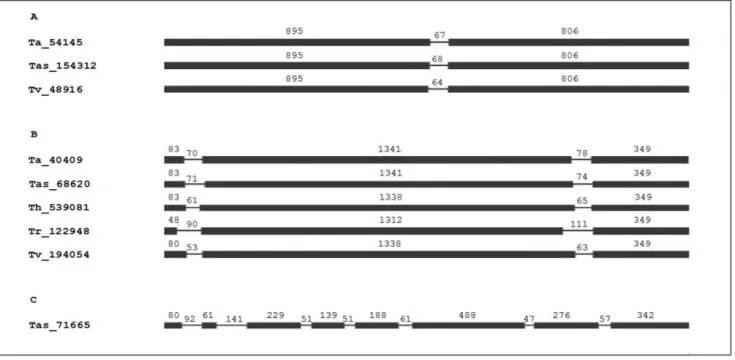
The nucleotide sequence length of Trichoderma spp. laccase encoding genes varies between 1765 (Tas_154312) and 2303 bp (Tas_71665). The GC percentage of these genes ranges from 46% (Tas_71665) to 58% (Tr_122948). The number and position of introns in laccase encoding genes in fungi have been employed to classify them into subfamilies [11,14]. The structure of the genes found in the five species ofTrichoderma separates them into three subfamilies. In each subfamily, the number of introns is preserved, and their positions are similar (Fig. 1). The first subfamily exhibits an intron between 64 and 68 bp in size and is made up of a gene of each one of the speciesT. atroviride, T. asperellum and T. virens
(Fig. 1A). The second subfamily is characterized by having two introns of between 53 and 111 bp, with one of these genes being found in the five species ofTrichoderma (Fig. 1B). In general, the subdivision of Trichoderma laccase encoding genes in these two
Table 1.Observed signature sequences in laccases.
Signature Reference
Axial coordination Leu or Phe [32]
L1 H-W-H-G-X9-D-G-X5-QCPI [29]
L2 G-T-X-W-Y-H-S-H-X3-Q-Y-C-X-D-G-L-X-G-X-(FLIM)
L3 H-P-X-H-L-H-G-H
L4 G-(PA)-W-X-(LFV)-HCHI-DAE-X-H-X3-G-(LMF)-X3-(LFM)
SDS gate Ser143, Ser511 and Asp561 in TaLcc1 [42]
C-termini Asp-Ser-Gly-(Leu/Ile/Val) [31]
In the fungal laccase signature sequences L1–L4, an X represents an undefined residue, whereas the multiple residues within brackets represent a partially conserved residue.
subfamilies is in agreement with previous reports of a low number of introns in this type of genes in the ascomycetes. In two varieties ofG. gramminis,the gene sequences ofLAC1andLAC2present 2 introns [33], just as in the single gene for extracellular laccase reported for the aquatic speciesMyrioconiumsp. [34]. The presence of 3 introns has been reported in thelac2gene [35] in the case ofP. anserinaand in theBclcc1andBclcc2genes ofB. cinerea[4]. In the yeastH. acidophila,3 introns have been documented in the gene that encodes for the extracellular enzyme and 2 for the intracellular enzyme gene [7]. A unique case is that of the Tas_ 71665 gene ofT. asperellum, with a structure of seven introns and for which no orthologous genes were found in the other analyzed
Trichoderma species (Fig. 1C). The only ascomycetes laccase gene that surpasses this number is thelac-1gene ofC. parasitica, which possesses 12 introns [36]. These two last genes represent intermediaries in the structure of introns/exons between the majority of ascomycetes and the basidiomycetes C. cinereaandL. bicolor, in which two subfamilies of genes have been found whose number of introns varies between 13 and 15 [11,14]. The introns found in theTrichoderma laccase genes preserve the splicing sites that comply with the GT/AG rule [37]. Previous data show the usefulness of the number and location of introns for generating subfamilies of ascomycetes laccase genes. This information could be useful for the identification of genes with distinct functions within a single species or for recognizing the same gene in different species. This analysis is beyond the scope of the present study and should be conducted in greater detail in the future. Interestingly, as a demonstration of the usefulness of gene family classification based on the presence of introns, the subfamilies of laccase genes formed inTrichodermaagree with classes established according to laccase protein structural characteristics (see below).
Structural Characteristics ofTrichodermaLaccases In general, the identification of laccases according to their amino acid sequence involves the recognition of the four segments L1–L4 [29]. However, such regions are common to all MCO family members including ascorbate oxidases, ferroxidases and
bilirubin oxidases, meaning that their presence is not sufficient to confirm that a protein sequence corresponds to a laccase sensu stricto. Because of this finding, in the present study we also considered the SDS-gate and the C-terminus end, which are distinctive characteristics of ascomycetes laccases (Table 1). The set of structural characteristics found in the amino acid sequence ofTrichodermalaccases is detailed below.
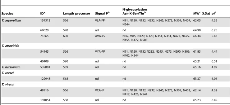
The analysis of the amino acid sequences of the 9 putative laccases that were found shows that their lengths are similar to the typical laccases of fungi (566–600 aa), and the calculated molecular mass for the protein sequences is between 61.83 and 66.84 kDa with acidic isoelectric points (Table 2). These results are in agreement with what has previously been reported for fungal laccases, which regularly have molecular weights between 60 and 70 kDa and which have isoelectric points (pI) that vary between pH 4.0–6.0 [3]. In particular, within the genus
Trichoderma, purified laccases from the wild strains WL1 of T. harzianum and CTM 10476 of T. atroviride presented, in their glycosylated form, molecular weights of 79 and 80 kDa, re-spectively [23,24]. Analysis in SWISS-MODEL of the proteins found in the genomes reviewed in this study showed that each one of the sequences is formed by three cupredoxin domains that are ordered in sequential form and are common to all the MCO family members [30]. As expected, the residues of amino acids that bind to copper T1 were located in domain I, whereas the residues that coordinate coppers T2/T3 were distributed between domains I and III (Fig. 2).
Laccases are distinguished by the presence of four consensus sequences, L1–L4, which possess a length of between 8 and 24 amino acid residues and are distributed along the polypeptide chain. Within these regions, one finds amino acid residues that serve as ligands for the copper atoms, as well as other preserved or partially preserved residues, which are critical for maintaining the conformational folding of the enzyme. Such characteristic sequences are found based on the multiple alignment of more than 100 laccases of plants and fungi [29] and represent the distinctive mark for the identification of putative new laccases. Table 2.Accession numbers and structural characteristics of laccasessensu strictofromTrichodermaspp.
Species IDa Length precursor Signal Pb
N-glycosylation
Asn-X-Ser/Thrb MWc(kDa) pId
T. asperellum 154312 566 VLA-FP N91, N120, N132, N232, N245, N273, N309, N409, N544
62.05 4.35
68620 590 nd nd 64.90 6.25
71665 600 AVA-LS N36, M85, N129, N320, N351, N351, N421, N425,
N455, N472, N508
66.34 5.43
T. atroviride
54145 566 VYA-FP N91, N120, N132 N232, N245, N273, N290, N309,
N402, N544
61.83 4.44
40409 590 nd nd 65.21 6.51
T. harzianum 539081 589 nd nd 65.16 4.97
T. reesei
122948 568 nd nd 63.37 6.06
T. virens
48916 566 VCA-IP N91, N120, N132, N232, N245, N273, N309, N402,
N412, N426, N544
62.14 4.32
194054 588 nd nd 65.23 6.49
aID, identification number in the
Trichodermaspecies genome;bDenotes the location of signal peptide cleavage sites in amino acid sequences from
Trichoderma laccases; nd, non detected.cMW, estimated molecular weight.dpI, predicted isoelectric point.
Regions L1–L4 in theTrichodermasequences have a high degree of similarity with the consensus designated by [29] (Fig. 2). However, some amino acid residues differ from the consensus. This finding is especially evident in L2, where Thr is replaced by a Ser in the second position. This change of amino acid is found in the Lac2 of
G.graminisvar.triticiandG. graminisvar.graminis[33], as well as in the Lac1 ofB. fuckeliana(anamorph =B. cinerea) [4], both of which are ascomycete fungi. In this same segment, in Trichoderma,
changes in the consensus QYCDGL are observed: Tyr is replaced by Ala, Cys by Ser/Ala/Trp, Asp by Gly/Glu and Leu by Val (Fig. 2). Thus, the results obtained here show that although laccases of basidiomycetes fully comply with the consensus designated by L2 [15], this region varies considerably in ascomycete laccases. In segments L1 and L4 of the Trichoderma,
gene changes that involve residues of amino acids with pro-pensities toward similar conformations or similar hydropathic indices are observed. For example, in L1, Trp is replaced by Phe (Fig. 2). This same change is observed inC. parasiticaLac3 [38]. In segment L4, the amino acid located 10 residues downstream from the preserved Cys corresponds to the axial position of copper T1. This residue is usually a Met in the MCOs; however, in the laccases, the Met residue is replaced by a Leu or Phe [39], as in the
Trichodermaspp. putative laccases (Fig. 2).
Axial coordination is one of the factors affecting the redox potential (E0) of laccases [39]. It has been previously suggested that
laccases with highE0(700–800 mV) possess a Leu or Phe in axial
coordination, whereas laccases with a Met residue have lowE0
(500 mV). Based on the substitution of this residue, laccases are classified into three classes: Lac 1 (M, Met), Lac 2 (L, Leu) and Lac 3 (F, Phe) [32]. Taking this characteristic into account, the putative laccases Ta_40409, Tas_68620, Th_539081, Tr_122948 and Tv_194054, are classified as Lac 2, whereas Tas_71665, Tas_154312, Ta_54145 and Tv_48916 are classified as Lac 3. These results are consistent with the hypothesis that Lac 1 laccases are primarily present in plants [32]. This classification has been
performed in one isoenzyme of each of the ascomycetes: C. parasitica, N. crasa, and P. anserina, belonging to class 2 [32]. Although this classification is still used to elucidate the functional relatedness between laccases, it is important to take into account that crystallographic data obtained from native high potential laccase of T. versicolor (TvL) have helped to describe other structural characteristics that might contribute to highE0in these
enzymes [40]. Such structural features include a reduction of electron density in the metal and the ligating amino acid. In high redox potential laccases, the distance between Cu+2
and one of the histidines from the T1 binding pocket is longer compared with that in enzymes of the middle potential group; the hydrogen bond between Glu-460 and Ser-113 in TvL seems to be responsible for this [40]. Interestingly, this particular serine is conserved in
Trichodermalaccases, but the Glu residue is not present. Further-more, it is well known that several factors can affect theE0value of
metalloproteins, including electrostatic intramolecular interac-tions, and solvation effects [41]. It is important to conduct further studies that allow for determining those factors involved in the modulation of redox potential inTrichodermalaccases.
Laccases have been the subject of intense investigation directed at understanding both their catalytic mechanism and the molecular determinants that modulate their broad range of E0
[40]. Although the laccase reaction scheme is not entirely understood, it is known that both binding and the oxidation of the substrate occurs in the T1 site and the electrons are transferred to the T2/T3 center, where the reduction of molecular oxygen takes place [40]. The reduction of a dioxygen molecule to two water molecules requires four electrons and four protons. The electron transfer pathway to the trinuclear center corresponds to the preserved motif Hys-Cys-Hys located at L4 [39], which is present inTrichodermalaccases (Fig. 2). Conversely, proton transfer is assisted by the so-called SDS-gate [30]. This gate is formed by two residues of Ser and one of Asp and is conserved in ascomycete laccases but has not been detected in basidiomycete laccases. InT.
Figure 1. Intron positions within the laccase genes ofTrichodermaspp. define three gene subfamilies.The thin lines indicate intron positions and the black lines indicate the exons. The first subfamily (A) contains only one intron, the second subfamily (B) contains two introns and the final (C) subfamily contains seven introns.
arenarialaccase TaLcc1, this gate is formed by Ser143, Ser511 and Asp561 [42]. Multiple alignment with TaLcc1 identified the SDS-gate amino acids inTrichodermaspp. laccases (Fig. 2). However, in Tas_154312 and Ta_54145, the amino acid that corresponds to Ser143 in TaLcc1 is replaced by Gly and in Tv_48916, by Ala. Additionally, in Th_539081, the residue that corresponds to Ser511 in TaLcc1 is replaced by a Thr (Fig. 2). This result suggests that Trichoderma laccases have adopted various strategies to facilitate the transfer of protons to the trinuclear site, thus modifying its catalytic activity.
An essential aspect of the catalytic activity of laccases is the mode of interaction and reaction with various substrates. The availability of a chemical compound to be used as a laccase substrate depends on both the nature and position of the substituents in the phenolic ring of the compound and also on the chemical environment of the sustrate binding site. The
amino acid residues that constitute the substrate cavity form loops and are founds in domains II and III. In the laccases of various organisms, the loops have different amino acid compositions, which results in diversity in the size and shape of the substrate binding site [43]. The nine Trichoderma spp. proteins present the substrate binding sites that are described in the tridimensional structure of crystallized laccases. As in other laccases, the sequences that form loops I-IV are little conserved in comparison with MaL and TaLcc1 (wild proteins of M. albomycesand T. arenaria, respectively), except in loop IV in C7– C8, which is also part of the L4 segment (Fig. 2). It has been determined that Pro192 in MaL (Ala193 in TaLcc1) in the loop I/B1–B2 interacts with the organic substrate [31]. This residue is present in five Trichoderma spp. laccases, although in this position, Th_539081 and Tv_194054 possess a Gln residue whereas Tas_68620 and Tas_71665 have Thr and Ser, Figure 2. Alignment of laccase sequences fromTrichodermaspp.The alignment was constructed with the Clustal X multiple-sequence alignment program. The accession number of each sequence in the JGI GeneBank is indicated on the left of the alignment. An asterisk indicates that the residues at a position are identical in all sequences in the alignment; a colon indicates that conserved substitutions have been observed and a period indicates semiconserved substitutions. Putative signal sequences are indicated by italics and the mitochondrial targeting peptides are enclosed in boxes. The conserved residues involved in copper binding are in red, and the complete L1–L4 regions are indicated by a double line under the alignment. The sequences of potential substrate loops were identified based on loops I-IV of the rMaL [44] and TaLcc1 [42], laccases ofM. albomycesandT. arenaria, respectively, and are underlined with a bold line. Amino acids shaded in yellow indicate residues in contact with the substrate. The residues forming the SDS gate are shaded in green in color, and the amino acid shaded in blue classified the laccases as class 1 (Met), class 2 (Leu) or class 3 (Phe). The conserved C-termini are in dark blue.
respectively (Fig. 2). In B4–B5, fungal laccases typically have Glu or Asp as the substrate ligand [31,42]. Directed mutagen-esis studies performed on MaL have shown that the Glu235 (Asp 236 in TaLcc1) carboxy group is of great importance for substrate binding because it stabilizes the cationic radical that is formed when Hys508 initiates the catalytic cycle [44]. However, in Tas_71665, Tr_122948 and Tv_194054, this residue is replaced by Ala. The multiple alignment of these sequences with other members of the MCO family revealed that in the same position, Ala is only present in a laccase of the dermatophyte fungus A. gypseum (anamorph =Microsporum gyp-seum). In the recombinantM. albomyceslaccase (rMaL) expressed in T. reesei, the Cys residue of the tripeptide Ala297-Cys298-Gly299 (Leu297-Cys298-Ala297-Cys298-Gly299 in TaLcc1) located in loop II/ B7-B8 is conserved inTrichodermaspp. laccases. This amino acid is also involved in substrate binding and was found inN. crassa, P. anserina and C. parasitica laccases [44]. Furthermore, the involvement of rMaL Phe427 (Val428 in TaLcc1) of loop IV/ C4–C5 in aligning substrate molecules in the correct orientation for oxidation has been suggested [44]. In the laccases of various organisms, including Trichoderma spp., this residue varies considerably, although the majority of basidiomycete laccases display Pro in this position. The differences found in the loop sequences of Trichoderma spp. laccases suggest a low substrate specificity and, most likely, various catalytic capacities, which indicates that each possesses different physiological functions, which is reinforced by subcellular localization (see below).
A preserved segment of four residues of amino acids has been identified at the C-terminal end ofTrichodermaspp. laccases (Fig. 2), a highly conserved sequence in ascomycetes corresponding to the consensus Asp-Ser-Gly-(Leu/Ile/Val). In laccases of the ascomy-cetesM. albomyces [31],P. anserina[35],T. arenaria[42],N. crassa
[45] and M. thermophila [46] the C-terminal end is post-translationally processed, leaving the active protein with the sequence Asp-Ser-Gly-Leu (DSGL) as the final amino acid residues at this end. As in the Trichoderma spp. laccases, the majority of ascomycete laccases do not present this C-terminal extension, which is removed post-translationally. Determination of the three-dimensional structure of MaL and TaLcc1 revealed that the C-terminal end DSGL is packed within a tunnel that leads to the trinuclear site and forms a plug [31]. In the crystal structure of other known laccases, this cavity is open and allows molecular oxygen to access the catalytic site [42]. In MaL and TaLcc1, the C-terminus plug impedes the movement of molecular oxygen and other solvents to the enzyme. Furthermore, the C-terminus carboxyl group forms a hydrogen bridge with the lateral chain of Hys140 in MaL (Hys141 in TaLcc1), which also coordinates copper T2. Directed mutagenesis studies performed on MaL cDNA revealed that a change or deletion of the C-terminal end dramatically affects enzyme activity [47]. Given those findings, it was suggested that ascomycete laccases use the C-terminal DSGL plug to carry out their catalytic function, forming a proton transfer pathway [42]. The C-terminus block appears to be a characteristic trait of ascomycete laccases because it has not been described in basidiomycete laccases. Nevertheless, Rigidopurus lignosus R1L laccase presents the C-terminus sequence DSGLA. Among basidiomycete laccases, R1L is the most closely related phyloge-netically with ascomycete laccases; for this reason, it was initially suggested that this sequence of the C-terminal end was more an evolutionary relic than a functional characteristic of the enzyme [39]. However, more recent observations documented in this study have shown that the C-terminal end DSGL is not an evolutionary relic in fungi, as it provides important functions for the ascomycete enzyme. Furthermore, even when laccases of basidiomycetes lacks
DSGL motif, it was recently established2using directed evolution
approach 2 that the C-terminus would play a role in enzyme
performance by influencing optimal pH and Km values for phenolic compounds [48]. This evidence requires further comparative studies between ascomycetes and basidiomycetes regarding the evolution of the C-terminal end and its functional role.
In the course of the analysis to identify laccasessensu stricto in
Trichoderma and to compare them with those reported in other ascomycetes, the particular case of the 6 enzymes reported for
Aspergillus niger[8] emerged, specifically those designated as Mco G, Mco J and Mco M. When we perform an analysis to find the signatures of laccasessensu stricto(Table 1), we find that the Mco G protein lacks the DSGL motif. Furthermore, Mco J and Mco M have a deletion of approximately 50 aa immediately after the L4 segment, which includes the DSGL motif. Interestingly, it was found that these two last ‘‘truncated’’ enzymes oxidize a limited number of substrates, whereas the former attacks all probed substrates. This suggests that some laccases from ascomycetes can be partially functional even if not presenting all the elements to be considered as laccasessensu stricto. These beings a special case and beyond the objectives of the present work, further analyses of these three enzymes were not performed and they were excluded from the phylogenetic analysis (see below).
Over the course of the evolutionary process, laccases have maintained a high degree of similarity in terms of amino acid sequence and three-dimensional structure. Generally, the laccase sequences of members of a group of fungi exhibit levels of amino acid identity of 50% or more, whereas the identity levels between sequences of members of different groups is approximately 30% [49]. These identity values are met, in general, forTrichodermaspp. laccases. The identity percentage among the 9 laccases varied between 30 and 88% (Table 3). The Ta_54145/Tas_514312/ Tv_48916 triad is more similar among themselves with an identity value above 83%. The rest of the laccases possess identity values between 51 and 86%. An identity percentage comparison between laccases of a single species has not been conducted in ascomycetes. In the case of basidiomycetes, the percentages of identity of the 8
C. cinerealaccases varied between 46 and 77% [50], whereas in the case ofP. ostreatus,these values were between 45 and 89% between the 6 enzymes found [49]. Thus, Trichoderma laccases present a greater interval of identity than those found in basidiomycete species, indicating a strong selective pressure on the various genes. With respect to their relationship to the laccases of other fungal species, Tas_154312, Ta_54145 and Tv_48916 proteins share 53% identity withG. graminis var. triticiandG. graminis var. graminis
Lac2 [33] and an identity of between 63 and 65% withF. oxysporum
Lcc4 laccase [10]. The group of enzymes Tas_68630, Ta_40409, Th_539081, Tr_122948 and Tv_194054 possess an identity greater than 63% withF. oxysporumlacc1 [10] and less than 30% with other ascomycete laccases. Interestingly, F. oxysporum lcc1
laccase is an intracellular laccase like the Trichoderma laccases, which show a stronger phylogenetic relation with it (see below). The Tas_71665 protein has an identity of between 51.4 and 56.2% with the putative laccases ofB. fuckeliana,S. sclerotiorumand
Prediction of Subcellular Localization ofTrichodermaspp. Laccases and Possible Physiological Functions
The majority of known fungal laccases are monomeric proteins with extracellular activity, although intracellular laccases have also been identified, particularly in white rot fungi [3]. The localization of laccases is associated with their physiological function and determines the range of substrates available for the enzyme. In fungi, the functions of extracellular laccases related to the degradation of lignocellulose material, recycling of organic material, reduction of oxidative stress and pathogenesis toward plants and animals have been extensively studied [3,4]. Of the nine Trichoderma spp. laccases, it was determined that four correspond to extracellular laccases: one each inT. atrovirideand
T. virens,and two inT. asperellum.The rest are intracellular proteins found in the five species analyzed (Table 2). The putative signal peptide of the extracellular laccases corresponds to the first 18 residues and presents the typical characteristics of signal peptides, that is, a highly hydrophobic region and Ala and Val residues in positions -1 and -3, respectively, relative to the cleavage site [51]. The mature forms of the laccases Tas_71665, Tas_154312, Ta_54145 and Tv_48916 possess between 9 and 11 putative N-glycosylation sites (Table 2). The average N-glycosylation is usually between 10 and 25%, although laccases with a carbohydrate content of greater than 30% have been detected [2]. Glycosylation influences enzyme secretion, and it has been suggested to play an important role in catalytic center stabilization, protection against hydrolysis, copper retention, and laccase thermal stability [52]. Extracellular laccase activity has previously been reported in
Trichodermaspp. [18,21], arriving in certain cases at purification of the protein [23,24]. Levasseur et al. [25] reported homologous overexpression of theT. reeseigene 124079 in the strain Rut-C30, and even when the recombinant protein presented biochemical properties similar to those reported for other laccases, the structural analysis carried out in this study suggests that the enzyme TrLAC1 studied by those authors corresponds to a pigment synthesis MCO and not to a laccase in sensu stricto. That is, the amino acid sequence obtained for this enzyme does not present either the SDS-gate or the C-terminal end DSGL/I/ V. Recently, Catalanoet al.[26] evaluated the participation of the extracellular T. virens LCC1 laccase (corresponding to laccase Tv_48916 analyzed in this study) in mycoparasitism towards sclerotia of the phytopathogens B. cinereaand S. sclerotiorum. The hypothesis in that study was that LCC1 is capable of attacking the sclerotia melanin of the studied fungi. However, although their results show that the enzyme can participate in mycoparasitism by
T. virensagainst S. sclerotiorum, the same result was not obtained againstB. cinereasclerotia, making it necessary to conduct more studies in this area. It would also be important to experimentally evaluate other functions of extracellularTrichodermalaccases, such as those cited at the beginning of this section.
The analysis carried out with various bioinformatic packages suggests the intracellular localization of laccases Ta_40409, Tr_122948, Tv_194054, Th_539081 and Tas_68620. Unlike extracellular laccases, little is known about the activity of intracellular laccases, both for the genusTrichodermaand for other fungal species. In several reports, the presence of laccase activity associated with the membrane or periplasmic space during the maturation ofT. atroviride, T. virideandT. harzianumconidia [21,22] has been suggested. The results of the present study show that
Trichodermaspp. do not possess laccases associated with the plasma membrane, such laccase activity associated with conidia reported in those studies is possibly due to the extracellular enzyme that remained trapped in the periplasmic space or the cell wall during the process of maturation of these structures. In this sense, fungal proteins have been reported to be located either adhering to the cell wall or in the extracellular medium, among which are included hydrophobins and adhesins [53,54], as well as hydrolytic enzymes [55]. Although, in certain cases, the interaction of the protein with the cell wall is only transitory, occurring while the protein reaches the extracellular medium, in other cases, specific mechanisms have been proposed for the retention of the proteins in the wall and their simultaneous localization in the extracellular medium [56]. There has also been documentation of A.oryzae
enzymes that are secreted into the medium when the fungus is grown in a solid substrate but are retained in the cell wall when the fungus grows in submerged cultures [57]. This finding indicates that it is possible that someTrichodermalaccases remain trapped in the periplasmic space or cell wall during conidia maturation, which is a possibility that should be investigated more thoroughly in the future. In ascomycetes, the activity of intracellular laccase has been detected in H. acidophila and has been related to the synthesis of melanin [7]. In the human pathogenic fungus
Cryptotoccus neoformans, laccase activity is found to be associated with the membrane and constitutes a virulence factor [56], whereas the phytopathogenic fungus F. oxysporum possesses two intracellular laccases, Lcc1 and Lcc3, which may be involved in the protection of the fungus against oxidative stress and toxic compounds [10]. In the basidiomycetes L. bicolor [11] and P. ostreatus[15], these isoenzymes are involved in the development of the fruiting body. In addition, it is possible that the intracellular Table 3.Identity (%) among protein sequences of the members of theTrichodermalaccase family.
Protein 2 3 4 5 6 7 8 9
1 Tas_68620 85.4 76.9 77.7 77.1 53.1 31.7 32.1 30.9
2 Ta_40409 75.2 76.1 76.1 53.1 33.6 33.4 32.7
3 Th_539081 86.7 75.7 52.1 30.5 31.5 30.5
4 Tv_194054 77.3 51.9 32.1 32.4 31.0
5 Tr_122948 51.0 30.6 31.0 30.8
6 Tas_71665 30.9 31.2 30.4
7 Tas_154312 88.5 86.2
8 Ta_54145 83.7
9 Tv_48916
laccases of fungi participate in the transformation of low molecular weight phenolic compounds produced in the cell [3]. Laccases associated with conidia are linked to the synthesis of pigments and other substances that protect the cell from stress factors, such as enzymatic lysis, temperature and UV light [3,10]. It is possible that
Trichodermaintracellular laccases are related to any of the processes described above; further experimental work is needed to confirm any of these functions.
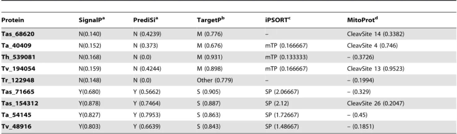
Surprisingly, when using various bioinformatics programs to analyze four of the laccases classified as intracellular by the package SignalP 4.0, the laccases presented a signal peptide and processing characteristics of mitochondrial localization (Fig. 2; Table 4). The MitoProt program shows processing sites congruent with the mitochondrially located peptide of the other packages (Table 4). These results should be viewed cautiously, as currently there are no reports of mitochondrial localization of laccases either in fungi or in any other biological group in which this enzymatic activity has been found.
With the unexpected finding of signs mitochondrial localization of laccase inTrichoderma, the first question that arises is whether this is feasible for enzymes that in fungi have only been reported as either cytoplasmic or associated with the plasma membrane or extracellular. Possible hypotheses, assuming that the prediction of mitochondrial localization is correct, arise from what has been documented for proteins in other fungi. There are examples in fungi of subcellular localization changes or the presence in two distinct cellular compartments of the same enzymatic activity. The latter case has been referred to as dual localization, dual targeting or dual distribution [58]. InS. cerevisiae,it has been found that up to a third of the proteins considered to be mitochondrial can present an alternative subcellular localization [59]. Among the best-studied examples of dual mitochondria-cytoplasmic localiza-tion in fungi are aconitase [60] and fumarase [61]. Moreover, in these two examples, the proteins present in mitochondria and cytoplasm come from the same gene, which generates a single transcript and a single translation product, that is, they are not isoenzymes in distinct compartments. However, in these two examples, the ‘‘original’’ localization of the protein is mitochon-drial, and the ‘‘new’’ localization is cytoplasmic, which is the opposite of what would be occurring with the products of
Trichodermalaccase proteins. Nevertheless, it is possible to consider the relocalization of a secretion protein to mitochondria in fungi. Recently it has been documented that tryptophan-rich sensory protein/peripheral-type benzodiazepine receptor (referred to as
TspO/MBR) that is found in the Golgi-associated secretory pathway in plants is directed toward mitochondria when expressed heterologously in S. cerevisiae [62]. The modification of the subcellular localization of a protein may be caused by a change of a single amino acid in the signal peptide, which can be generated by a single nucleotide mutation [63].
The second question that arises pertains to the function of a laccase in mitochondria. Experimental evidence suggests that a protein that reaches a new subcellular location can develop new functions [63]. Although it is difficult to speculate about the possible function of a mitochondrial laccase in fungi, it is feasible to establish hypotheses based on our understanding of the structural aspects of this enzyme. As mentioned above, laccases are enzymes that can bind four copper atoms, which are important for catalytic activity. Intracellular Cu+2
can have damaging effects because it induces the formation of reactive oxygen species (ROS); therefore, there are mechanisms that regulate its concentration [64]. Further, it has been documented that mitochondria possess a pool of copper that responds to changes in copper levels in the cytoplasm [65]. It is possible that the mitochondrial location of
Trichoderma laccases contributes to the homeostasis of mitochon-drial Cu+2
under particular circumstances, which is a possibility that would be important to evaluate experimentally in the future. The prediction of the subcellular localization of eukaryotic proteins is a complicated task in that there is always a certain degree of uncertainty; therefore, protocols and bioinformatics packages have been designed that have optimized the certainty of the prediction [66]. Although speculative hypotheses are proposed in agreement with bioinformatics and experimental evidence collected in eukaryotes in general and in fungi in particular, it would be most prudent to assume that the four Trichoderma
enzymes can be considered to be intracellular, which is supported by the phylogenetic analysis (see following paragraph). The here described role of putative mitochondrial signal peptides in the subcellular localization ofTrichoderma laccases can be experimen-tally verified in the future. One way to do this, is to design an expression vector in which the putative mitochondrial target sequences are fused to the green fluorescent protein (gfp) gene and monitoring the localization of the expressed recombinant protein.
Phylogenetic Analysis ofTrichodermaspp. Laccases A phylogenetic analysis performed according to two distinct criteria separatedTrichodermalaccases into two distinct clades with a bootstrap value of 99%. Interestingly, all intracellular laccases
Table 4.Signal peptide sequences fromTrichodermalaccases predicted by different software programs.
Protein SignalPa PrediSia TargetPb iPSORTc MitoProtd
Tas_68620 N(0.140) N (0.4239) M (0.776) – CleavSite 14 (0.3382)
Ta_40409 N(0.152) N (0.373) M (0.676) mTP (0.166667) CleavSite 4 (0.746)
Th_539081 N(0.168) N (0.0) M (0.931) mTP (0.133333) – (0.3726)
Tv_194054 N(0.159) N (0.4244) M (0.898) mTP (0.166667) CleavSite 13 (0.9523)
Tr_122948 N(0.148) N (0.0) Other (0.779) – – (0.1994)
Tas_71665 Y(0.680) Y (0.5662) S (0.905) SP (2.06667) – (0.329)
Tas_154312 Y(0.878) Y (0.7464) S (0.887) SP (2.12) CleavSite 26 (0.2047)
Ta_54145 Y(0.827) Y (0.7953) S (0.863) SP (1.72667) – (0.45)
Tv_48916 Y(0.803) Y (0.6639) S (0.843) SP (1.48667) – (0.1851)
The values between parentheses are the probability that the prediction is accurate.aN, non present; Y, present.bM, mitochondrial; S, secretory.cmTP, mitochondrial
targeting peptide; SP, secretory signal peptide.dCleavSite, location of signal peptide cleavage sites.
are grouped in the first of these clades. The remaining three extracellular laccases are included in the second clade with the exception of Tas_71665 (Fig. 3a), which grouped in the clade of intracellular proteins but in a different terminal branch.
AlthoughTrichodermaextracellular laccases exhibit close phylo-genetic relationships withF. oxysporum,G. gramminis,A. nigerandC. parasiticaorthologs, intracellular laccases show relationships withS. thermophile, N. crassa, S. macrospora, P. anserina, F. oxysporumandC. globosum orthologs. One may suggest the hypothesis that the phylogenetic closeness between these isoenzymes involves struc-tural similarities in terms of the regions and amino acids discussed above forTrichodermalaccases. It is important to emphasize that all the proteins included in the phylogenetic analysis of Figure 3a were ‘‘curated’’ according to the same criteria as those of
Trichoderma (Table 1) to ensure that they included only laccases
sensu stricto. Because of this approach, the phylogram generated here excludes proteins that were used in previous phylogenetic analyses of ascomycete laccases [12,25], which are possibly another members of MCO family. In the future, it will be important to consider this aspect of treatment to obtain more robust phylogenetic patterns that will provide a clearer idea of the evolutionary process of this enzymatic function in ascomycetes. This approach will allow for a better definition of laccases with similar functions among distinct species of this group of fungi.
The phylogenetic separation of the subfamilies of laccases is fully congruent with the analysis of sites or characteristic sequences of each subfamily of genes discussed above. Both the structure and number of genes found in the species of Trichoderma and the phylogenetic analysis suggest that the common ancestor of said genus possessed two laccases, with one being intracellular and the other extracellular.
Comparative genomic analysis together with the results of molecular phylogeny leads to the conclusion that the ancestral state ofTrichoderma/Hypocreawas mycoparasitical and that it later acquired saprophytic characteristics that helped it to pursue its prey through lignified substrates [27]. These studies have shown thatT. atrovirideis the earliest species within the genus, whereasT. virens and T. reesei appeared later. Furthermore, during the evolutionary process, T. reesei lost a significant quantity of the genetic information present inT. atroviride, which was retained in
T. virens[27]. The number of Trichoderma laccase genes and the phylogenetic analysis of the proteins coded by these genes are congruent with the described scenario (Fig. 3b). With the generated data, it is possible to support the hypothesis that during the process of speciation, species such asT. atrovirideandT. virens
maintained the original number of laccase genes present in the common ancestor, whileT. harzianumandT. resseirepresent species that lost copies of extracellular laccase. On the other hand, several lines of evidence strongly suggest that the Tas_71665 gene ofT. asperellumwas acquired through a horizontal gene transfer event. This gene has the following structural features, which are discussed in the previous sections: i) it has the lowest GC-content (46%) of all known laccase genes; ii) it has a unique structure with seven introns; iii) the genome context of this gene differs from those of other laccase genes inT. asperellum; iv) no orthologous genes were found in the other analyzedTrichoderma species; v) compared to
laccases in other genera of ascomycetes, this protein shows higher identity values (51.4–52.6%) with laccases of B. fuckeliana, S. sclerotiorumandP. tritici-repentis; vi) in the phylogenetic analysis, the protein encoded by this gene does not cluster toghether with other laccases from Trichoderma. Most of these features have been recognized as evidence of horizontal gene transfer in fungi [67]. It has been previously suggested that inS. macrosporaandP. anserina– belonging to the same Order as Trichoderma (Sordariales) – the acquisition of laccase genes occured through horizontal trans-ference – [12]; the author postulating the possibility of gene transfer fromS. sclerotiorum to S. macrospora.Taken together, our results strongly suggest thatT. asperellumacquired the Tas_71665 gene through horizontal gene transfer, either from one of the known necrotrophic phytopathogensB. fuckeliana,S. sclerotiorumand
P. tritici-repentis, or from a related fungus. This is noteworthy because there is experimental evidence showing mycoparasitic activity of Trichoderma, althought not specifically T. asperellum, againstB. fuckelianaandS. sclerotiorum[26], which indicates that this gene could have been acquired during the mycoparasitic process. This pattern of retention/loss/gain of laccase genes by groups of species inTrichodermamay reflect phenomena of selective pressure associated with the lifestyle of each of these species.
The phylogeny obtained shows that the hypothesis of two laccase genes in the commonTrichodermaancestor with later events of retention, loss or gain can be extended to the Ascomycetes group. In fact, this asymmetrical pattern of change in gene families has been reported for the case of family 28 of ascomycetes glycosyl hydrolases [68], where duplications and losses of genes in distinct groups of fungi within this subdivision are observed. Detailed studies of ascomycete genomes to locate more laccase isoenzymes and carry out a broader phylogenetic analysis would allow for the corroboration of this hypothesis.
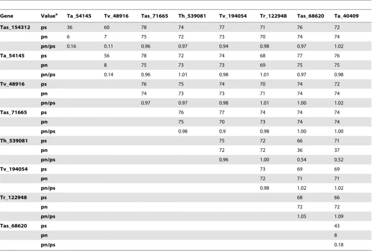
Analysis of the evolutionary patterns of Trichoderma laccases shows that the proportion of synonymous substitutions of the majority of enzymes varies between 56 and 78%, indicating saturation (Table 5). The exceptions were Tas_154312 and Ta_54145 (36%), as well as Tas_68620 and Ta_40409 (43%). The majority of the non-synonymous substitutions show values higher than 70%, indicating saturation. A low percentage of non-synonymous substitution is found in 36–37%, between 6 and 8%. These values, together with the percentage non-synonymous/ percentage synonymous (pn/ps) proportions, show that the majority of Trichoderma extracellular laccases evolved under a process of purifying selection with the exception of Ta_71665, which has values that indicate neutral modifications (Table 5). In the case of the intracellular laccases, the majority of values indicate neutral changes, although in the case of the pair Tas_68620 and Ta_40409, purifying selection is shown.
Levasseur et al. [25] found that the TrLACgene, identified by those authors as aT. reeseilaccase, evolved under positive selection. However, as mentioned above, our data suggest that this gene is not a laccasesensu strictobut rather is another member of the MCO family. To this date, there are no studies that would allow us to compare the evolutionary patterns ofTrichodermalaccases found in this study with those of ascomycete laccases.
Figure 3. Phylogenetic analysis of laccases and ofTrichodermaspecies.(A) Neighbor-joining tree of the deduced amino acid sequences of the fiveTrichodermalaccases and selected laccases of other ascomycetes, basidiomycetes and plants.The tree is calculated using the Jones-Taylor-Thornton (JTT) model in Mega Ver 5.05 based on a ClustalX alignment. Bootstrap values are from 1000 replications. The scale bar indicates a distance equivalent to 0.2 amino acid substitutions per site. Species and strains are indicated in the experimental procedures. Upward triangles indicate Trichodermaintacellular laccases and downward triangles extracellular ones. Green circles denote the laccases fromM. albomyces[44] andT. arenaria [42]. (B) Phylogenetic tree ofTrichodermaspecies showing gain, loss and retention of laccase genes.
Conclusions
The search for laccasessensu strictoin ascomycetes involves the location in the amino acid sequence of motifs that are not present in basidiomycete laccases. The characterization of such motifs allows for better description of the possible functional properties of the proteins. The identification of ascomycete laccasessensu strictois important to conduct robust phylogenetic analyses of this enzymatic function. The genusTrichodermahas preserved a limited number of functional laccase genes in the course of the evolutionary process. Structural and phylogenetic evidence suggests that the common ancestor of Trichoderma spp. had two laccase genes, with one being intracellular and the other extracellular. Species within the genusTrichodermatend to preserve
intracellular laccase activity, whereas evolution patterns of extracellular activity are variable. In the case of T. asperellum, there is strong evidence of horizontal gene transference through the mycoparasitic proceses. The herein presented data will contribute to the understanding of the functional role of laccases in the genus Trichoderma and to the optimization of their biotechnological applications.
Author Contributions
Conceived and designed the experiments: GVM MSVG. Performed the experiments: SVCG. Analyzed the data: GVM SVCG MSVG. Contrib-uted reagents/materials/analysis tools: MSVG GVM. Wrote the paper: SVCG GVM.
References
1. Quintanar L, Stoj C, Taylor AB, Hart PJ, Kosman DJ, et al. (2007) Shall we dance? How a multicopper oxidase chooses its electron transfer partner. Acc Chem Res 40: 445–452.
2. Giardiana P, Faraco V, Pezzella C, Piscitelli A, Vanhulle S, et al. (2010) Laccases: a never-ending story. Cell Mol Life Sci 67: 369–385.
3. Baldrian P (2006) Fungal laccases – occurrence and properties. FEMS Microbiol Rev 30: 215–242.
4. Schouten A, Wagemakers L, Stefanato FL, van der Kaaij RM, van Kan JAL (2002) Resveratrol acts as a natural profungicide and induces self-intoxication by a specific laccase. Mol Microbiol 43: 883–894.
5. Mate´ D, Garcı´a-Ruı´z E, Camarero S, Alcalde M (2011) Directed evolution of fungal laccases. Curr Genomics 12: 113–122.
6. Shraddha, Shekher R, Sehgal S, Kamthania M, Kumar A (2011) Laccase: microbial sources, production, purification, and potential biotechnological applications. Enzyme Res doi:10.4061/2011/217861.
7. Tetsch L, Bend J, Ho¨lker U (2006) Molecular and enzymatic characterization of extra- and intracellular laccases from the acidophilic ascomycete Hortaea acidophila. Anton Leeuw 90: 183–194.
8. Tamayo-Ramos JA, Barends S, Verhaert RMD, de Graaff LH (2011) The
Aspergillus niger multicopper oxidase family: analysis and overexpression of laccase-like encoding genes. Microb Cell Fact 10: 78.
9. Hoshida H, Nakao M, Kanazawa H, Kubo K, Hakukawa T, et al. (2001) Isolation of five laccase gene sequences from the white-rot fungus Trametes sanguineaby PCR, and cloning, characterization and expression of the laccase cDNA in yeast. J Biosci Bioeng 92: 372–380.
Table 5.Synonymous and non-synonymous substitution rates (%) between lacccase genes ofTrichodermaspp.
Gene Valuea Ta_54145 Tv_48916 Tas_71665 Th_539081 Tv_194054 Tr_122948 Tas_68620 Ta_40409
Tas_154312 ps 36 60 78 74 77 71 76 72
pn 6 7 75 72 73 70 74 74
pn/ps 0.16 0.11 0.96 0.97 0.94 0.98 0.97 1.02
Ta_54145 ps 56 78 72 74 68 77 76
pn 8 75 73 73 69 75 75
pn/ps 0.14 0.96 1.01 0.98 1.01 0.97 0.98
Tv_48916 ps 76 75 74 70 74 72
pn 74 73 73 71 74 74
pn/ps 0.97 0.97 0.98 1.01 1.00 1.02
Tas_71665 ps 76 77 74 74 74
pn 75 70 73 74 74
pn/ps 0.98 0.9 0.98 1.00 1.00
Th_539081 ps 75 72 66 71
pn 72 72 36 37
pn/ps 0.96 1.00 0.54 0.52
Tv_194054 ps 73 69 69
pn 72 71 71
pn/ps 0.98 1.02 1.02
Tr_122948 ps 68 66
pn 72 72
pn/ps 1.05 1.09
Tas_68620 ps 43
pn 8
pn/ps 0.18
Genes designed as in the JGI GeneBank.aps, percentage of synonymous substitutions; pn, percentage of non-synonymous substitutions; pn/ps, ratio of synonymous,
10. Cordoba CD, Roncero MIG (2008) Functional analyses of laccase genes from
Fusarium oxysporum. Mycology 98: 509–518.
11. Courty PE, Hoegger PJ, Kilaru S, Kohler A, Bue´e M, et al. (2009) Phylogenetic analysis, genomic organization, and expression analysis of multi-copper oxidases in the ectomycorrhizal basidiomyceteLaccaria bicolor. New Phytol 182: 736–750. 12. Po¨ggeler S (2011) Evolution of multicopper oxidase genes in coprophilous and non-coprophilous members of the order sordariales. Curr Genomics 12: 95–103. 13. Hoegger PJ, Kilaru S, James TY, Thacker JR, Ku¨es U (2006) Phylogenetic comparison and classification of laccase and related multicopper oxidase protein sequences. FEBS J 273: 2308–2326.
14. Kilaru S, Hoegger PJ, Ku¨es U (2006) The laccase multi-gene family inCoprinopsis cinereahas seventeen different members that divide into two distinct subfamilies. Curr Genet 50: 45–60.
15. Lettera V, Piscitelli A, Leo G, Birolo L, Pezzella C, et al. (2010) Identification of a new member ofPleurotus ostreatuslaccase family from mature fruiting body. Fungal Biol 114: 724–730.
16. Kubicek CP, Bissett J, Druzhinina I, Kullnig-Gradinger C, Szakacs G (2003) Genetic and metabolic diversity ofTrichoderma: a case study on South-East Asian isolates. Fungal Genet Biol 38: 310–319.
17. Schuster A, Schmoll M (2010) Biology and biotechnology ofTrichoderma. Appl Microbiol Biotechnol 87: 787–799.
18. Assavanig A, Amornkitticharoen B, Ekpaisal N, Meevootisom V, Flegel TW (1992) Isolation, characterization and function of laccase fromTrichoderma. Appl Microbiol Biotechnol 38: 198–202.
19. Krastanov AI, Gochev VK, Girova TD (2007) Nutritive medium dependent biosynthesis of extracellular laccase fromTrichodermaspp. Bulg J Agric Sci 13: 349–355.
20. Gochev VK, Krastanov AI (2007) Isolation of laccase producingTrichodermaspp. Bulg J Agric Sci 13: 171–176.
21. Ho¨lker H, Dohse J, Ho¨fer M (2002) Extracellular laccases in ascomycetes
Trichoderma atrovirideandTrichoderma harzianum. Folia Microbiol 47: 423–427. 22. Pokorny´ R, Vargovicˇ P, Ho¨lker U, Janssen M, Bend J, et al. (2005)
Developmental changes inTrichoderma virideenzymes abundant in conidia and the light-induced conidiation signalling pathway. J Basic Microbiol 45: 219–229. 23. Sadhasivam S, Savitha S, Swwaminathan K (2009) Redox-mediated de-colorization of recalcitrant textile dyes byTrichoderma harzianumWL1 laccase. World J Microbiol Biotechnol 25: 1733–1741.
24. Chakroun H, Mechichi T, Martinez MJ, Dhouib A, Sayadi S (2010)Trichoderma atroviride: application on bioremediation of phenolic compounds. Process Biochem 45: 507–513.
25. Levasseur A, Saloheimo M, Navarro D, Andberg M, Pontarotti P, et al. (2010) Exploring laccase-like multicopper oxidase genes from the ascomycete
Trichoderma reesei: a functional, phylogenetic and evolutionary study. BMC Biochem 11: 32.
26. Catalano V, Vergara M, Hauzenberger JR, Seiboth B, Sarrocco S, et al. (2011) Use a non-homologous end-joining-deficient strain (delta-ku70) of the biocontrol fungusTrichoderma virensto investigate the function of the laccase gene lcc1 in sclerotia degradation. Curr Genet 57: 13–23.
27. Kubicek CP, Herrera-Estrella A, Seidl-Seiboth V, Martı´nez DA, Druzhinina IS, et al. (2011) Comparative genome sequence analysis underscores mycoparasitism as the ancestral life style forTrichoderma. Genome Biol 12: R40.
28. Martı´nez D, Berka RM, Henrissat B, Saloheimo M, Arvas M, et al. (2008) Genome sequencing and analysis of the biomass-degrading fungusTrichoderma reesei(syn.Hypocrea jecorina). Nat Biotechnol 26: 553–560.
29. Kumar S, Phale P, Durani S, Wangikar PP (2003) Combined sequence and structure analysis of the fungal laccase family. Biotechnol Bioeng 83: 386–394. 30. Hakulinen N, Andberg M, Kallio J, Koivula A, Kruus K, et al. (2008) A near atomic resolution structure of aMelanocarpus albomyceslaccase. J Struct Biol 162: 29–39.
31. Hakulinen N, Kiiskinen LL, Kruus K, Saloheimo M, Paananen A, et al. (2002) Crystal structure of a laccase fromMelanocarpus albomyceswith an intact trinuclear copper site. Nature Struct Biol 9: 601–605.
32. Eggert C, LaFayette PR, Temp U, Eriksson KEL, Dean JFD (1998) Molecular analysis of a laccase gene from the white rot fungusPycnoporus cinnabarinus. Appl Environ Microbiol 64: 1766–1772.
33. Litvintseva AP, Henson JM (2002) Cloning, characterization, and transcription of three laccase genes fromGaeumannomyces graminisvar.tritici, the take-all fungus. Appl Environ Microbiol 68: 1305–1311.
34. Martin C, Pecyna M, Kellner H, Jehmlich N, Junghanns C, et al. (2007) Purification and biochemical characterization of a laccase from the aquatic fungusMyrioconiumsp. UHH 1–13–18–4 and molecular analysis of the laccase-encoding gene. Appl Microbiol Biotechnol 77: 613–624.
35. Ferna´ndez LJ, Stahl U (1996) Isolation and characterization of a laccase gene fromPodospora anserina. Mol Gen Genet 252: 539–551.
36. Choi GH, Larson TG, Nuss DL (1992) Molecular analysis of the laccase gene from the chestnut blight fungus and selective suppression of its expression in an isogenic hypovirulent strain. Mol Plant Microbe Interact 5: 119–128. 37. Shapiro MB, Senepathy P (1987) RNA splice junctions of different classes of
eukaryotes: sequence statistics and functional implications in gene expression. Nucleic Acids Res 15: 7155–7174.
38. Chung HJ, Kwon BR, Kim JM, Park SM, Park JK, et al. (2008) A tannic acid-inducible and hypoviral-regulated laccase3 contributes to the virulence of the chestnut blight fungusCryphonectria parasitica. Mol Plant Microbe In 21: 1582– 1590.
39. Garavaglia S, Cambria MT, Miglio M, Ragusa S, Lacobazzi V, et al. (2002) The structure ofRigidoporus lignosuslaccase containing a full complement of copper ions, reveals an asymmetrical arrangement for the T3 copper pair. J Mol Biol 342: 1519–1531.
40. Piontek K, Antorini M, Choinowsky T (2002) Crystal structure of a laccase from the fungusTrametes versicolorat 1.90-A˚ resolution containing a full complement of coppers. J Biol Chem 277: 37663–37669.
41. Li H, Webb SP, Ivanic J, Jensen JH (2004) Determinants of the reactive reduction potentials of type-1 copper sites in proteins. J Am Chem Soc 126: 8010–8019.
42. Kallio JP, Gasparetti C, Andberg M, Boer H, Koivula A, et al. (2011) Crystal structure of an ascomycete fungal laccase fromThielavia arenaria – common structural features of asco-laccases. FEBS J 278: 2283–2295.
43. Ge H, Gao Y, Hong Y, Zhang M, Xiao Y, et al. (2010) Structure of native laccase B fromTrametessp. AH28–2. Acta Cryst F66: 254–258.
44. Kallio JP, Auer S, Ja¨nis J, Anderberg M, Kruus K, et al. (2009) Structure-function studies of a Melanocarpus albomyces laccase suggest a pathway for oxidation or phenolic compounds. J Mol Biol 392: 895–909.
45. German UA, Muller G, Hunziker PE, Lerch K (1988) Characterization of two allelic forms ofNeurospora crassalaccase amino- and carboxyl-terminal processing of a precursor. J Biol Chem 263: 885–896.
46. Bulter T, Alcalde M, Sieber V, Meinhold P, Schlachtbauer C, et al. (2003) Functional expression of a fungal laccase inSaccharomyces cerevisiaeby directed evolution. Appl Environ Microbiol 69: 987–995.
47. Andberg M, Hakulinen N, Auer S, Saloheimo M, Koivula A, et al. (2009) Essential role of the C-terminus in Melanocarpus albomyceslaccase for enzyme production, catalytic properties and structure. FEBS J 276: 6285–6300. 48. Pardo I, Vicente AI, Mate DM, Alcalde M, Camareno S (2012) Development of
chimeric laccases by directed evolution. Biotechnol Bioeng doi 10.1002/ bit.24588.
49. Pezzella C, Autore F, Giardina P, Piscitelli A, Sannia G, et al. (2009) The
Pleurotus ostreatuslaccase multi-gene family: isolation and heterologous expression of new family members. Curr Genet 55: 45–57.
50. Hoegger PJ, Navarro-Gonza´lez M, Kilaru S, Hoffmann M, Westbrook ED, et al. (2004) The laccase gene family inCoprinopsis cinerea(Coprinus cinereus). Curr Genet 45: 9–18.
51. Nielsen H, Engelbrecht J, Brunak S, von Heijne G (1997) Identification of prokaryotic and eukaryotic signal peptides and prediction of their cleavage sites. Protein Eng 10: 1–6.
52. Vite-Vallejo O, Palomares LA, Danta´n-Gonza´lez E, Ayala-Castro HG, Martı´nez-Anaya C, et al. (2009) The role of N-glycosylation on the enzymatic activity of aPycnoporus sanguineuslaccase. Enzyme Microb Technol 45: 233–239. 53. Martin F, Laurent P, de Carvalho D, Voiblet C, Balestrini R, et al. (1999) Cell wall proteins of the ectomycorrhizal basidiomycetePisolithus tinctorius: identifi-cation, function and expression in symbiosis. Fungal Genet Biol 27: 161–174. 54. Brandhorst T, Klein B (2000) Cell wall biogenesis ofBlastomyces dermatitidis.
Evidence for a novel mechanism of cell surface localization of a virulence-associated adhesin via extracellular release and reassociation with cell wall chitin. J Biol Chem 275: 7925–7934.
55. Soragni E, Bolchi A, Balestrini R, Gambaretto C, Percudani R, et al. (2001) A nutrient-regulated, dual localization phospholipase A2in the symbiotic fungus
Tuber borchii. The EMBO J 20: 5079–5090.
56. Zhu X, Williamson PR (2004) Role of laccase in the biology and virulence of
Cryptococcus neoformans. FEMS Yeast Res 5: 1–10.
57. Oda K, Kakizono D, Yamada O, Lefuji H, Akita O, et al. (2006) Proteomic analysis of extracellular proteins fromAspergillus oryzaegrown under submerged and solid-state culture conditions. Appl Environ Microbiol 72: 3448–3457. 58. Yogev O, Pines O (2011) Dual targeting of mitochondrial proteins: mechanism,
regulation and function. Biochim Biophys Acta 1808: 1012–1020.
59. Ben-Menachem R, Tal M, Shadur T, Pines O (2011) A third of the yeast mitochondrial proteome is dual localized: a question of evolution. Proteomics 11: 4468–4476.
60. Regev-Rudzki N, Karniely S, Ben-Haim NN, Pines O (2005) Yeast aconitase in two locations and two metabolic pathways: seeing small amounts is believing. Mol Biol Cell 16: 4163–4171.
61. Yogev O, Naamati A, Pines O (2011) Fumarase: a paradigm of dual targeting and dual localized functions. FEBS J 278: 4230–4242.
62. Vanhee C, Guillon S, Masquelier D, Degand H, Deleu M, et al. (2011) A TSPO-related protein localizes to the early secretory pathway inArabidopsis, but is targeted to mitochondria when expressed in yeast. J Exp Bot 62: 497–508. 63. Byun-McKay SA, Geeta R (2007) Protein subcellular relocalization: a new
perspective on the origin of novel genes. TRENDS Ecol Evol 22: 338–344. 64. Stohs SJ, Bagchi D (1995) Oxidative mechanisms in the toxicity of metal ions.
Free Radic Biol Med 18: 321–336.
65. Cobine PA, Ojeda LD, Rigby KM, Winge DR (2004) Yeast contain a non-proteinaceous pool of copper in the mitochondrial matrix. J Biol Chem 279: 14447–14455.
66. Emanuelsson O, Brunak S, von Heijne G, Nielsen H (2007) Locating proteins in the cell using TargetP, SignalP and related tools. Nat Protoc 2: 953–971. 67. Fitzpatrick DA (2012) Horizontal gene transfer in fungi. FEMS Microbiol Lett
329: 1–8.