J of Evolution of Med and Dent Sci/ eISSN- 2278-4802, pISSN- 2278-4748/ Vol. 3/ Issue 19/May 12, 2014 Page 5161
CLINICAL OUTCOME AND FLOUROSCOPIC COMPARISON OF TWO
DIFFERENT APPROACHES OF EPIDURAL STEROID IN LOW BACKACHE
PATIENTS
Nayyaamat Sandhu1, Ruchi Gupta2, Saru Singh3, Kuljit Singh Aujla4, Maninder Kaur5, Tripat Bindra6
HOW TO CITE THIS ARTICLE:
Nayyaamat Sandhu, Ruchi Gupta, Saru Singh, Kuljit Singh Aujla, Maninder Kaur, Tripat Bindra. Clinical Outcome and Flouroscopic Comparison of two different Approaches of Epidural Steroid in Low Backache Patients. Journal of Evolution of Medical and Dental Sciences 2014; Vol. 3, Issue 19, May 12; Page: 5161-5171, DOI: 10.14260/jemds/2014/2568
ABSTRACT: Epidural steroid injections have a long history of efficacy and safety in treating low back pain and radiculitis still their use remains controversial. OBJECTIVES: To compare and correlate clinical outcome with flouroscopic findings of interlaminar midline with caudal approaches for epidural steroid injections in low backache patients. METHODS: In a randomised manner, 40 patients with chronic low backache with or without leg radiation, either received midline interlaminar (Group A; n=20) or caudal epidural block (Group B; n=20) under fluoroscopic guidance and epidurography was noted. A two weekly follow up till 3 month done, recording VAS (Visual Analogue Scale) and Modified NASS patient satisfaction index score (NASSPSI). RESULTS: Demographic and preinjection clinical data was comparable between groups (p>0.05). Improvement in VAS score was seen in both groups compared to baseline (p<0.001); comparable amongst groups (p>0.05). Dorsal spread was seen in all patients, ventral spread was seen in 30% patients in group A and 20% patients in group B. Nerve root spread was seen in 70% patients in Group A and 60% patients in Group B. All findings were statistically comparable between groups. Clinical outcome in terms of percentage improvement in VAS at 3 months did not correlate with ventral spread (p< 0.05). Nor did it correlate with nerve root spread (p>0.05). More than 70% patients in both groups were satisfied (NASSPSI 1 & 2) with their results on last follow up. CONCLUSION: Presence of ventral spread of contrast did not correlate well with VAS improvement. Both approaches were comparable for improving pain and providing patient satisfaction.
KEYWORDS: epidural steroid injections, interlaminar lumbar, caudal, epidurography, low backache.
INTRODUCTION: Low back ache is the most devastating form of chronic pain which has not only physical implications but also affects mental, social, economical and occupational status of the patient. Being a symptom of multifaceted etiologies, it has multiple treatment options available. In addition to pharmacotherapy, epidural steroid injections remain the mainstay.1-3 Different
approaches in multiple studies have emphasized the importance of drug delivery in ventral epidural space, with paramedian interlaminar and transforaminal approaches reaching target of pain generation appropriately, however minimal studies correlating the ventral spread with actual clinical improvement are described in literature.4, 5 We conducted a pilot study to find the clinical outcome of
J of Evolution of Med and Dent Sci/ eISSN- 2278-4802, pISSN- 2278-4748/ Vol. 3/ Issue 19/May 12, 2014 Page 5162
MATERIAL AND METHODS:
STUDY DESIGN: This preliminary prospective randomized study was conducted in 40 patients suffering from chronic low back pain with or without radiculitis for at least 3 months duration not responding to any conservative treatment. After taking informed consent patients underwent preprocedure assessment of pain with VAS (Visual analogue scale 0-100) along with complete neurological examination. Magnetic resonance imaging (MRI) scans done to confirm lumbar disc disease. Patients with history of previous spinal surgery, LESI in last 6 months, cauda equina syndrome, coagulation disorder, allergy to drugs used, concurrent use of systemic steroid medications, severe depression, opioid habituation & pregnancy were excluded from the study.
The ethical committee did not clear the placebo control group on the basis that it is unethical to make patients suffer in pain, therefore after clearance; the patients were randomly allocated into two groups of 20 each by computer assisted software. In Group A lumbar interlaminar midline approach and in Group B caudal epidural approach was used to give epidural steroid injections.
All procedures were done under fluoroscopic guidance with patient placed prone on operating table. After sterile skin preparation and local anesthesia, 18 gauge Tohuey epidural needle was either inserted in midline interlaminar L4-L5 or L5-S1 level (Group A) or advanced in sacral hiatus (Group B) guided by loss of resistance and confirmed upon epidurography on injecting 2 ml nonionic contrast in increments of 0.5 ml. The lateral and anterior posterior view were taken to determine the exact needle placement and assess the dorsal, ventral, cephalic, caudal spread and nerve root filling of contrast.
The drug solution comprising of 0.5% bupivacaine 3ml, injection triamcinolone 80 mg and injection hyaluronidase 1500 I.U. made upto a volume of 14 ml was injected in both the groups. If needed additional 10 ml saline was injected through caudal route. Any complications seen were noted. The author R.G. and S.S. did the procedure and N.S. blinded to the procedure followed up the cases. Fluoroscopic findings were recorded by authors unaware of the clinical data outcome. Data was compiled at the end of study.
OUTCOMES: The primary outcome VAS percentage improvement was noted at one hour post-procedure and thereafter at 2 weekly till 3 months. Secondary outcome assessment included, Patient satisfaction score calculated using North American Spine Society Patient Satisfaction Index (NASSPSI) which was judged at two weeks follow up as first reading, thereafter noted 2 weekly till last 3 months follow up. Fluoroscopic spread of contrast in dorsal, ventral, nerve root, segmental, cephalad and caudal direction was also studied. At the end correlation of mean VAS percentage improvement at 3months with flouroscopic ventral and nerve root spread of contrast was done.
STATISTICAL ANALYSIS: The correlation between VAS percentage improvement at 3 months with ventral and nerve root spread of contrast agent was done using spearman correlation coefficient. Chi square test for non- parametric data and student t test for parametric data. Paired t test was used for intragroup comparison and student t test for intergroup comparison. A p value less than 0.05 was considered significant and less than 0.001 highly significant.
J of Evolution of Med and Dent Sci/ eISSN- 2278-4802, pISSN- 2278-4748/ Vol. 3/ Issue 19/May 12, 2014 Page 5163 Significant mean VAS percentage improvement of 70.25% in Group A and 75.22% in Group B was seen from pre-procedure (baseline) value at last follow up (p <0.001) but comparison between groups was insignificant (p >0.05).Thus indicating both approaches of epidural steroid injection were equally effective in improving the pain (VAS). [Fig. 1]
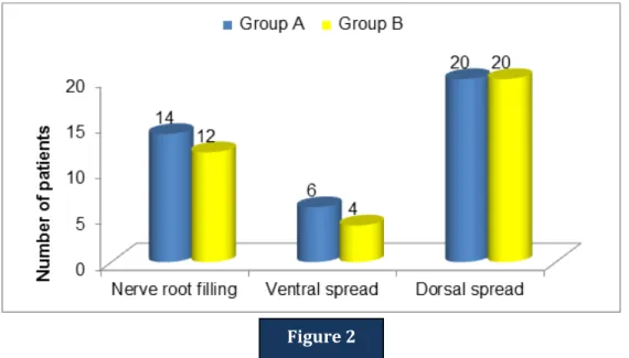
Epidurography showed dorsal spread in all patients of both the groups. The ventral spread seen in 30% patients of Group A and 20% in Group B. [Fig. 2] It was seen at last follow up that the patients showing ventral spread epidurographically had clinically higher mean percentage VAS improvement (88% in group A & 92.01% in Group B) compared to no ventral spread (62.6% in Group A & 71.03% in Group B) but this was statistically insignificant (p > 0.05). [Fig. 3]There was no correlation of the VAS improvement at 3 months to ventral epidurographic spread of contrast agent in either group (p >0.05). [Fig. 4a & 4b] Patients improved despite absence of ventral spread of contrast with significant VAS improvement in all patients till the last follow up.
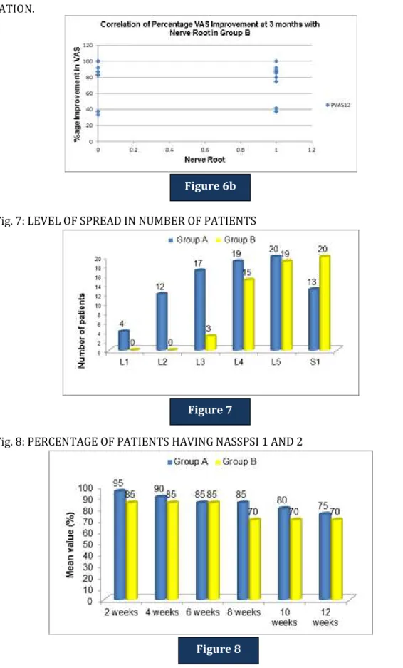
The nerve root spread was noted in 70% patients in group A versus 60% patients in group B (p >0.05). [Fig. 5] Pain relief was present at 3 months whether nerve root spread seen or not (p>0.05), thus showing a lack of correlation between mean VAS improvement and nerve root filling. [Fig. 6a & 6b].
Both the approaches were similar in their spread of contrast at L4-L5 & L5-S1 levels (p>0.05) but group A had significant cephalic spread (L1-L3) compared to group B (L3-S1) (p <0.05). However S1 nerve root delineation was more with caudal approach (p<0.05). [Fig. 7] Group B patients showed better cephalad segmental spread compared to Group A (3.65 versus 1.87 segments) (p <0.001).
The patient satisfaction was judged by NASSPSI showed similar improvement in both the groups at all follow up. At 3 months 75% patients in group A and 70% in group B were satisfied with their results having NASSPSI 1 or 2. [Fig. 8]
No major complication was seen in both the groups. Only minor complaints of heaviness in low back region were reported by 2 patients in each group and dural puncture in 1 patient in interlaminar midline group. (p >0.05)
DISCUSSION: Epidural steroid injections are one of the most commonly performed invention for the treatment of low back pain. It has shown to reduce inflammation by inhabiting either synthesis or release of number of pro-inflammatory mediator. Various approaches have been described, out of which interlaminar & caudal are the most common.Both these approaches are useful in delivering the steroids near the problem site in low back pain patients which is usually L4-5 & L5 S1. For achieving
such affectivity, various studies have evaluated the role of epidural steroid injections in low back ache with and without fluoroscopy. The use of fluoroscopy confirms the needle placement thereby improving drug delivery to appropriate site; thus reducing complications.6-11
Further advancement in techniques of epidural injections shifted the focus on targeted drug delivery at the level of pathology as well as for ventral and nerve root filling to improve the clinical outcome by decreasing inflammation at these sites.The ventral epidural space being a potential site where inflammation may be present due to herniated disc; therefore postulated that a good ventral spread of drugs would imply better pain relief. Thereafter many authors aimed this space via different techniques.4,5,12,13 However minimal literature available to correlate clinical outcome with
J of Evolution of Med and Dent Sci/ eISSN- 2278-4802, pISSN- 2278-4748/ Vol. 3/ Issue 19/May 12, 2014 Page 5164 In the present study VAS percentage improvement seen in both groups equally at all follow ups may be due to steroid diluted to higher volume could adequately reach via both the routes to the site of pathology.15, 16
Therefore, failure in correlation of the VAS improvement at 3 months to ventral epidurographic spread of contrast agent in either group (p >0.05). Significant pain improvement seen in all patients could be attributed to the dilution of substance P or the slow seepage of drug over time, or the higher volume of drug that reached the pathological areas. Whatever is the reason but preinjection ventral spread presence or absence could not correlate well with the clinical improvement.
There was no relationship found between the nerve root delineation and clinical outcome. The reason may be firstly multiple pain generators other than nerve root like disc, zygophyseal joint. Moreover similar lack of correlation between leg pain and nerve root filling had been described by Manchikanti L et al. The presence of dermatome overlapping has also been reported.15, 17
Patient satisfaction being a very important factor was evaluated in this study using modified NASSPSI. Both the groups showed comparable satisfaction at short term follow up of 3 months (75 % vs. 70%; Group A vs. Group B). The patient satisfaction which is ultimate aim of treatment in chronic pain is an important subjective assessment and adding a sensitive and holistic touch at the same. This assessment has been used previously by multiple authors.18, 19
There were some limitations in this study. Only a preinjection epidurography was noted, the post injection film could have shown improvement in contrast dispersion following the drug injection. A longer follow up might have shown different results. However, this being a preliminary study and more & large scale research which is needed to validate our observations has been in progress.
Thus to conclude midline interlaminar and caudal approaches were statistically comparable and clinically efficacious in improving pain in patients with chronic low backache irrespective to the presence or absence of ventral and nerve root spread in preinjection film leaving more than 70% patients satisfied with their results.
REFERENCES:
1. Chou R, Qaseem A, Snow V, Casey D, Cross Jr. JT, Shekelle P et al. Clinical guidelines-diagnosis and treatment of low back pain: a joint clinical practice guideline from the American college of physicians and the American pain society.Annals of Internal Medicine. 2007; 147: 478-91. 2. Manchikanti L, Staats PS, Singh V, Schultz DM, Vilims BD, Jasper JF. Evidence-based practice
guidelines for interventional techniques in the management of chronic spinal pain. Pain Physician. 2003; 6:3-81.
3. Shen FH, Samartzis D, Andersson GBJ. Nonsurgical management of acute and chronic low back pain. Journal of the Americanacademy of orthopaedic surgeons. 2006; 14: 477-87.
J of Evolution of Med and Dent Sci/ eISSN- 2278-4802, pISSN- 2278-4748/ Vol. 3/ Issue 19/May 12, 2014 Page 5165 5. Botwin K, Natalicchio J, and Brown LA. Epidurography contrast patterns with fluoroscopic
guided lumbar transforaminal epidural injections: a prospective evaluation. Pain Physician. 2004; 7: 211-5.
6. Barham G, Hilton A. Caudal epidurals: the accuracy of blind needle placement and the value of a confirmatory epidurogram. European Spine Journal. 2010; 19: 1479-83.
7. Collighan N, Gupta S. Epidural steroids. Continuing education in anaesthesia, critical care and pain [Internet]. 2010 [cited 2009 Dec 15]; 10: [5p]. Available from: doi: 10.1093/ bjaceaccp/ mkp043.
8. Botwin K P, Gruber R D. Lumbar epidural steroid injections in the patient with lumbar spinal stenosis. Physical Medicine and Rehabilitation clinics of North America. 2003; 14: 121-41. 9. Mehta M. Extradural block. Confirmation of the injection site by x-ray monitoring. Anesthesia.
1985; 40: 1009–12.
10.Renfrew DL, Moore TE, Kathol MH, el-Khoury GY, Lemke JH, Walker CW. Correct placement of epidural steroid injections: fluoroscopic guidance and contrast administrations. AJNR Am J Neuroradiol. 1991; 12: 1003–7.
11.Sitz M, Sommer H. Accuracy of blind versus fluoroscopically guided caudal epidural injections. Spine.1999; 13: 1371–6.
12.Choi YK, Barbella JD. Evaluation of epidurographic contrast patterns with fluoroscopic-guided lumbar interlaminar ventral epidural injection. Pain Pract. 2009; 9: 275-81.
13.Candido KD, Raghavendra MS, Chinthagada M, Badiee S, Trepashko DW. A prospective evaluation of iodinated contrast flow patterns with fluoroscopically guided lumbar epidural steroid injections: the lateral parasagittal interlaminar epidural approach versus the transforaminal epidural approach. Anesth Analg. 2008; 106 (2):638-44.
14.Ackerman WE, Ahmad M. The efficacy of lumbar epidural steroid injections in patient with lumbar disc herniation. Anesthesia & Analgesia. 2007; 104: 1217-22.
15.Manchikanti L, Cash KA, Pampati V, McManus CD, BSN, Damron KS. Evaluation of fluoroscopically guided caudal epidural injections. Pain Physician. 2004; 7:81-92.
16.Cleary M, Keating C, Poynton AR. The flow patterns of caudal epidural in upper lumbar spinal pathology. European Spine Journal. 2011; 20: 804–07.
17.Wolff AP, Groen GJ, Wilder-Smith OHG, Richardson J, Van Egmond J, Cru BJP. Do diagnostic segmental nerve root blocks in chronic low back pain patients with radiation to the leg lack distinct sensory effects? A preliminary study. British Journal of Anaesthesia. 2006; 96: 253–8. 18.Barré L, Lutz GE, Southern D, Cooper G. Fluoroscopically guided caudal epidural steroid
injections for lumbar spinal stenosis: A retrospective evaluation of long term efficacy. Pain Physician. 2004; 7: 187-93.
J of Evolution of Med and Dent Sci/ eISSN- 2278-4802, pISSN- 2278-4748/ Vol. 3/ Issue 19/May 12, 2014 Page 5166
GROUP A N=20
GROUP B N=20
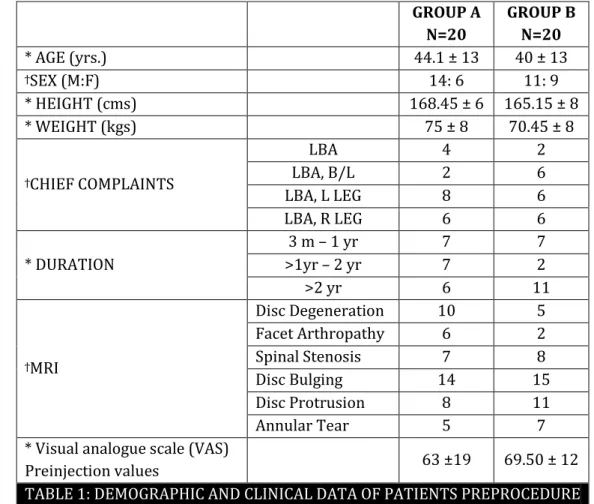
* AGE (yrs.) 44.1 ± 13 40 ± 13
ϯSEX (M:F) 14: 6 11: 9
* HEIGHT (cms) 168.45 ± 6 165.15 ± 8
* WEIGHT (kgs) 75 ± 8 70.45 ± 8
ϯCHIEF COMPLAINTS
LBA 4 2
LBA, B/L 2 6
LBA, L LEG 8 6
LBA, R LEG 6 6
* DURATION
3 m – 1 yr 7 7
>1yr – 2 yr 7 2
>2 yr 6 11
ϯMRI
Disc Degeneration 10 5
Facet Arthropathy 6 2
Spinal Stenosis 7 8
Disc Bulging 14 15
Disc Protrusion 8 11
Annular Tear 5 7
* Visual analogue scale (VAS)
Preinjection values 63 ±19 69.50 ± 12
TABLE 1: DEMOGRAPHIC AND CLINICAL DATA OF PATIENTS PREPROCEDURE * The values are given as the mean with the standard deviation
ϯ The values are given as number of patients
LBA- Low back ache, B/L- Bilateral, L –Left, R- Right
Fig. 1: % IMPROVEMENT IN VAS
J of Evolution of Med and Dent Sci/ eISSN- 2278-4802, pISSN- 2278-4748/ Vol. 3/ Issue 19/May 12, 2014 Page 5167 Fig. 2: VENTRAL, DORSAL AND NERVE ROOT SPREAD
Fig. 3: CORRELATION OF VENTRAL SPREAD WITH MEAN PERCENTAGE (%) IMPROVEMENT IN VAS SCORE AT 3 MONTHS.
Figure 2
J of Evolution of Med and Dent Sci/ eISSN- 2278-4802, pISSN- 2278-4748/ Vol. 3/ Issue 19/May 12, 2014 Page 5168 Fig. 4a: SCATTER PLOT SHOWING NO CORRELATION OF MEAN PERCENTAGE VAS IMPROVEMENT AT 3 MONTHS WITH VENTRAL SPREAD IN GROUP A USING SPEARMAN CORRELATION.
Fig. 4b: SCATTER PLOT SHOWING NO CORRELATION OF MEAN PERCENTAGE VAS IMPROVEMENT AT 3 MONTHS WITH VENTRAL SPREAD IN GROUP B USING SPEARMAN CORRELATION.
Figure 4a
J of Evolution of Med and Dent Sci/ eISSN- 2278-4802, pISSN- 2278-4748/ Vol. 3/ Issue 19/May 12, 2014 Page 5169 Fig. 5: CORRELATION OF NERVE ROOT SPREAD WITH MEAN PERCENTAGE IMPROVEMENT IN VAS AT 3 MONTHS.
Fig. 6a: SCATTER PLOT SHOWING NO CORRELATION OF MEAN PERCENTAGE VAS IMPROVEMENT AT 3 MONTHS WITH NERVE ROOT SPREAD IN GROUP A USING SPEARMAN CORRELATION.
Figure 5
J of Evolution of Med and Dent Sci/ eISSN- 2278-4802, pISSN- 2278-4748/ Vol. 3/ Issue 19/May 12, 2014 Page 5170 Fig. 6b: SCATTER PLOT SHOWING NO CORRELATION OF MEAN PERCENTAGE VAS IMPROVEMENT AT 3 MONTHS WITH NERVE ROOT SPREAD IN GROUP B USING SPEARMAN CORRELATION.
Fig. 7: LEVEL OF SPREAD IN NUMBER OF PATIENTS
Fig. 8: PERCENTAGE OF PATIENTS HAVING NASSPSI 1 AND 2 Figure 6b
Figure 7
J of Evolution of Med and Dent Sci/ eISSN- 2278-4802, pISSN- 2278-4748/ Vol. 3/ Issue 19/May 12, 2014 Page 5171
AUTHORS:
1. Nayyaamat Sandhu 2. Ruchi Gupta 3. Saru Singh 4. Kuljit Singh Aujla 5. Maninder Kaur 6. Tripat Bindra
PARTICULARS OF CONTRIBUTORS:
1. IIIrd Year Post Graduate Student, Department
of Anaesthesia, SGRDIMSR, Amritsar. 2. Professor and Head, Department of
Anaesthesia, SGRDIMSR, Amritsar. 3. Associate Professor, Department of
Anaesthesia, SGRDIMSR, Amritsar. 4. Professor, Department of Anaesthesia,
SGRDIMSR, Amritsar.
5. IIIrd Year Post Graduate Student, Department
of Anaesthesia, SGRDIMSR, Amritsar. 6. Associate Professor, Department of
Anaesthesia, SGRDIMSR, Amritsar.
NAME ADDRESS EMAIL ID OF THE CORRESPONDING AUTHOR:
Dr. Nayyaamat Sandhu, H. No. 64, Sector -71, Mohali-160071, Punjab.
Email: [email protected]