DNA Metabolism in Balance: Rapid Loss of a
RecA-Based Hyperrec Phenotype
Irina V. Bakhlanova1,2, Alexandra V. Dudkina1, Elizabeth A. Wood3, Vladislav A. Lanzov1, Michael M. Cox3*, Dmitry M. Baitin1,2*
1Petersburg Nuclear Physics Institute, NRC Kurchatov Institute, Gatchina, 188300, Russia,2Peter the Great St. Petersburg Polytechnic University, Saint-Petersburg, 195251, Russia,3Department of
Biochemistry, University of Wisconsin-Madison, Madison, Wisconsin, 53706–1544, United States of America
*[email protected](MMC);[email protected](DMB)
Abstract
The RecA recombinase ofEscherichia colihas not evolved to optimally promote DNA pair-ing and strand exchange, the key processes of recombinational DNA repair. Instead, the recombinase function of RecA protein represents an evolutionary compromise between necessary levels of recombinational DNA repair and the potentially deleterious conse-quences of RecA functionality. A RecA variant, RecA D112R, promotes conjugational recombination at substantially enhanced levels. However, expression of the D112R RecA protein inE.coliresults in a reduction in cell growth rates. This report documents the conse-quences of the substantial selective pressure associated with the RecA-mediated hyperrec phenotype. With continuous growth, the deleterious effects of RecA D112R, along with the observed enhancements in conjugational recombination, are lost over the course of 70 cell generations. The suppression reflects a decline in RecA D112R expression, associated pri-marily with a deletion in the gene promoter or chromosomal mutations that decrease plas-mid copy number. The deleterious effects of RecA D112R on cell growth can also be negated by over-expression of the RecX protein fromNeisseria gonorrhoeae. The effects of the RecX proteinsin vivoparallel the effects of the same proteins on RecA D112R filaments in vitro. The results indicate that the toxicity of RecA D112R is due to its persistent binding to duplex genomic DNA, creating barriers for other processes in DNA metabolism. A sub-stantial selective pressure is generated to suppress the resulting barrier to growth.
Introduction
DNA metabolism is a set of seemingly distinct processes that are tightly interlinked. The genome must be protected, replicated, expressed, organized, and segregated. All of the pro-cesses of DNA metabolism must share the same chromosomal substrate. Spontaneous DNA lesions are ubiquitous, hundreds of thousands appearing daily in a typical human cell, several thousand in each cell within an aerobic bacterial culture [1–3]. The nucleotide excision repair, base excision repair, mismatch repair, and other repair operations that counter these insults typically leave a transient break in the DNA strand undergoing repair. If a replication fork
a11111
OPEN ACCESS
Citation:Bakhlanova IV, Dudkina AV, Wood EA, Lanzov VA, Cox MM, Baitin DM (2016) DNA Metabolism in Balance: Rapid Loss of a RecA-Based Hyperrec Phenotype. PLoS ONE 11(4): e0154137. doi:10.1371/journal.pone.0154137
Editor:Fenfei Leng, Florida International University Bimolecular Sciences Institute, UNITED STATES
Received:January 7, 2016
Accepted:April 9, 2016
Published:April 28, 2016
Copyright:© 2016 Bakhlanova et al. This is an open access article distributed under the terms of the Creative Commons Attribution License, which permits unrestricted use, distribution, and reproduction in any medium, provided the original author and source are credited.
Data Availability Statement:All relevant data are within the paper.
appears before the break is sealed, the fork collapses [4]. The resulting double strand break is perhaps the most dangerous of all DNA damage events [5,6].
Homologous genetic recombination, or more appropriately recombinational DNA repair, evolved to repair double strand breaks [7–12]. Recombination systems ensure DNA replication success in all cells, facilitating reconstitution of the replication forks that have collapsed upon encounters with those transient template strand discontinuities [7–12]. The capacity to repair double strand breaks was a likely prerequisite to the evolution of large genomes. The enzymatic capabilities inherent to recombinational DNA repair of double strand breaks have been re-pur-posed by evolution to permit recombination in the context of eukaryotic meiosis, bacterial con-jugation, and a host of other functions [13].
The enzymes at the center of recombinational DNA repair systems are the RecA family recombinases. These include the RecA protein of bacteria, the RadA protein of archaea, and the Rad52 and Dmc1 proteins of eukaryotes [14–18]. These proteins function as filaments typi-cally formed on single-stranded DNA (ssDNA). The nucleoprotein recombinase filaments align the bound ssDNA with complementary sequences in an intact duplex DNA, and then promote DNA pairing and strand exchange. Additional enzymes resolve the branched DNA structures created by recombinases. The end product of recombinational DNA repair of a a dis-integrated replication fork is a reconstituted fork structure, after replication collapses upon an encounter with a template strand discontinuity,
Whereas recombinational repair is a critical cellular asset, (spurious) homologous recombi-nation is not and in fact can be detrimental. There are at least three ways that recombirecombi-nation systems could harm a cell. First, promiscuous recombination involving repeated chromosomal sequences could re-order or eliminate genes, creating genomic chaos. Second, the nucleopro-tein filaments formed by RecA family recombinases are potentially the largest structures that assemble on a bacterial chromosome, and could represent formidable barriers to replication or transcription if not removed from the DNA. Third, unresolved recombination intermediates in DNA could cause genome instability, imparting a different kind of barrier to DNA metabolism and/or chromosomal segregation. In bacteria, recombinase binding to DNA can have addi-tional deleterious effects via the potential induction of the SOS response and its associated halt in cell division and mutagenesis [19].
For these reasons, cellular recombination systems are subject to multiple layers of regulation [20–25]. Here, we focus on bacteria and the RecA recombinase. RecA is expressed only at levels appropriate to the metabolic situation. RecA access to DNA is proscribed under normal cir-cumstances by a very limited capacity to nucleate filament formation on duplex DNA or on ssDNA that is bound by SSB [18,26–33]. A C-terminal autoregulatory flap in RecA mediates the block to RecA filament nucleation on these DNA substrates [34–37]. The RecA binding protein PsiB sequesters RecA protein under some conditions [38,39]. Multiple regulatory pro-teins (RecFOR, DprA, and RecBCD) direct RecA filament formation on SSB-coated single-stranded DNA when required and appropriate [20,27,30–33,40–48]. The regulatory protein DinI modulates RecA filament stability and function after assembly [49–51]. When RecA func-tion is no longer needed, RecA filament removal from the DNA is ensured by addifunc-tional bacte-rial regulatory proteins such as RecX protein and DinD protein [50,52–58], as well as the UvrD, Rep, and PcrA helicases [59–65]. Genetic modulation or ablation of these different con-trols often has deleterious consequences for the cell [20,60,61,66–70].
Another layer of control on recombination systems has been imposed by evolution. RecA family recombinases have not evolved to promote DNA pairing and strand exchange opti-mally. Instead, RecA functionality represents an evolutionary compromise that reflects a bal-ance between the critical functions and deleterious consequences of recombination. This balance may be subtly different in each bacterial species. Substantial increases in RecA function design, data collection and analysis, decision to
publish, or preparation of the manuscript.
can be obtained via mutation [71,72]. The RecA D112R variant is perhaps the best example of functional enhancement to date, substantially increasing the efficiency of conjugational recom-bination [71].
The RecA D112R mutant protein was originally isolated as part of an investigation of amino acid residues at the subunit-subunit interface of RecA protein [73]. Additional mutations at codon 112 arose in a study of RecA mutant proteins that were inhibited less by high concentra-tions of the UmuD and UmuC proteins [74,75]. In vitro, and relative to wild type RecA, RecA D112R exhibits improvements in loading onto SSB-coated ssDNA, in resistance to inhibitors such as RecX and PsiB, and in pairing homologous DNAs [71]. The enhanced functionality of the D112R mutant protein and other RecA variants gives rise to a broader question. If substan-tial improvements in RecA functionality are possible, why have they not appeared during evo-lution? The answer must lie in the potentially deleterious consequences of enhanced RecA recombinase function, which we explore in this report.
Materials and Methods
Strains and plasmids
Note: for all protein names, Ec =Escherichia coliand Ng =Neisseria gonorrhoeae. Donor KL227 (HfrP4xmetB) and recipients: AB1157 (thr-1 leuB6 ara14 proA2 hisG4 argE3 thi-1 supE44 rpsL31) and recombination deficient JC10289 (as AB1157 butΔ[recA-srlR306]::Tn10 =
ΔrecA306) were from A.J. Clark’s collection. An additional∆araBAD mutation, deleting the entire operon and its control region, was introduced into AB1157 and its derivatives. A plasmid with amino acid substitution D112R in the interface of subunit interactions in the EcRecA fila-ment (pD112R) and a plasmid with generecAEc(pT420) were constructed in K.L. Knight’s lab-oratory [76]. GenesrecXEcorrecXNgwere cloned in pT420 and in pD112R along with the expressedrecAgenes already present. Thus, pEAW958 isrecXNgwith an araBAD promoter in pD112R; pEAW959 isrecXNgwith an araBAD promoter in pT420(wt); pEAW858 isrecXEc with an araBAD promoter in pD112R; and pEAW847 isrecXEcwith an araBAD promoter in pT420(wt). MG1655 with RecA D112R expressed on the chromosome at the normal recA locus is EAW166.
Proteins
The wild typeE.coliRecA, RecA D112R, RecX [38,77] and SSB [27] proteins were purified as previously described. Their concentrations were determined using native extinction coeffi-cients:ε
280= 2.23 x 10⁴M-1cm-1for all RecA protein variants[78], andε280= 2.38 x 10⁴M-1
cm-1for SSB protein [79]. The concentration ofE.coliRecX was determined from the absor-bance at 280 nm using the native extinction coefficient 2.57 × 10⁴m−1cm−1. TheN. gonor-rhoeaeRecX protein was purified as described [53]. Antibodies raised against the purified RecX protein were from "Genetel Lab" (Мadison, Wisconsin, USA).
ssDNA dependent ATP hydrolysis (ATPase) Assays
A coupled enzyme, spectrophotomeric assay [80,81] was used to measure RecA-mediated ATP hydrolysis. The ADP generated by hydrolysis was converted back to ATP by a regenera-tion system of pyruvate kinase and phosphoenolpyruvate (PEP). The resultant pyruvate was converted to lactate by lactate dehydrogenase using NADH as a reducing agent. The conver-sion of NADH to NAD+was monitored as a decrease in absorbance at 380 nm. The amount of ATP hydrolyzed over time was calculated using the NADH extinction coefficientε380= 1.21
temperature controller and a 12-position cell changer. The path length was 0.5 cm or 1 cm, the band pass was 2 nm. All ssDNA dependent ATPase assays contained a reaction solution of 25 mM Tris.OAc (pH 7.5, 80% cation), 10 mM MgOAc (except where noted), 3 mM potassium glutamate, 5% w/v glycerol, 1 mM dithiothreitol (DTT), 3 mM PEP, 30 U/ml pyruvate kinase, 30 U/ml lactate dehydrogenase, 4.5 mM NADH and 5μM M13mp18 cssDNA. The wild-type
RecA, RecAD112R (3μM) were preincubated with ATP and 5μm M13mp8 circular ssDNA
for 5 min. SSB protein (0.5μM) was then added, followed by another 5-min incubation. The
time point of the RecX addition is shown by arrow (5 min). For reactions involving ATPase during DNA strand exchange, linear duplex M13mp18 DNA (10μM) was added at the
indi-cated time. Where Ec or NgRecX proteins were added, The final concentration is indiindi-cated in the figure and legend.
Cell Culture
Each day, each of multiple stationary phase cultures grown overnight were diluted 50X and again grown to stationary phase in L-broth at 37°C (5–6 cell generations). A sample was taken at mid-log phase and used to measure conjugation proficiency. Cells were again diluted 50X into fresh L-broth. A sample was again taken at mid-log phase for the conjugation assay, and the culture was then allowed to grow overnight. The procedure was repeated for 6 days, or a total of approximately 70 cell generations.
UV radiation sensitivity
Cells (EAW 166 (RecA D112R on the chromosome) and MG1655) were grown, serially diluted, and 100μl of appropriate dilutions were spread onto LB plates. Dilutions for samples/
treatments were empirically determined. The plates were then exposed to UV in a calibrated Spectrolinker XL-1000 UV crosslinker (Spectronics Corp) to the dose indicated. After incubat-ing at 37°C overnight, the colonies were counted and divided by the dilution factor to get cfu/ ml. For percent survival, colony counts on the treated plates were divided by the counts on untreated plates.
Conjugationwas carried out essentially as described [82]. Both Hfr and F-strains were grown, crossed and selected for recombinants at 37°C in mineral salts 56/2 medium supplied with all necessary growth factors at pH 7.5. TherecAgenes were not induced by IPTG butrecX genes were induced by indicated concentration of arabinose. The ratio between donors and recipients in the mating mixture was 1:10, 2–4 x10⁷donors and 2–4 x 10⁸recipients per 1 ml. The yields of Ara+Strrrecombinants in all independent crosses were 0.1–7.8% relative to donors.
FRE value calculationswere carried out as described [82]. During mating, the donor Hfr KL227 transfers markers into recipients in the orderleu+,ara+andthr+. Recombination exchanges in this region of theE.colimap are adequately described mathematically by the Hal-dane formula [82]. In the slightly rearranged form,λ= 2l/ln(2μ-1), this formula relates the
average distance between two neighboring genetic exchanges (in minutes) to the linkage of selected and unselected markers (μ), and the distance (l), in minutes between markers on theE.
colimap. The distance,l, in minutes between thethr(0.05) andleu(1.75) markers is 1.7, and between theara(1.47) andleu(1.75) markers is 0.28. Donor KL227 transfersleu+andthr+as a proximal and distal marker, respectively. The frequency of recombinational exchanges is expressed as FRE, the average number of exchanges per oneE.coligenome equivalent (100 minutes), and thus equals 100/λ. For wild typeE.coli, FRE = 5.0 [82]. In this study, we are
under investigation.ΔFRE can also be calculated by use of the formula:ΔFRE = ln(2μ1−1)/ln
(2μ2−1), whereμ1is the linkage observed with either a modified RecA or the wild type RecA
under particular conditions of HR, andμ2is the linkage observed with wild type EcRecA under
standard conditions [83].
Alterations in FRE (ΔFRE) promoted by theRecA D112RandRecXEcorRecXGcgenes or
by theEcRecAandRecXEcorRecXGcgenes relative to the FRE value promoted by the wild typeEcRecAgene were calculated using the following formula:ΔFRE = ln(2μ1−1)/(2μ2−1),
whereμ1is the linkage of selectedara+and unselectedleu+markers in a cross using wild type
E.colistrain AB1157 andμ2is the similar linkage in the cross being analyzed. Calculations of
uncertainty of relative FRE values were determined as deviations from the average values by making use of the program Excel-97 with formula [= 2STDEV] and by inputting the values
from independent repeats of three experiments.
Cell competition assays
Wild type cells and cells expressing the D112R variant of RecA protein at the normalrecA chromosomal locus, were modified to carry a neutral Ara−mutation (which confers a red color
on colonies when grown on tetrazolium arabinose (TA) indicator plates) to permit color based scoring of mixed populations [84]. Cells from a freezer stock of each strain were streaked on LB plates [85] and incubated at 37° C. After growth overnight, competition cultures were started by inoculating 3 mL fresh LB broth with an isolated colony of competition Ara+ or Ara−strains in triplicate and grown overnight at 37° C with shaking. Equal amounts of strains to be compared were mixed in triplicate. A sample of the mixture was taken, diluted by a factor of 10−6, and plated on tetrazolium arabinose indicator plates. Then, 3 mL fresh LB broth was inoculated with 30μL of each mixture, and grown overnight. The plating, inoculation, and
growth cycle was repeated two more times. White and red colonies were counted on plates con-taining 40–300 colonies, and the % of cells expressing mutant RecA proteins was determined. Normally for counting colonies, plates with fewer than 20 colonies of either competitor are excluded to reduce the effect of outliers caused by low counts [86]. However, at the 48 hour time point, because the differences in fitness is so great between wt recA and recA D112R, it was impossible to retrieve at least 40 colonies of the recA D112R in a range that could also be used to calculate the density of the wt recA strains.
Visualizing intracellular RecX amounts by Western blot
E.colicells were grown up to mid-log phase in LB medium at 37°C. A cell pellet containing 5 x10⁷cells was lysed by boiling with sodium dodecyl sulfate, electrophoresed through sodium dodecyl sulfate-10% polyacrylamidеgels. The RecA or RecA D112R amounts were detected by
immunoblotting using polyclonal chicken antibodies to RecA protein from“Genetel Lab”
(Madison, Wisconsin, USA) in a standard procedure described earlier [87]. Primary antibody binding was visualized with secondary antibodies coupled to horseradish peroxydase (Genetel Lab).
Strand Exchange reaction
Three-strand exchange reactions were carried out in 25 mm Tris-OAc buffer (80% cation), 1 mm dithiothreitol, 5% (w/v) glycerol, 3 mM potassium glutamate, 2 mM ATP and 10 mM of Mg(OAc)2. Reactions also contained an ATP regeneration system of 30 units/ml pyruvate
for 5 min. SSB protein (0.5μM) was then added, followed by another 5-min incubation. The
reactions were initiated by the addition of M13mp8 linear dsDNA to 10μM. The time point of
dsDNA addition is shown by arrow. DsDNA was added at 5 min on the graph. The RecX was added at 30 min after dsDNA or 35 min on the graph (shown by arrow). On the some graph RecX was added at 10 min after dsDNA.
Growth Curves
LB media (25 ml) was inoculated with 0.25 ml of a fresh (to avoid de-evolution) culture of a strain to be tested and grown for the indicated times at 37°C with shaking at 200 rpm. The con-centration of arabinose was 0.00002% for RecX induction but IPTG was not added. Culture ali-quots were removed every hour, and growth was monitored by absorption of the sample at a wavelength of 600 nm, using a Cary 100 spectrophotometer. Solutions were diluted as needed to bring the absorption into the linear range of the spectrophotometer.
Comparative Genome Sequencing (microarray analysis)
DNA samples were analyzed to detect genomic alterations using the Comparative Genome Sequencing Service provided by NimbleGen Systems, Inc. as described earlier [88].
Direct DNA sequencing
Standard Sanger sequencing was carried out at the University of Wisconsin Biotechnology Center, after PCR amplification of the target genomic DNA sequence.
Results
Expression of RecA D112R confers a growth disadvantage
The RecA D112R variant provides a substantial increase in function with respect to conjuga-tional recombination, utilizing two different conjugation assays [71,72]. To explore the delete-rious consequences of this increase in functionality, we first examined cellular growth rates. As shown inFig 1A, cells expressing the RecA D112R variant grow somewhat more slowly than do cells expressing the wild type RecA protein. The difference appears small, but it proved highly reproducible over the course of 4 trials. To provide a more sensitive measurement of any positive or deleterious effects of therecAmutations on cell growth and survival, we carried out direct competition assays between strains expressing wild type or the D112RrecAprotein, each present at the same normal chromosomal forrecA[84]. Wild type cells were modified to carry a neutral Ara−mutation (which confers a red color on colonies when grown on
tetrazo-lium arabinose (TA) indicator plates) to permit color based scoring of mixed populations [84]. Overnight cultures of cells expressing the D112RrecAvariant were mixed in a 50/50 ratio with isogenic wild type cells, with one strain carrying the Ara−mutation. The ratio of wild type to mutant strains was then followed over the course of several days, with stationary phase cultures diluted and grown out each day as described in Methods. Results are presented inFig 1B. Based on earlier work, the Ara−mutation itself should not affect growth rates [84,89]. In all
We also tested the effect of RecA D112R on cellular sensitivity to ultraviolet radiation (Fig 1C. With the RecA variant expressed from the normal chromosomal locus, there was little evi-dent effect.
Growth of cells expressing RecA D112R leads to de-evolution of the
hyperrec phenotype
In this study and as described in Methods, we are reporting the frequency of recombinational exchanges (FRE) during the conjugational cross, defined as the average number of exchanges per oneE.coligenome equivalent (100 minutes). For wild typeE.coli, FRE = 5.0 [82]. In previ-ous work, we demonstrated that FRE is increased by over 50 fold when RecA D112R replaces the wild type RecA protein inE.coli. Cells expressing RecA D112R thus exhibit a hyperrec phe-notype [71]. We wished to determine whether the hyperrec phenotype was stable.
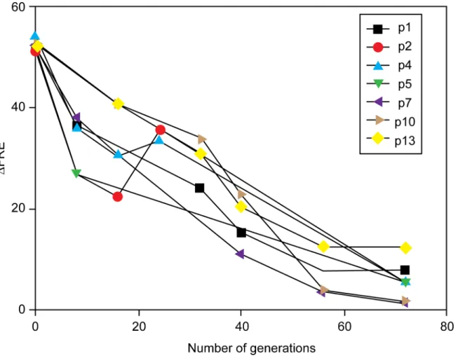
Continuous cycles of growth and dilution of a strain expressing RecA D112R results in a decline in the measuredΔFRE value (the ratio of FRE values of the strain being measured
rela-tive to that observed for wild type cells) (Fig 2). Over 70 cell generations, a marked decline in
ΔFRE was seen for all 16 cultures ofΔrecAstrain JC10289, with a plasmid expressing RecA
D112R (pRecA [D112R]). The molecular basis for the decline generally fell into three catego-ries: (a) a deletion in pRecA [D112R] that reduced expression of the RecA variant, (b) a muta-tion in the host chromosome that impacted RecA D112R expression, or (c) a combinamuta-tion of the two.
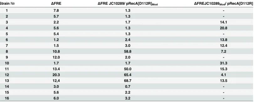
The three effects can be seen in the results tabulated inTable 1. The finalΔFRE for the 16
separate cultures are listed in the first data column. In each case, this measure of conjugation function has been reduced from over 50 to a range of 1.2–20.3.
When the plasmids derived from the culture endpoints were isolated and used to transform strain JC10289 again, the plasmid preps produced lowΔFRE values indicative of loss of the
hyperrec phenotype in 12 of 16 cultures. This indicated that the genetic alteration that pro-duced the loss of phenotype was present on the plasmid in those 12 cases. The entire protocol featuring continuous cycles of growth and dilution of the strain expressing RecA D112R was repeated for an additional 6 cultures, and a plasmid alteration was again detected in 3 of them. Sequencing of the plasmids derived from the 3 new cultures revealed a common 633 bp dele-tion in the promoter region for therecA D112Rgene, produced by apparent recombination between two 37 bp direct repeats (Fig 3A).
In four of the cases highlighted inFig 2andTable 1, (cultures 8, 11, 12, and 13), the isolated plasmids still produced a highΔFRE value, suggesting that the mutations affecting the loss of
the hyperrec phenotype were chromosomal. Strains 8 and 12 were subjected to microarray analysis to identify mutations relative to theE.coligenome reference sequence [90], and chro-mosomal mutations were confirmed by direct sequencing. Most of the mutations detected were present in the founder strain JC10289 used as the host for these experiments, as well as the de-evolved strains. Notably, in both strains 8 and 12 there was a mutation that may have inactivated thepcnBgene, important for the maintenance of colE1-based plasmids [91–94]. The two differentpcnBmutations (which introduce stop codons at either codon 250 or 264) were moved into an otherwise wild type background, and the resulting strains were tested for the mutant RecA protein are plotted as a function of the daily growth cycle of the experiment. (C) Resistance to ultraviolet light. Experiments were carried out as described in Materials and Methods. The strains compared are MG1655 and EAW166 (RecA D112R expressed on the chromosome at the normalrecA locus).
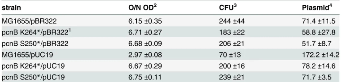
maintenance of plasmids pUC19 and pBR322, which share the replication origin of pRecA [D112R].
As shown inFig 3B, the plasmids pUC19 and pBR322 appeared to be maintained at signifi-cantly lower levels in cells with either of thepcnBmutations. This experiment was repeated three times with identical results. Data ancillary toFig 3Bis presented inTable 2. Colony form-ing units (CFU) generally scaled with the optical density of the overnight cultures. Those O.D.s were similar except for MG1655 hosting pUC19, which was less than half of the density and less than half of the CFUs in an identical culture volume. Isolation of plasmid DNA as inFig 3Bproduced apparent plasmid DNA levels (as measured by optical density at 260 nm) that are reported inTable 2, although this measurement includes RNA contamination. The reduction in overall nucleic acid in strains with pUC19 when pcnB mutations are present are readily evi-dent in this assay. The changes in pBR322 levels are not evievi-dent, possibly because of the pres-ence of RNA or some other contaminant.
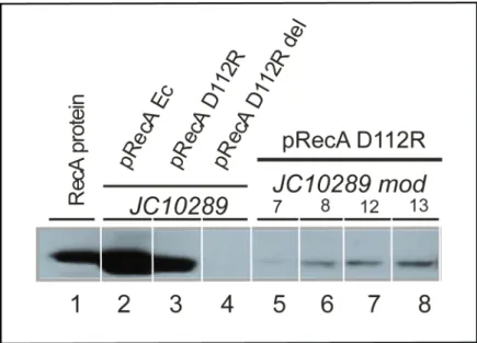
In a separate experiment, the levels of expression of RecA D112R were measured and were substantially reduced in strains 7, 8, 12, and 13 (Fig 4). Intracellular RecA D112R protein levels were reduced dramatically in all four strains. ThepcnBmutations provide one, but possibly not the only explanation for the reduction in RecA D112R expression.
Fig 2. Loss of hyperrec phenotype in cells expressing RecA D112R upon continuous culture.Multiple cultures of strain JС10289, expressing RecA D112R protein from plasmid pRecA[D112R] were grown as described in Materials and Methods for 70 generations.ΔFRE was determined for each culture at the indicated times, and is the ratio of the FRE value measured for JC10289 with pRecA[D112R] to the FRE value for JC10289 expressing wild type RecA protein.
Independently, we cured nine of the cultured strains of their resident plasmids, and trans-formed them with the original plasmid expressing RecA D112R. The results are tabulated in the final data column ofTable 1. In no case did this produce a strain that exhibited theΔFRE
level of the original strain. In strains 8, 11, 12, and 13, theΔFRE values were comparable to
those seen after 70 generations of growth in the original experiment, indicating that back-ground chromosomal mutations were dominant in diminishing the hyperrec phenotype. In strains 3, 4, 6, 7, and 10, the measuredΔFRE increased, but not to theΔFRE level of 50
+ observed in the original strain. The results suggest the presence of background alterations in the chromosome that moderate the effects of RecA D112R expression and/or function, in addi-tion to alteraaddi-tions on the plasmid.
The growth deficiency conferred by RecA D112R expression reflects
persistent binding to the
E
.
coli
chromosome
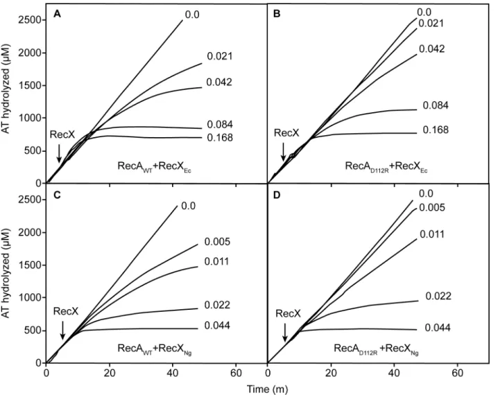
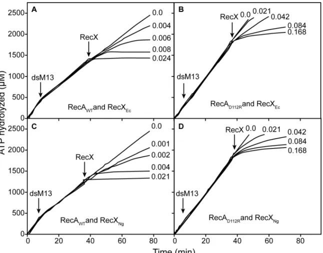
In vitro, the RecA D112R protein rapidly displaces SSB to nucleate on ssDNA, and also binds to duplex DNA more readily than the wild type RecA protein [71]. These properties could pro-duce more extensive binding to chromosomal DNA inE.coli, and lead to inhibition of normal chromosomal DNA replication. To explore this possibility, we utilized the RecX protein in the attempt to disrupt the RecA filaments and restore normal growth rates on theE.colicells. The E.coliRecX protein (EcRecX) works primarily by binding to the 3'-proximal end of the RecA filament, blocking RecA filament extension. Net dissociation of the filament then occurs at the 5'-proximal end [52]. The RecX protein derived from the bacteriumNeisseria gonorrhoeae (NgRecX) has a more robust activity, creating internal filament discontinuities that trigger more rapid filament disassembly [53]. Both proteins were tested with RecA D112R.
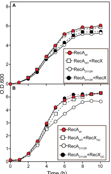
As shown inFig 5A, the expression or overexpression of EcRecX has little effect on the growth rates ofE.colicells expressing either wild type RecA or RecA D112R. In contrast,
Table 1. Loss of the hyperrec phenotype in strains expressing RecA D112R protein.The second column documentsΔFRE, the relative increase in the frequency of genetic exchange per chromosomal DNA unit length (ΔFRE is a ratio of the FRE observed with the indicated strain divided by FRE for compara-ble cells expressing the wild type RecA protein), after continuous culture for 70 cell generations. The third column shows results obtained after the plasmids from the cultured strains were isolated and used to again transform strain JC10289. Four of the plasmids (from cultures 8, 11, 12, and 13) still produce an ele-vatedΔFRE. In the final column, the strains were cured of their resident plasmid, and re-transformed with pRecA D112R. The lowerΔFRE scores in some of these strains reflect the effects of chromosomal mutations.
Strain№ ΔFRE ΔFRE JC10289/ pRecA[D112R]Mod ΔFREJC10289Mod/ pRecA[D112R]
1 7.8 1.3
-2 5.7 1.3
-3 2.2 1.7 14.1
4 5.6 1.3 20.8
5 5.4 1.3
-6 1.2 2.4 13.8
7 1.5 3.0 12.4
8 10.8 58.8 7.2
9 12.0 2.0
-10 1.7 1.7 31.3
11 13.4 50.0 15.3
12 20.3 65.4 4.1
13 12,4 68.7 13.5
14 3.0 0.7
-15 5.6 2.2
-16 6.0 3.2
expression of the more robust NgRecX from a plasmid restores normal growth rates to the cells (Fig 5B). These experiments were repeated 3 times with consistent results.
The differential effects of the two RecX proteins are readily seen in vitro (Fig 6). To produce a given level of inhibition of the RecA or RecA D112R ATPase, about 4 fold less NgRecX was required than was EcRecX (Fig 6). About 2 fold more of either RecX protein is needed for the RecA D112R protein than is required for wild type RecA. The resistance of RecA D112R pro-tein to RecX increases during DNA strand exchange. If the RecX propro-teins are added when RecA is actively promoting DNA strand exchange, the amount of either RecX needed to obtain a given level of inhibition of RecA D112R was about 20–40 fold greater than seen for the wild type RecA protein (Fig 7). Again, NgRecX was more effective than EcRecX, by a factor of at least 2.
The effects of the two RecX proteins were determined with more resolution by measuring their effects on FRE in the various constructs. The genes encoding theE.coliRecX protein or the Neisseria RecX (RecXNG) were expressed from the arabinose promoter. The RecA proteins
were expressed from thetacpromoter, but no IPTG inducer fortacwas added. The level of the RecX proteins was varied by increasing the concentration of arabinose added to the media, and FRE levels were monitored (Table 3). As expression of the wild type RecX protein fromE.coli was increased to high levels by arabinose addition, FRE levels declined in cells expressing either the wild type RecA protein or RecA D112R. However, the decline was greater when RecXNG
was expressed. In general, the RecA D112R mutant protein was significantly less sensitive to either RecX protein than was the wild type RecA protein.
Discussion
The hyperrec phenotype conferred by expression of the RecA D112R variant confers a readily demonstrable cell growth deficiency. Whereas the difference in the rate of growth appears
Fig 3. Plasmid and genomic changes detected in de-evolution trials.(A) The 633 base pair deletion (green) was detected in the promoter upstream of the gene expressing RecA D112R as described in the text. The 37 bp repeats that appear to facilitate the deletion are shown in blue. The beginning of the gene expressing RecA D112R is shown in red. (B) Effect ofpcnBmutations on the maintenance of plasmids pBR322 and pUC19. Cells with or without the indicatedpcnBmutations were grown and harvested, and plasmid DNA was extracted. The plasmid DNA shown in each lane represents the amount isolated from 1.5 ml of stationary phase cells at OD600= 6.6–6.9, except for MG1655 (WT RecA) containing pUC19, which represents 3 ml of stationary phase cells at OD600= 3.0. All OD600measurements were carried out after 1/10 dilution of the cultures listed so that readings remained in the linear range of the
spectrophotometer. The very high copy number of pUC19 in strain MG1655 may limit cell growth. The*indicates that the mutation represents the introduction of a stop codon at the residue position K264 or S250 indicated. The experiment was repeated twice with consistent results.
doi:10.1371/journal.pone.0154137.g003
Table 2. Data reflecting plasmid levels in pcnB mutants.
strain O/N OD2 CFU3 Plasmid4
MG1655/pBR322 6.15±0.35 244±44 71.4±11.5
pcnB K264*/pBR3221 6.71±0.27 183±22 58.8±27.8
pcnB S250*/pBR322 6.68±0.09 206±21 51.7±8.7
MG1655/pUC19 2.97±0.08 70±13 172.2±14.2
pcnB K264*/pUC19 6.67±0.29 200±16 78.2±14.6
pcnB S250*/pUC19 6.75±0.11 239±21 71.7±3.5
1. The*denotes a stop codon at the position indicated.
2. Total optical density of an overnight culture measured (after dilution of 1/20) at 600 nM (n = 4). 3. Colony forming units found after spreading 50μl of a 10−6dilution of the overnight culture (n = 3). 4. Plasmid DNA (plus any contaminating nucleic acid; in ng/μl) isolated from 3 ml of an overnight culture as determined by optical density at 260 nm.
small, the defect provides a substantial selective disadvantage. Upon continuous culturing, rapid loss of the hyperrec phenotype occurs producing mutations that decrease the expression of RecA D112R. We conclude that an enhancement of RecA function alters an existing balance between different processes in DNA metabolism, giving rise to a strong selection pressure to restore that balance.
The growth deficiency most likely reflects an increase in binding of RecA D112R to the bac-terial chromosome, impeding other processes in DNA metabolism. The growth deficiency is alleviated by expression of a potent RecA inhibitor, the NgRecX protein, which facilitates a robust removal of RecA protein from DNA. The NgRecX protein is also more effective in the inhibition of RecA D112R functions in vitro. The NgRecX protein provides an artificial path to restoring that balance. The only known function of RecX protein is the removal of RecA pro-tein from the DNA, and the NgRecX propro-tein has a particularly robust capacity to carry out that function [53]. The capacity of NgRecX to eliminate the growth deficiency in cells expressing RecA D112R implies that this RecA variant simply binds to chromosomal DNA too persis-tently, impeding other processes in DNA metabolism. In the absence of NgRecX protein pro-vided artificially, the balance is restored via adaptations that reduce RecA D112R expression. Functional enhancement of RecA is not well-tolerated byEscherichia coli.
It has become clear that RecA protein properties can limit replication and cell growth rates [72,95]. The present work reinforces this conclusion, but also provides a first look at how a bacterial cell responds to the limitations. Even the modest changes in cell growth rate imposed by a RecA filament that is resistant to removal from DNA by RecX create a selective pressure sufficient to select for adaptive alterations in the expression of the responsible RecA variant. In the cases explored here, the cells adapted by limiting expression of the RecA variant, either by alterations to the RecA variant promoter or by chromosomal alterations in thepcnBgene that
Fig 4. RecA protein expression in cells that have lost the hyperrec phenotype.Intracellular RecA protein was measured by Western blotting as described in Materials and Methods. Each lane reflects the RecA protein content of 5 X 10⁷cells. Lane 1 is a marker. Lanes 2 and 3 represent expression of either EcRecA (lane 2) or RecA D112R (lane 3) from their respective plasmids controlled by thetacpromoter in the absence of induction. Lane 4 shows an empty vector control. Lanes 5–8 show results from de-evolved strains 7, 8, 12, and 13 respectively as labeled.
reduced plasmid copy number. We have not yet searched exhaustively for genomic changes that could permit cell adaptation to a RecA variant that binds to DNA too persistently.
It is conventional to assume that each cellular protein has evolved to optimize its molecular function. In the case of proteins involved in DNA metabolism, this is not necessarily the case. Instead, evolution has refined a balance between the different aspects of DNA metabolism that must all share the same chromosomal substrate. Thus, the RecA protein existing in each bacte-rial species is not optimized for the promotion of DNA pairing and strand exchange per se. Recombinase function can be altered in a variety of ways, increasing cellular resistance to ioniz-ing radiation [96] or proficiency in conjugation [71,72]. If RecA protein filament disassembly is slowed, cell growth rates are slowed [95]. It is possible to make a better recombinase, but increased functionality does not always result in enhanced fitness.
Fig 5. Effects of RecX proteins on the growth curves of cells expressing wild type RecA protein or RecA D112R protein.Cells containing pRecA Ec or pRecA [D112R] were grown in LB with Ampicillin 100mkg/ml. Where indicated, the cells contained plasmid pEAW847, expressing the RecX and RecA proteins fromE.coli, pEAW858–RecX and RecA D112R proteins fromE.coli, pEAW959–RecA fromE.coli and RecXNG, or pEAW958–RecA D112R and RecXNG.
Conclusion
All processes in DNA metabolism–replication, repair, recombination, transcription, packag-ing–share the same chromosomal DNA substrate. Conflicts are common and are mediated by a wide range of mechanisms honed by evolution. The RecA protein filament represents the largest contiguous structure assembled on a bacterial chromosome, a potentially substantial barrier to replication. RecA has not evolved to promote its DNA strand exchange reaction opti-mally. Instead, evolution has crafted a functional compromise appropriate to cellular require-ments. Functional enhancements in RecA protein are possible that produce a hyperrec
phenotype. However, they lead to a growth disadvantage and a substantial selection pressure to reverse the adverse effects. A RecA-based perturbation of the DNA metabolic balance creates a strong selective pressure that leads to rapid loss of the hyperrec phenotype. In the cases we doc-ument, this involves chromosomal or plasmid alterations that result in a reduction in the expression of therecAgene variant.
Fig 6. Inhibition of RecA-mediated and DNA-dependent ATP hydrolysis by RecX proteins.ATPase reactions were carried out and monitored as described in Materials and Methods, with 5μM RecA or RecA D112R protein as indicated and RecX protein (fromE.coli) or RecXNG, as indicated. RecX proteins were added at the time indicated to a final concentrations indicated for each line (inμM).
Fig 7. Inhibition of RecA-mediated ATP hydrolysis by RecX proteins during DNA strand exchange.ATPase reactions were carried out and monitored as described in Materials and Methods, with 5μM RecA or RecA D112R protein as indicated and RecX protein (fromE.coli) or RecXNG, as indicated. Duplex linear M13mp18 DNA was added at the time indicated by“dsM13”. RecX proteins were added at the time indicated to a final concentrations indicated for each line (inμM).
doi:10.1371/journal.pone.0154137.g007
Table 3. Effect of RecX protein expression on measured FRE levels in strains expressing RecA or RecA D112R proteins.The strain used is JC10289 (AB1157ΔrecA) engineered with a deletion of the araBAD operon as described in Materials and Methods. Plasmids were constructed to express both the indicated RecA (from thetacpromoter, uninduced) and the indicated RecX (from thearapromoter, with arabinose added as an inducer as indicated) proteins.
RecXEc RecXGc None
RecA wt D112R wt D112R wt D112R
0.0% ara
2.4%*0.935±0.016(600) **FRE = 24.8***
5.3% 0.548±0.017 (600)FRE = 416.7
0.67% 0.943±0.005 (900)FRE = 21.7
0.26% 0.554±0.032 (900)FRE = 400.0
4.5% 0.939±0.007 (800)FRE = 23.2
0.82% 0.560±0.006 (600)FRE = 384.6
0.01% ara
4.6% 0.928±0.011(1200)
FRE = 27.8
4.1% 0.642±0.020 (900)FRE = 227.3
0.68% 0.960±0.003 (1200)FRE = 14.8
0.85% 0.763±0.073 (600)FRE = 114.7
0.1% ara
5.5% 0.981±0.009(900)
FRE = 6.9
7.8% 0.843±0.032 (900)FRE = 67.3
0.48% 0.993±0.007 (500)FRE = 2.5
0.61% 0.900±0.020 (600)FRE = 41.1
1% ara 4.5% 0.986±0.008(1200)
FRE = 5.0
4.3% 0.944±0.017 (1220)FRE = 21.3
011% 0.996±0.004 (900)FRE = 1.5
0.09% 0.984±0.008 (700)FRE = 5.8
*Yield of Thr+Strr recombinants (% to donors)
**Linkage (m) between selectedthr+ and unselectedleu+ markers ***Frequency of recombination exchanges per DNA unit length (FRE)
Acknowledgments
The work was carried out with financial support from the Russian Science Foundation (grant No. 14-34-00023 to Dmitry Baitin, partly RecA biochemistry), the Russian Foundation for Basic Research (Grant 14-04-00817Аto Irina Bakhlanova, FRE data), and the National
Insti-tutes of General Medical Sciences (USA) grant GM32335 (to MMC).
Author Contributions
Conceived and designed the experiments: IVB AVD VAL MMC DMB. Performed the experi-ments: IVB AVD EAW VAL DMB. Analyzed the data: IVB AVD EAW VAL MMC DMB. Contributed reagents/materials/analysis tools: IVB AVD EAW VAL MMC DMB. Wrote the paper: IVB AVD EAW MMC DMB.
References
1. Park EM, Shigenaga MK, Degan P, Korn TS, Kitzler JW, Wehr CM, et al. Assay of excised oxidative DNA lesions: isolation of 8-oxoguanine and its nucleoside derivatives from biological fluids with a monoclonal antibody column. Proc Natl Acad Sci USA. 1992; 89:3375–3379. PMID:1565629
2. Richter C, Park JW, Ames BN. Normal oxidative damage to mitochondrial and nuclear DNA is exten-sive. Proc Natl Acad Sci USA. 1988; 85:6465–6467. PMID:3413108
3. Cox MM. Recombinational DNA repair of damaged replication forks inEscherichia coli: questions. Annu Rev Genet. 2001; 35:53–82. PMID:11700277
4. Kuzminov A. Single-strand interruptions in replicating chromosomes cause double-strand breaks. Proc Natl Acad Sci USA. 2001; 98:8241–8246. PMID:11459959
5. Iliakis G. The role of DNA double strand breaks in ionizing radiation-induced killing of eukaryotic cells. Bioessays. 1991; 13:641–648. PMID:WOS:A1991GX90000003.
6. Resnick MA. Similar responses to ionizing radiation of fungal and vertebrate cells and importance of DNA double strand breaks. J Theoret Biol. 1978; 71:339–346. doi:10.1016/0022-5193(78)90164-9 PMID:WOS:A1978EW47500005.
7. Cox MM. Historical overview: Searching for replication help in all of the rec places. Proc Natl Acad Sci USA. 2001; 98(15):8173–8180. PMID:11459950
8. Cox MM, Goodman MF, Kreuzer KN, Sherratt DJ, Sandler SJ, Marians KJ. The importance of repairing stalled replication forks. Nature. 2000; 404(6773):37–41. PMID:10716434
9. Kowalczykowski SC. Initiation of genetic recombination and recombination-dependent replication. Trends Biochem Sci. 2000; 25:156–165. PMID:10754547
10. Kuzminov A. DNA replication meets genetic exchange: Chromosomal damage and its repair by homol-ogous recombination. Proc Natl Acad Sci USA. 2001; 98:8461–8468. PMID:11459990
11. Michel B. Replication fork arrest and DNA recombination. Trends Biochem Sci. 2000; 25:173–178. PMID:10754549
12. Michel B, Boubakri H, Baharoglu Z, LeMasson M, Lestini R. Recombination proteins and rescue of arrested replication forks. DNA Repair. 2007; 6:967–980. PMID:ISI:000248092000010.
13. Haber JE. Genome Stability: DNA repair and recombination. 1st ed. New York, NY USA: Garland Sci-ence; 2014.
14. Cox MM. Motoring along with the bacterial RecA protein. 1Nature Rev Mol Cell Biol. 2007; 8(2):127–38. PMID:ISI:000247564900002.
15. Sehorn MG, Sung P. Meiotic recombination—An affair of two recombinases. Cell Cycle. 2004; 3 (11):1375–1377. PMID:ISI:000225591100009.
16. Kowalczykowski SC, Eggleston AK. Homologous pairing and DNA strand-exchange proteins. Annu Rev Biochem. 1994; 63:991–1043. PMID:7979259
17. Galletto R, Kowalczykowski SC. RecA. Curr Biol. 2007; 17:R395–R397. doi:10.1016/j.cub.2007.03. 009PMID:WOS:000247196100008.
18. Cox MM. The RecA Protein. In: Higgins NP, editor. The Bacterial Chromosome. Washington, D. C.: American Society of Microbiology; 2004. p. 369–388.
20. Cox MM. Regulation of bacterial RecA function. Crit Rev Biochem Mol Biol. 2007; 42:41–63. PMID: 17364684
21. Baudat F, Imai Y, de Massy B. Meiotic recombination in mammals: localization and regulation. Nature Rev Genet. 2013; 14(11):794–806. doi:10.1038/nrg3573PMID:WOS:000325923900011.
22. Daley JM, Niu HY, Sung P. Roles of DNA helicases in the mediation and regulation of homologous recombination. In: Spies M, editor. DNA Helicases and DNA Motor Proteins. Advances in Experimental Medicine and Biology. 9732013. p. 185–202.
23. Heyer WD, Ehmsen KT, Liu J. Regulation of homologous recombination in eukaryotes. In: Campbell A, Lichten M, Schupbach G, editors. Annu Rev Genet, Vol 44. Annu Rev Genet. 442010. p. 113–139.
24. Holthausen JT, Wyman C, Kanaar R. Regulation of DNA strand exchange in homologous recombina-tion. DNA Repair. 2010; 9:1264–1272. doi:10.1016/j.dnarep.2010.09.014PMID:
WOS:000285656900007.
25. Krejci L, Altmannova V, Spirek M, Zhao XL. Homologous recombination and its regulation. Nuc Acids Res. 2012; 40:5795–818. doi:10.1093/nar/gks270PMID:WOS:000306970700009.
26. Cox MM. Recombinational DNA repair in bacteria and the RecA protein. Prog Nuc Acids Res Mol Biol. 2000; 63:311–366.
27. Hobbs MD, Sakai A, Cox MM. SSB protein limits RecOR binding onto single-stranded DNA. J Biol Chem. 2007; 282:11058–11067. PMID:ISI:000245941500026.
28. Pugh BF, Cox MM. Stable binding of RecA protein to duplex DNA. Unraveling a paradox. J Biol Chem. 1987; 262:1326–1336. PMID:3543002
29. Pugh BF, Cox MM. General mechanism for RecA protein binding to duplex DNA. J Mol Biol. 1988; 203:479–493. PMID:3058986
30. Sakai A, Cox MM. RecFOR and RecOR as Distinct RecA Loading Pathways. J Biol Chem. 2009; 284:3264–3272. doi:10.1074/jbc.M807220200PMID:ISI:000262700900069.
31. Arnold DA, Kowalczykowski SC. Facilitated loading of RecA protein is essential to recombination by RecBCD enzyme. J Biol Chem. 2000; 275:12261–12265. PMID:10766864
32. Churchill JJ, Kowalczykowski SC. Identification of the RecA protein-loading domain of RecBCD enzyme. J Mol Biol. 2000; 297:537–542. PMID:10731409
33. Spies M, Kowalczykowski SC. The RecA binding locus of RecBCD is a general domain for recruitment of DNA strand exchange proteins. Mol Cell. 2006; 21:573–580. PMID:16483938
34. Kuzminov A. A mechanism for induction of the SOS response inEscherichia coli—insights into the reg-ulation of reversible protein polymerization in vivo. J Theoret Biol. 1995; 177:29–43. doi:10.1006/jtbi. 1995.0222PMID:WOS:A1995TE23400003.
35. Eggler AL, Lusetti SL, Cox MM. The C terminus of theEscherichia coliRecA protein modulates the DNA binding competition with single-stranded DNA-binding protein. J Biol Chem. 2003; 278:16389–
16396. PMID:12598538
36. Lusetti SL, Shaw JJ, Cox MM. Magnesium ion-dependent activation of the RecA protein involves the C terminus. J Biol Chem. 2003; 278:16381–16388. PMID:12595538
37. Lusetti SL, Wood EA, Fleming CD, Modica MJ, Korth J, Abbott L, et al. C-terminal deletions of the Escherichia coliRecA protein—Characterization of in vivo and in vitro effects. J Biol Chem. 2003; 278:16372–16380. PMID:12598539
38. Petrova V, Chitteni-Pattu S, Drees JC, Inman RB, Cox MM. An SOS Inhibitor that binds to free RecA protein: the PsiB protein. Mol Cell. 2009; 36:121–130. doi:10.1016/j.molcel.2009.07.026PMID: 19818715
39. Petrova V, Satyshur KA, George NP, McCaslin D, Cox MM, Keck JL. X-ray Crystal structure of the bac-terial conjugation factor PsiB, a negative regulator of RecA. J Biol Chem. 2010; 285:30615–30621. doi: 10.1074/jbc.M110.152298PMID:ISI:000282135500030.
40. Bork JM, Cox MM, Inman RB. The RecOR proteins modulate RecA protein function at 5' ends of single-stranded DNA. EMBO J. 2001; 20:7313–7322. PMID:11743007
41. Handa N, Morimatsu K, Lovett ST, Kowalczykowski SC. Reconstitution of initial steps of dsDNA break repair by the RecF pathway ofE.coli. Genes Dev. 2009; 23:1234–1245. PMID:19451222. doi:10. 1101/gad.1780709
42. Marsin S, Mathieu A, Kortulewski T, Guérois R, Radicella JP. Unveiling the other RecO family of homol-ogous recombination proteins. PLoS Genet. 2008; 4:e1000146. doi:10.1371/journal.pgen.1000146 PMID:18670631
44. Umezu K, Chi NW, Kolodner RD. Biochemical interaction of theEscherichia coliRecF, RecO, and RecR proteins with RecA protein and single-stranded DNA binding protein. Proc Natl Acad Sci U S A. 1993; 90:3875–3879. PMID:8483906
45. Webb BL, Cox MM, Inman RB. Recombinational DNA repair—the RecF and RecR proteins limit the extension of RecA filaments beyond single-strand DNA gaps. Cell. 1997; 91:347–356. PMID:9363943
46. Mortier-Barriere I, Velten M, Dupaigne P, Mirouze N, Pietrement O, McGovern S, et al. A key presynap-tic role in transformation for a widespread bacterial protein: DprA conveys incoming ssDNA to RecA. Cell. 2007; 130:824–836. doi:10.1016/j.cell.2007.07.038PMID:ISI:000249581500015.
47. Smeets LC, Becker SC, Barcak GJ, Vandenbroucke-Grauls C, Bitter W, Goosen N. Functional charac-terization of the competence protein DprA/Smf inEscherichia coli. FEMS Microbiol Lett. 2006; 263:223–228. doi:10.1111/j.1574-6968.2006.00423.xPMID:ISI:000241383100015.
48. Tadesse S, Graumann PL. DprA/Smf protein localizes at the DNA uptake machinery in competent Bacillus subtiliscells. BMC Microbiol. 2007; 7. 105 doi:10.1186/1471-2180-7-105PMID: ISI:000252735300001.
49. Galkin VE, Britt RL, Bane LB, Yu X, Cox MM, Egelman EH. Two modes of binding of DinI to RecA fila-ment provide a new insight into the regulation of SOS response by DinI protein. J Mol Biol. 2011; 408:815–824. doi:10.1016/j.jmb.2011.03.046PMID:WOS:000291066200002.
50. Lusetti SL, Drees JC, Stohl EA, Seifert HS, Cox MM. The DinI and RecX proteins are competing modu-lators of RecA function. J Biol Chem. 2004; 279:55073–55079. PMID:15489505
51. Lusetti SL, Voloshin ON, Inman RB, Camerini-Otero RD, Cox MM. The DinI protein stabilizes RecA pro-tein filaments. J Biol Chem. 2004; 279(29):30037–46. PMID:15138263
52. Drees JC, Lusetti SL, Chitteni-Pattu S, Inman RB, Cox MM. A RecA filament capping mechanism for RecX protein. Mol Cell. 2004; 15(5):789–98. PMID:15350222
53. Gruenig MC, Stohl EA, Chitteni-Pattu S, Seifert HS, Cox MM. Less is more:Neisseria gonorrhoeae RecX protein stimulates recombination by inhibiting RecA. J Biol Chem. 2010; 285:37188–37197. doi: 10.1074/jbc.M110.171967PMID:WOS:000284424000012.
54. Pages V, Koffel-Schwartz N, Fuchs RP.recX, a new SOS gene that is co-transcribed with therecA gene inEscherichia coli. DNA Repair. 2003; 2:273–284. PMID:12547390
55. Ragone S, Maman JD, Furnham N, Pellegrini L. Structural basis for inhibition of homologous recombi-nation by the RecX protein. EMBO J. 2008; 27(16):2259–2269. Epub 2008/07/25. emboj2008145 [pii] doi:10.1038/emboj.2008.145PMID:18650935; PubMed Central PMCID: PMC2500204.
56. Stohl EA, Brockman JP, Burkle KL, Morimatsu K, Kowalczykowski SC, Siefert HS.Escherichia coli RecX inhibits RecA recombinase and coprotease activities in vitro and in vivo. J Biol Chem. 2003; 278:2278–2285. PMID:12427742
57. Uranga LA, Balise VD, Benally CV, Grey A, Lusetti SL. TheEscherichia coliDinD protein modulates RecA activity by inhibiting postsynaptic RecA filaments. J Biol Chem. 2011; 286:29480–29491. doi:10. 1074/jbc.M111.245373PMID:WOS:000294046600005.
58. Shvetsov AV, Lebedev DV, Chervyakova DB, Bakhlanova IV, Yung IA, Radulescu A, et al. Structure of RecX protein complex with the presynaptic RecA filament: Molecular dynamics simulations and small angle neutron scattering. FEBS Lett. 2014; 588:948–955. doi:10.1016/j.febslet.2014.01.053PMID: WOS:000333088800016.
59. Centore RC, Sandler SJ. UvrD limits the number and intensities of RecA-Green fluorescent protein structures inEscherichia coliK-12. J Bacteriol. 2007; 189:2915–2920. PMID:ISI:000245842000037.
60. Flores MJ, Sanchez N, Michel B. A fork-clearing role for UvrD. Mol Microbiol. 2005; 57:1664–1675. PMID:16135232
61. Lestini R, Michel B. UvrD controls the access of recombination proteins to blocked replication forks. EMBO J. 2007; 26:3804–3814. Epub 2007/07/21. 7601804 [pii] doi:10.1038/sj.emboj.7601804PMID: 17641684; PubMed Central PMCID: PMC1952219.
62. Petrova V, Chen SH, Molzberger ET, Tomko EJ, Chitteni-Pattu S, Jia HF, et al. Active displacement of RecA filaments by UvrD translocase activity. Nuc Acids Res. 2015; 43:4133–4139.
63. Veaute X, Delmas P, Selva M, Jeusset J, Le Cam E, Matic I, et al. UvrD helicase, unlike Rep helicase, dismantles RecA nucleoprotein filaments inEscherichia coli. EMBO J. 2005; 24:180–189. PMID: 15565170
65. Park J, Myong S, Niedziela-Majka A, Lee KS, Yu J, Lohman TM, et al. PcrA helicase dismantles RecA filaments by reeling in DNA in uniform steps. Cell. 2010; 142:544–555. doi:10.1016/j.cell.2010.07.016 PMID:WOS:000281115900009.
66. Centore RC, Leeson MC, Sandler SJ. UvrD303, a hyperhelicase mutant that antagonizes RecA-depen-dent SOS expression by a mechanism that depends on its C terminus. J Bacteriol. 2009; 191:1429–
1438. Epub 2008/12/17. JB.01415-08 [pii] doi:10.1128/JB.01415-08PMID:19074381; PubMed Cen-tral PMCID: PMC2648194.
67. Bierne H, Seigneur M, Ehrlich SD, Michel B. UvrD mutations enhance tandem repeat deletion in the Escherichia colichromosome via SOS induction of the RecF recombination pathway. Mol Microbiol. 1997; 26:557–567. PMID:9402025
68. Linn S, Imlay JA. Toxicity, mutagenesis and stress responses induced inEscherichia coliby hydrogen peroxide. J Cell Sci Suppl. 1987; 6:289–301. PMID:3308921
69. Petranovic M, Zahradka K, Zahradka D, Petranovic D, Nagy B, Salaj-Smic E. Genetic evidence that the elevated levels ofEscherichia colihelicase II antagonize recombinational DNA repair. Biochimie. 2001; 83:1041–1047. PMID:11879732
70. Waleh NS, Stocker BAD. Effect of hostlex,recA,recF, anduvrDgenotypes on the ultraviolet light-pro-tecting and related properties of plasmid R46 inEscherichia coli. J Bacteriol. 1979; 137:830–838. PMID:370103
71. Bakhlanova IV, Dudkina AV, Baitin DM, Knight KL, Cox MM, Lanzov VA. Modulating cellular recombi-nation potential through alterations in RecA structure and regulation. Mol Microbiol. 2010; 78:1523–
1538. doi:10.1111/j.1365-2958.2010.07424.xPMID:WOS:000285156600016.
72. Kim T, Chitteni-Pattu S, Cox BL, Wood EA, Sandler SJ, Cox MM. Directed evolution of RecA variants with enhanced capacity for conjugational recombination. PLoS Genet. 2015; 11:e1005278. doi:10. 1371/journal.pgen.1005278PMID:26047498
73. Logan KM, Forget AL, Verderese JP, Knight KL. ATP-mediated changes in cross-subunit interactions in the RecA protein. Biochemistry. 2001; 40:11382–11389. doi:10.1021/bi011081uPMID:
WOS:000171205300011.
74. Bailone A, Devoret R, Sommer S. New RecA Mutants Resistant to UmuD'C Proteins. J Cell Biochem. 1995:Suppl 21A: , 346. PMID:WOS:A1995QT86401256.
75. Sommer S, Boudsocq F, Devoret R, Bailone A. Specific RecA amino acid changes affect RecA-UmuD'C interaction. Mol Microbiol. 1998; 28:281–291. PMID:9622353
76. Eldin S, Forget AL, Lindenmuth DM, Logan KM, Knight KL. Mutations in the N-terminal region of RecA that disrupt the stability of free protein oligomers but not RecA-DNA complexes. J Mol Biol. 2000; 299:91–101. PMID:318NC-0006.
77. Shan Q, Cox MM, Inman RB. DNA strand exchange promoted by RecA K72R. Two reaction phases with different Mg2+ requirements. J Biol Chem. 1996; 271:5712–5724. PMID:8621437
78. Craig NL, Roberts JW. Function of nucleoside triphosphate and polynucleotide inEscherichia coli recA protein-directed cleavage of phage lambda repressor. J Biol Chem. 1981; 256:8039–8044. PMID: 6455420
79. Lohman TM, Green JM, Beyer RS. Large-scale overproduction and rapid purification of theEscherichia coli ssbgene product. Expression of thessbgene under l PLcontrol. Biochemistry. 1986; 25:21–25. PMID:3006753
80. Morrical SW, Lee J, Cox MM. Continuous association ofEscherichia colisingle-stranded DNA binding protein with stable complexes of RecA protein and single-stranded DNA. Biochemistry. 1986; 25:1482–
1494. PMID:2939874
81. Lindsley JE, Cox MM. Assembly and disassembly of RecA protein filaments occurs at opposite filament ends: relationship to DNA strand exchange. J Biol Chem. 1990; 265:9043–9054. PMID:2188972
82. Lanzov VA, Bakhlanova IV, Clark AJ. Conjugational hyperrecombination achieved by derepressing the LexA regulon, altering the properties of RecA protein and inactivating mismatch repair inEscherichia coliK-12. Genetics. 2003; 163:1243–1254. PMID:12702672
83. Bakhlanova IV, Ogawa T, Lanzov VA. Recombinogenic activity of chimericrecAgenes (Pseudomonas aeruginosa/Escherichia coli): A search for RecA protein regions responsible for this activity. Genetics. 2001; 159:7–15. PMID:11560883
84. Lenski RE, Rose MR, Simpson SC, Tadler SC. Long-term experimental evolution inEscherichia-coli.1. Adaptation and divergence during 2,000 generations. American Naturalist. 1991; 138:1315–1341. doi: 10.1086/285289PMID:WOS:A1991HB55000001.
86. Breed RS, Dotterrer WD. The number of colonies allowable on satisfactory agar plates. J Bacteriol. 1916; 1:321–331. PMID:16558698
87. Baitin DM, Bakhlanova IV, Kil YV, Cox MM, Lanzov VA. Distinguishing characteristics of hyperrecombi-nogenic RecA protein fromPseudomonas aeruginosaacting inEscherichia coli. J Bacteriol. 2006; 188:5812–5820. PMID:ISI:000239715200016.
88. Harris DR, Pollock SV, Wood EA, Goiffon RJ, Klingele AJ, Cabot EL, et al. Directed evolution of radia-tion resistance inEscherichia coli. J Bacteriol. 2009; 191:5240–5252. doi:10.1128/JB.00502-09PMID: 19502398
89. Byrne RT, Klingele AJ, Cabot EL, Schackwitz WS, Martin JA, Martin J, et al. Evolution of extreme resis-tance to ionizing radiation via genetic adaptation of DNA Repair eLife. 2014; 3:e01322.
90. Blattner FR, Plunkett Gr, Bloch CA, Perna NT, Burland V, Riley M, et al. The complete genome sequence ofEscherichia coliK-12. Science. 1997; 277(5331):1453–74. PMID:9278503
91. Lopilato J, Bortner S, Beckwith J. Mutations in a new chromosomal gene ofEscherichia coliK-12, pcnB, reduce plasmid copy number of PBR322 and its derivatives. Mol Gen Genet. 1986; 205:285–
290. doi:10.1007/bf00430440PMID:WOS:A1986F034800015.
92. March JB, Colloms MD, Hartdavis D, Oliver IR, Masters M. Cloning and characterization of an Escheri-chia coligene,pcnB, affecting plasmid copy number. Mol Microbiol. 1989; 3:903–910. doi:10.1111/j. 1365-2958.1989.tb00239.xPMID:WOS:A1989AG69800007.
93. Masters M, Colloms MD, Oliver IR, He L, Macnaughton EJ, Charters Y. ThepcnBgene ofEscherichia coli, which is require for colE1 copy number maintenance, is dispensible. J Bacteriol. 1993; 175:4405–
4413. PMID:WOS:A1993LM26000017.
94. Sarkar N, Cao GJ, Jain C. Identification of multicopy suppressors of thepcnBplasmid copy number defect inEscherichia coli. Mol Genet Genom. 2002; 268:62–69. doi:10.1007/s00438-002-0723-0 PMID:WOS:000178876400008.
95. Cox JM, Li H, Wood EA, Chitteni-Pattu S, Inman RB, Cox MM. Defective dissociation of a "Slow" RecA mutant protein imparts anEscherichia coligrowth defect. J Biol Chem. 2008; 283:24909–24921. doi: 10.1074/jbc.M803934200PMID:ISI:000258820000054.
![Fig 1. Growth disadvantage conferred by RecA D112R. (A) Growth curves for E. coli cells expressing either the wild type RecA protein (red circles) from plasmid pRecA or the RecA D112R variant (black squares) from plasmid pRecA[D112R]](https://thumb-eu.123doks.com/thumbv2/123dok_br/18359898.354038/7.918.297.677.109.957/growth-disadvantage-conferred-expressing-protein-plasmid-variant-squares.webp)