Alteration of the Microbiota and Virulence
Gene Expression in
E
.
coli
O157:H7 in Pig
Ligated Intestine with and without AE
Lesions
Bianfang Liu1☯, Xianhua Yin2☯, Hai Yu2, Yanni Feng3, Xin Ying4, Joshua Gong2, Carlton L. Gyles5*
1College of Food Science and Engineering, Northwest A&F University, Yangling, Shaanxi Province, P. R. China,2Guelph Food Research Centre, Agriculture and Agri-Food Canada, Guelph, Ontario, Canada, 3College of Animal Science and Technology, Qingdao Agricultural University, Qingdao, P. R. China, 4China National Cereals, Oils and Foodstuff s Corporation, Beijing, P. R. China,5Department of Pathobiology, Ontario Veterinary College, University of Guelph, Guelph, Ontario, Canada
☯These authors contributed equally to this work.
Abstract
Background
Previously we found thatE.coliO157:H7 inoculated into ligated pig intestine formed attach-ing and effacattach-ing (AE) lesions in some pigs but not in others. The present study evaluated changes in the microbial community and in virulence gene expression inE.coliO157:H7 in ligated pig intestine in which the bacteria formed AE lesions or failed to form AE lesions.
Methodology/Principal Findings
The intestinal microbiota was assessed by RNA-based denaturing gradient gel electrophore-sis (DGGE) analyelectrophore-sis. The DGGE banding patterns showed distinct differences involving two bands which had increased intensity specifically in AE-negative pigs (AE- bands) and several bands which were more abundant in AE-positive pigs. Sequence analysis revealed that the two AE- bands belonged toVeillonella caviae, a species with probiotic properties, and Bacter-oidessp. Concurrent with the differences in microbiota, gene expression analysis by quantita-tive PCR showed that, compared with AE negaquantita-tive pigs,E.coliO157:H7 in AE positive pigs had upregulated genes for putative adhesins, non-LEE encodednleAand quorum sensing
qseF, acid resistance geneureD, and genes from the locus of enterocyte effacement (LEE).
Conclusions/Significance
The present study demonstrated that AE-positive pigs had reduced activities or populations ofVeillonella caviaeandBacterioidessp. compared with AE-negative pigs. Further studies are required to understand how the microbiota was changed and the role of these organ-isms in the control ofE.coliO157:H7.
a11111
OPEN ACCESS
Citation:Liu B, Yin X, Yu H, Feng Y, Ying X, Gong J, et al. (2015) Alteration of the Microbiota and Virulence Gene Expression inE.coliO157:H7 in Pig Ligated Intestine with and without AE Lesions. PLoS ONE 10(6): e0130272. doi:10.1371/journal. pone.0130272
Editor:Gayatri Vedantam, University of Arizona, UNITED STATES
Received:June 23, 2014
Accepted:May 19, 2015
Published:June 19, 2015
Copyright:© 2015 Liu et al. This is an open access article distributed under the terms of theCreative Commons Attribution License, which permits unrestricted use, distribution, and reproduction in any medium, provided the original author and source are credited.
Data Availability Statement:All relevant data are within the paper and its Supporting Information files.
Introduction
EnterohemorrhagicEscherichia coli(EHEC) O157:H7 is an enteric pathogen that causes
food-borne disease ranging from uncomplicated diarrhea to hemorrhagic colitis (HC) and
life-threatening hemolytic uremic syndrome (HUS) in humans [1]. Two major virulence factors
are involved in causing disease: products of a pathogenicity island named the locus of entero-cyte effacement (LEE), that are needed for intestinal colonization, and Shiga toxin (Stx) which
causes damage to tissues [2]. The LEE encodes a type III secretion system which secretes
pro-teins involved in signal transduction and subversion of host cell functions, and the adhesin
molecule intimin (encoded byeae) and its receptor (Tir) required for intimate host-cell
interac-tion [3]. LEE genes are required for the formation of the attaching and effacing (AE) lesion,
which is characterized by localized destruction of microvilli and intimate attachment of
bacte-ria to the apical enterocyte membrane [2,4]. Additional putative virulence factors, such as
putative adhesins Iha (IrgA homolog adhesin) and EhaA (EHEC autotransporter) [5,6], and
pO157 plasmid-encoded StcE [7], may also play roles in pathogenesis [8].
AE lesion formation and expression and secretion of LEE gene products are regulated by a variety of environmental clues, including nutrient availability, quorum sensing, global
regula-tors such as IHF, FIS, H-NS and RpoS, and pathogen-specific regularegula-tors such as Ler [2,5,9].
However, little is known about regulationin vivo. We have shown thatE.coliO157:H7 grown
in MacConkey broth and exposed to pH 2.5 had little or no adherence to intestinal epithelial
cellsin vitro, but caused significant adherence and AE lesions when inoculated into pig
intesti-nal loops [10,11], suggesting that the intestinal environment, including the microbiota, favors
the expression and secretion of virulence factors for infection and AE lesion [5,10,12]. This
hypothesis is supported by the observations that EHEC O157:H7 caused more robust AE
lesions on intestinal epithelial cells than on cultured cells [8,13]. A healthy intestinal
micro-biota, a microbial community consisting of eukaryotes, viruses and bacteria, is essential for the
health of the host and provides protection against enteric infection [14]. Alteration of the
microbial community has been shown to influence susceptibility of the host toSalmonella
infection [14,15]. We have shown previously that there was a large variation in adherence in
intestinal loops in littermate pigs challenged with EHEC O157:H7 bacteria; although the
bacte-rial inocula were identical, some pigs developed AE lesions while others did not [16,17]. It is
critical to understand why certain pigs are more prone to colonization and AE lesion develop-ment by EHEC than others. Alteration of microbiota was hypothesized to be involved.
There-fore, the present study analyzed effect of inoculatedE.coliO157:H7 strain 86–24 on the
metabolically active microbial population in the pig ligated ileum with or without AE lesion was evaluated by PCR-DGGE analysis of 16S rRNA genes, and compared the expression of
major virulence factors and putative virulence genes ofE.coliO157:H7 recovered from the pig
ligated intestine with and without AE lesions.
Materials and Methods
Ethics statement
The experimental protocols and care of the animals were approved by the University of Guelph Animal Care Committee (Approval ID #05R143).
Pig gut-loop experiments
E.coliO157:H7 strain 86–24 was grown in brain-heart infusion (BHI) broth plus NaHCO3
(final concentration 44 mM) (BHIN) at 37°C overnight statically, concentrated by
Foodstuff s Corporation provided support in the form of a salary for author Xin Ying, but did not have any additional role in the study design, data collection and analysis, decision to publish, or preparation of the manuscript.
centrifugation and resuspended in Eagle’s minimum essential medium (EMEM) containing
10% fetal bovine serum (FBS) to prepare an inoculum of approximately 5x10¹⁰cfu/mL of
bacteria.
A total of 34 female pigs (12 to 14 days old) were used, with two or three pigs from the same litter being used at one time; three of the pigs were used as control pigs. The pigs were obtained from the University of Guelph Swine Research Station. The pigs were housed together and had access to a balanced electrolyte solution (Vetoquinol, Lavaltrie, Quebec) but no food for 24 h before surgery. The pigs were premedicated with a mixture of ketamine (50 mg/mL), xylazine (10 mg/mL), and butorphenol (1 mg/mL), given intramuscularly at 0.2 mL/kg body weight. About 10 min later anesthesia was achieved by slow intravenous injection of sodium pentobar-bital (55 mg/100 mL). Following cleaning and disinfection of the abdomen, a ventral midline laparotomy was performed aseptically and the distal ileum was exteriorized. Loops (each about 10 cm long) were created with nylon ligatures in the distal ileum, beginning approxi-mately 10 cm from the ileocecal junction. Each loop was followed by a short intervening
seg-ment (2–3 cm) that was not inoculated. In each pig, 4 loops were used for this study; 2 loops
were forE.coliO157:H7 grown in BHIN, and 2 loops for EMEM. The loops were randomly
assigned and were inoculated with a 25 gauge needle; each received a 2-mL volume of either
E.coliO157:H7 strain 86–24 (1011CFU) or EMEM. After inoculation, the ileum was replaced
in the abdomen and the laparotomy incision was closed. Immediately following the surgery and at 4-h intervals thereafter, the pigs were injected intramuscularly with butorphenol (Wyeth Canada, St. Lautent, QC) at 0.4 mg/kg body weight. The pigs were euthanized by an
overdose of pentobarbital 15–16 h after inoculation of the loops. The ligated segments of
intes-tine were quickly excised and the loop contents were collected in RNAlater at 1:5 ratio (Ambion, TX), kept at 4°C overnight and stored at -80°C until RNA isolation.
Histological examination
Tissues taken from the loops were fixed immediately in 10% neutral buffered formalin at room temperature for at least 24 h. The fixed tissues were cut into smaller pieces (approximately 0.5x1.5 cm) and every second piece of tissue was chosen for a total of four pieces from each
loop that were processed by routine methods. After embedding the tissue in paraffin 1μ
m-thick sections were cut and stained with Giemsa. All villi in these sections were examined by
light microscopy to determine the percentage of villi with adherent bacterial clusters (5
bacte-ria). Selected fixed tissues likely to contain AE lesions as identified by the light microscopic examination were processed for electron microscopy (EM). Thin sections were stained with uranyl acetate and lead citrate and examined with a 100S transmission electronmicroscope (Joel, Japan).
RNA isolation
Ileal loop contents of 8 pigs with positive AE lesions and 4 pigs without AE lesions were selected for RNA isolation. In parallel, RNA was also isolated from the contents of the ileal loops from the 3 control pigs inoculated with EMEM.
Bacterial total RNA was isolated using the RiboPure-Bacteria kit protocol (Ambion, TX), with some modifications. Briefly, samples in RNAlater from pig loop contents were mixed with
equal volumes of PBS and centrifuged at 5000 ×gfor 10 min. The bacterial pellets were
resus-pended in RNAwiz (lysis buffer), transferred to 2 mL screw-capped tubes containing ~500μL
Zirconia beads, and subjected to bead-beating by a Mini-Beadbeater (Bioscience Products) for 90 s twice with cooling on ice for 2 min in between. Subsequent steps for RNA isolation and
The extracted RNA was treated several times with DNase I until it was confirmed to be free of genomic DNA contamination, as determined by PCR using RNA as the template. Total RNA was quantified by a NanoDrop ND-1000 spectrophotometer (NanoDrop Technologies), and RNA quality was confirmed by visualization on agarose gel.
Reverse transcription (RT)
First-strand cDNA was synthesized from a 1.0μg quantity of the DNase I-treated bacterial
total RNA using SuperScript II reverse transcriptase with 100 ng random primer pd(N)9 according to the procedures recommended by the supplier (Invitrogen, Carlsbad, CA, USA).
Bacterial 16S rRNA transcript amplification and denaturing gradient gel
electrophoresis (DGGE)
The V3 region of the 16S rRNA genes of Eubacteria was amplified using primers HDA1-GC (CGC CCG GGG CGC GCC CCG GGC GGG GCG GGG GCA CGG GGG GAC TCC TAC
GGG AGG CAG CAG T-3’; the GC clamp is in boldface) and HDA2 (5’-GTA TTA CCG CGG
CTG CTGGCA C-3’) [18]. PCR amplification was performed with the appropriate cDNA
tem-plate, consisting of initial heating at 94°C for 4 min followed by 30 cycles of denaturing (94°C, 30 s), annealing (56°C, 30 s) and extension (72°C, 2 min) and a final extension at 72°C for 10 min. DGGE was performed using the Bio-Rad DCode Universal Detection System (Bio-Rad, Mississauga, ON, Canada) to separate PCR products according to their GC content. The gel
contained 10% polyacrylamide (acrylamide/bisacrylamide 35.7:0.8) with a 35–65% gradient of
urea and formamide increasing in the direction of electrophoresis (a 100% denaturing solution consisting of 7M urea and 40% deionized formamide). The electrophoresis was conducted in 1×TAE buffer under 100 V at 60°C for 16 h. DNA bands in gels were visualized by silver
stain-ing [19]. Images of the gel were taken by a Nikon Camera (Japan).
Recovery of bands from DGGE gels and sequencing analysis
Specific bands of interest were excised from the DGGE gel as described [20]. The gel fragments
were washed twice with standard PCR buffer (New England Bio-labs, Ipswich, MA, USA) and then incubated in diffusion buffer [the PCR buffer plus 0.1% Triton-X100] at 4°C overnight. The eluted DNA was re-amplified by PCR under the same conditions as described above and cloned into pCR4-TOPO (Invitrogen, Carlsbad, CA, USA). Clones were selected and subjected to DGGE to confirm that migration of the cloned products on DGGE were the same as the original band positions. After confirmation, the cloned PCR products were sequenced and the sequences were compared to 16S rRNA gene data base by BLASTn (GenBank, NCBI) to deter-mine the closest sequence homologies for each band.
Quantitative PCR (qPCR)
qPCR was performed using a Stratagene Mx3005p thermal cycler and iTaq SYBR Green Super-mix with ROX (Bio-Rad, Mississauga, ON, Canada). Briefly, the program was 4 min at 95°C,
then 40 cycles of 95°C for 30 s, 53–59°C for 30 s, and 72°C for 30 s. Fluorescence was measured
after each annealing. The gene for 16S rRNA was used as an internal control [5]. Relative
mRNA levels of genes of interest were determined and normalized to 16S rRNA using a
modi-fied 2-ΔΔCtmethod [21]. The qPCR data are expressed as the changes in expression levels in AE
lesion positive loops compared with the levels in AE lesion negative loops. To test whether the RNA isolated from the contents of pig intestine contained inhibitors of enzymatic reactions, an
Statistical analysis
All analyses were performed with SAS for Windows version 8.02 (SAS Institute). Relative mRNA levels of genes of interest were similarly analysed for each treatment group of four to
six biological replicates. Tukey’s test was used for multiple comparisons.Pvalues0.05 were
considered significant.
Results
Colonization of
E
.
coli
O157:H7 in the pig ligated intestine
Of the 31 pigs, 26 pigs showedE.coliO157:H7 colonization (villi showing adherent bacterial
clusters5 bacteria). The adherent bacteria were confirmed to beE.coliO157 by
immunohis-tochemistry and electron microscopy of sections with adherent bacteria showed typical AE lesions (data not shown).
The degree of colonization ranged from 0.86 to 78% (percentage of villi with adherent
bacte-rial clusters5 bacteria) with an average of 30.2 ± 20.2% (mean ± SD). Eight pigs with a high
frequency ofE.coliO157:H7 colonization (>20%) (AE positive) and 4 pigs with noE.coli
O157:H7 colonization (AE negative) were chosen for RNA based DGGE analysis. The AE posi-tive pigs were from 6 different experiments, and the 4 AE negaposi-tive pigs were from 4 different experiments.
DGGE fingerprinting comparison and sequence analysis
The 16S rRNA gene fragment amplified fromE.coliO157:H7 strain 86–24 grownin vitroand
used for inoculation was run in parallel to the 3 samples from control pigs, followed by 4 sam-ples from pigs without AE lesions and 8 samsam-ples from pigs with AE lesions. The control pigs
were inoculated with EMEM with normal ileum [11]. The DGGE banding fingerprints showed
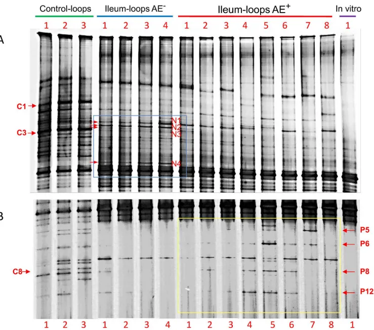
differences in ileal microbiota population of AE-negative, AE-positive, and control pigs. The band intensity was visually examined. Among the highlighted differences, two bands were
associated with AE negative pigs, band N2 and N4 (AE-bands), which were present in all 4 AE
negative samples and were present in either low intensity or absent from the control and
sam-ples from the AE-positive pigs (Fig 1). It is noteworthy that band N1 was present at higher
intensity in both AE-negative and AE-positive pigs compared to the controls; while band N3 in samples from the AE-negative pigs showed a markedly decreased abundance compared with the controls (corresponding to band C3), and was almost not detected in 7 of the 8 AE-positive pigs. Several bands were more abundant in some positive pigs compared to the
AE-negative pigs, i.e. bands P5, P6, P8 and P12 (Fig 1). These bands were almost non-detectable in
the AE negative pigs; although they were in similar abundance compared to the control pigs at
the corresponding positions (Fig 1). However, some of these bands showed higher abundance
than in the control, such as P5 in AE+pig #7 and P6 in AE+pig #5. The intensity of band P12
was higher in 3 of the 8 AE+pigs than in both AE-and control pigs (Fig 1). After challenge
with EHEC O157:H7 strain 86–24, band C1, present in higher abundance in the control pigs,
was sharply decreased in both AE-and AE+pigs. Band C8 migrated in the same position as
band P8 (Fig 1).
Prominent bands identified on DGGE profiles were excised, cloned and sequenced to
iden-tify species of origin. Results of the sequence analysis by BLASTn are listed inTable 1. Bands
N2 and N4 belonged toVeillonella caviaeand unculturedBacteroidessp. respectively. Bands
N1 and N3 belonged toStreptococcussp. andPasteurella aerogenes, respectively. Bands C3 and
C8 from the controls, with migration positions corresponding to bands N3 and P8, and were
Expression of virulence related genes in EHEC from AE positive and
negative ileal loops
To understand the genetic basis that might lead to the different colonization and AE lesion
phenotypes by EHEC O157:H7 strain 86–24 inoculated in pig ligated ileal loops, a set of 72
genes, including genes encoding LEE proteins and effectors, putative adhesins and virulence
factors, and regulatory proteins, were evaluated by qPCR (S1 table) and compared between the
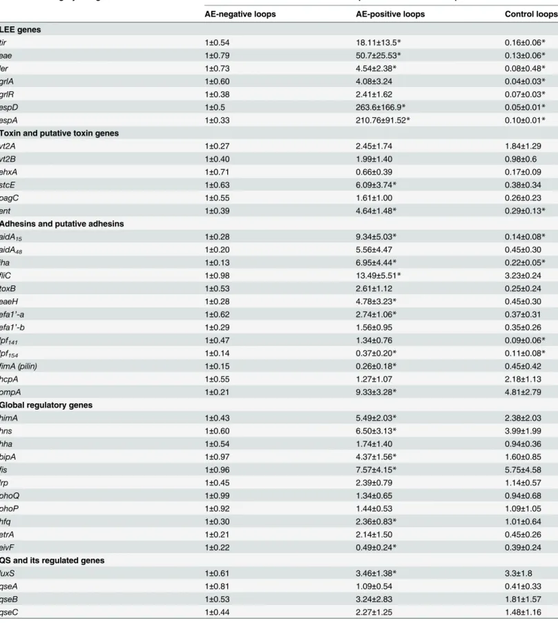
EHEC from AE positive and negative ileal loops. The expression data are summarized in
Table 2. A 3-fold change cutoff was considered as having a significant difference in bacterial gene expression provided there was statistical significance.
Fig 1. DGGE analysis of the bacterial community in the pig ileal loops with or without attaching and effacing (AE) lesion after inoculation ofE.coli
O157:H7 strain 86–24.The bacterial community profiles were generated by targeting 16S rRNA gene from reverse transcribed total intestinal bacterial RNA of the ileal contents. Figures A and B are overlapped top and lower portions of the same DGGE profiles but from different DGGE pictures with different resolutions. The profiles included control ileal loops inoculated with EMEM (control-loops), ileal loops without AE lesion and with AE lesion afterE.coliO157: H7 inoculation (ileum-loops AE-and ileum-loops AE+respectively), andE.coliO157:H7 strain 86
–24 grownin vitrobefore inoculation (In vitro).
1. LEE island and toxin genes
Given that LEE genes are necessary for AE lesion formation and intimate attachment, we assessed the relative levels of LEE gene expression in EHEC that caused colonization and AE
lesions in pig ligated ileal loops compared with those that did not. As expected,eaeandtir,
encoding the adhesin intimin and translocated intimin receptor (Tir), respectively, as well as
the key regulator of LEE,ler, were increased by 50, 18 and 4.5 fold respectively in EHEC from
AE lesion loops. The genesespAandespDwere sharply increased by 210- and 260–fold,
respec-tively, in EHEC from AE-positive loops.
The levels of transcripts for verotoxin genesvt2A andvt2B were not different in EHEC from
AE positive and negative loops (Table 2). Of note,ent/espL2, located in O island (OI)-122, was
increased in the EHEC from AE positive loops by ~4 fold (Table 2,P<0.05).
2. Putative adhesins, secreted proteins and stress genes
The gene encoding putative adhesinehaA(aidA15), located in OI-15, was increased by ~9.3 fold
in AE positive loops (Table 2,P<0.05). OI-48 genes, including putative adhesiniha, and genes
involved in stress responseterCandureDwere almost uniformly increased by approximately
5.0–7.3 fold, althoughureCwas increased by only 3.9-fold in AE positive loops (Table 2,
P<0.05).
ThehcpAgene encodes hemorrhagic coli pili (HCP), a type IV pilus (TFP) associated with
EHEC O157 pathogenicity. Our data showed that the expression ofhcpAgene was not different
in EHEC O157:H7 from AE positive loops compared with EHEC O157:H7 from AE-negative
loops (Table 2).
fliCtranscripts were 13.5-fold greater in EHEC from AE-positive than in EHEC from AE
negative loops (Table 2,P<0.05). Similarly,ompAtranscripts were 9.3-fold higher in EHEC
from AE positive loops (Table 2,P<0.05). There was no significant difference in expression of
genes encoding putative adherence factorstoxB,efa1’-a,efa1’-b,lpf141andlpf154between
EHEC from AE-positive and AE-negative loops, butfimAtranscripts showed a 3.8-fold
decrease in AE-positive loops (Table 2).
The non-LEE encoded (Nle) effector genenleAwas sharply increased 14.5-fold and there
was a significant increase in genenleB(4.7-fold) in the positive loops compared with
AE-negative loops. Putative virulence genestcEwas elevated 6.1- fold (Table 2,P<0.05).
Table 1. Sequence analysis of bands associated with AE-positive and AE-negative ligated ileal loops of pigs inoculated withE.coliO157:H7.
Band Closest isolate relative Identity (%) Accession No.
C1 Haemophilus parasuisstrain AZ2-1 100 GU226388.1
N1 Streptococcussp. 1561-2D2-04 100 FN908166.1
N2 Veillonella caviaestrain PV1 99 NR_025762.1
N3/C3 Pasteurella aerogenesstrain P592 99 AY465373.1
N4 UnculturedBacteroidessp. 99 GU905761.1
P5 Clostridium perfringens 100 AB910734.1
P6 Clostridium chauvoeistrain ASU55 99 KF372580.1
P8/C8 Clostridiumsp. clone JCC 100 HG726039.1
P12 UnculturedClostridiumsp. Clone PLYFP96 99 JN792362.1
Table 2. Gene expression profiles of O157:H7 in pig intestinal loops as determined by qPCR.
Functional category and gene Relative fold expression in intestinal loops
AE-negative loops AE-positive loops Control loops
LEE genes
tir 1±0.54 18.11±13.5* 0.16±0.06*
eae 1±0.79 50.7±25.53* 0.13±0.06*
ler 1±0.73 4.54±2.38* 0.08±0.48*
grlA 1±0.60 4.08±3.24 0.04±0.03*
grlR 1±0.38 2.41±1.62 0.07±0.03*
espD 1±0.5 263.6±166.9* 0.05±0.01*
espA 1±0.33 210.76±91.52* 0.10±0.01*
Toxin and putative toxin genes
vt2A 1±0.27 2.45±1.74 1.84±1.29
vt2B 1±0.40 1.99±1.40 0.98±0.6
ehxA 1±0.71 0.66±0.39 0.17±0.09
stcE 1±0.63 6.09±3.74* 0.38±0.34
pagC 1±0.55 1.61±1.00 0.26±0.23
ent 1±0.39 4.64±1.48* 0.29±0.13*
Adhesins and putative adhesins
aidA15 1±0.28 9.34±5.03* 0.14±0.08*
aidA48 1±0.20 5.56±4.47 0.45±0.30
iha 1±0.13 6.95±4.44* 0.22±0.05*
fliC 1±0.98 13.49±5.51* 3.23±0.24
toxB 1±0.53 2.61±1.12 0.25±0.24
eaeH 1±0.28 4.78±3.23* 0.45±0.30
efa1’-a 1±0.62 2.74±1.06* 0.37±0.31
efa1’-b 1±0.29 1.56±0.95 0.35±0.26
lpf141 1±0.47 1.34±0.76 0.09±0.06*
lpf154 1±0.14 0.37±0.20* 0.11±0.08*
fimA (pilin) 1±0.15 0.26±0.18* 0.45±0.42
hcpA 1±0.55 1.27±1.07 2.18±1.13
ompA 1±0.21 9.33±3.28* 4.81±2.79
Global regulatory genes
himA 1±0.43 5.49±2.03* 2.38±2.03
hns 1±0.60 6.50±3.13* 3.99±1.99
hha 1±0.54 1.74±1.40 0.94±0.36
bipA 1±0.97 4.37±1.56* 1.60±0.85
fis 1±0.96 7.57±4.15* 5.75±4.58
lrp 1±0.45 2.39±0.79 1.14±0.57
phoQ 1±0.99 1.34±0.65 0.94±0.68
phoP 1±0.92 1.44±0.53 1.09±1.05
hfq 1±0.30 2.36±0.83* 1.01±0.64
etrA 1±0.21 2.14±1.50 0.45±0.26
eivF 1±0.22 0.49±0.24* 0.39±0.24
QS and its regulated genes
luxS 1±0.61 3.46±1.38* 3.3±1.8
qseA 1±0.81 1.09±0.54 0.41±0.33
qseB 1±0.53 3.24±2.83 1.81±1.57
qseC 1±0.44 2.27±1.25 1.48±1.16
Table 2. (Continued)
Functional category and gene Relative fold expression in intestinal loops
AE-negative loops AE-positive loops Control loops
qseE 1±0.37 2.41±1.94 0.72±0.56
qseF 1±0.11 3.57±2.03* 1.31±0.86
flhD 1±0.46 4.03±3.02 3.76±3.57
kdpE 1±0.71 2.72±1.84 2.58±1.34
rcsB 1±0.63 3.02±1.61 2.01±1.65
Acid response and stress genes
gadA 1±0.22 2.40±0.78* 0.25±0.21*
gadC 1±0.34 4.85±4.80 0.26±0.26*
gadE 1±0.64 1.47±0.71 1.02±0.39
gadX 1±0.30 1.76±0.93 0.60±0.50
gadW 1±0.84 0.89±0.66 1.01±0.71
adiA 1±0.61 2.35±1.6 2.3±2.23
ureC 1±0.59 3.86±1.62* 0.49±0.47
ureD 1±0.50 4.97±2.51* 1.09±0.84
mnmE 1±0.30 2.04±1.29 3.19±1.64
ydeO 1±0.46 1.44±1.07 0.7±0.32
evgA 1±0.73 0.39±0.28 0.91±0.69
katP 1±0.25 2.80±2.10 0.05±0.04*
sodA 1±0.89 3.54±2.04 2.33±2.20
chuA 1±0.14 8.72±5.52* 0.77±0.25
terC 1±0.52 7.35±1.79* 0.37±0.14*
terF 1±0.18 8.39±4.95* 0.47±0.33
rpoA 1±0.33 7.21±2.60* 1.31±0.49
rpoS 1±0.40 6.35±4.41* 4.23±2.01
Secreted proteins
espP 1±0.72 2.52±0.95 0.67±0.58
espJ 1±0.19 1.11±0.39 0.27±0.23*
espFu 1±0.48 1.82±0.81 0.74±0.34
nleA 1±0.38 14.5±5.59* 0.57±0.25
nleB 1±0.67 4.74±2.72* 0.34±0.3
nleD 1±0.41 1.72±1.11 0.04±0.04*
Genes with unknown function
yhbM 1±0.20 2.73±1.97 0.78±0.51
Z1006 1±0.42 2.21±1.28 1.03±0.63
Z3276 1±0.87 1.54±1.24 0.57±0.30
a, Data are presented as relative fold expression (RFE) and represent the changes in extent of transcription compared to that of the bacteria from
AE-negative loops (assigned a value of 1.0).
b, Data are expressed as the means±SD for RNA extracted in 4
–6 biological replicates. c, Control loops were inoculated with EMEM medium only.
*indicatesp<0.05 as compared to bacteria from AE-negative loops.
3. Regulatory genes
LEE genes and other virulence factors are tightly controlled by environmental cues and co-ordinately regulated by several regulatory systems such as global regulators and quorum
sens-ing [9,22], however, the importance of these regulatory systemsin vivoare not clear.
Expres-sion of global regulatorsrpoS,himA(encoding IHF),bipA,fis, was significantly upregulated in
EHEC from AE positive loops. Interestingly, transcripts for most quorum sensing genes were
not significantly different in EHEC from both AE positive and negative loops, exceptluxSand
qseF, which showed 3.5 and 3.6-fold increases (Table 2,P<0.05).
Discussion
Enteric pathogens, including EHEC O157:H7,Vibrio choleraeandCitrobacter rodentium, have
been reported to exhibit enhanced infectivity upon passage through host intestine [8,23,24].
The mechanism of the enhanced infectivity phenotype is not clear, however, available data sug-gest that the intestinal environment favors the expression and secretion of virulence factors for infection and AE lesion by EHEC in pigs (8). This may result in selection of bacteria that read-ily form AE lesions. As the intestinal microbiota comprises an important part of the host intes-tinal environment, alteration of the normal intesintes-tinal microbiota and the resulting microbiota-dependent activation of virulence gene expression may play a critical role in infectivity by
enteric pathogens. It has been shown thatSalmonellainfection disrupted the microbial
compo-sition of a murine gastrointestinal tract and alteration of the microbiota was associated with
increased host susceptibility toSalmonellainfection in a mouse model [25,26].
In the present study, we used RNA-based DGGE to compare the microbiota of the ileum with AE and without AE lesion. One advantage of using RNA-based microbiota analysis is that it identifies active members of the bacterial community, and changes in banding pattern and intensity in DGGE indicate changes in active microbial populations, potentially linking
promi-nent microbial populations to specific functions [27,28]. In the present study, we observed that
AE-positive ileal loops resulting from inoculation of pig intestinal loops with EHEC O157:H7
strain 86–24 was associated with differences in gene expression of EHEC O157:H7 and in
intes-tinal microbial composition compared with AE-negative ileal loops. Two DGGE bands
belong-ing toVeillonella caviaeand Bacteroides sp. were more prominent in AE-loops compared to
the controls, and were almost non-detectable in AE+loops. Species ofVeillonella caviaeare
con-sidered probiotic. These species are lactate utilizers and have been used as probiotics for poultry
due to their inhibitory activities against pathogens, such asListeria monocytogenesand
Salmo-nellaTyphimurium [29,30].Bacteroidessp. such asB.fragilisandB.vulgatushave probiotic
activities in relieving neurodevelopmental disorders and againstE.coli-induced colitis in
gnoto-biotic mice respectively [31,32]. A reduced level of intestinalB.vulgatuswas suggested to be
involved in intestinal inflammation in cystic fibrosis [33]. In our present study, we do not know
whether the observed increase in activities ofVeillonella caviaeandBacterioidessp. was a
response toE.coliO157:H7 or might have existed before inoculation of the ileal loops.
There-fore, a functional relationship between the colonization and virulence ofE.coliO157:H7 and
presence or activity ofVeillonella caviaeandBacterioidessp. requires further investigation.
In AE+pigs, in addition to a reduced population or activity ofVeillonella caviaeand the
Bac-terioidessp., some pigs appeared to have increased populations/activities ofClostridiumsp.
such asC.chauvoeiandC.perfringens.C.chauvoeiis known as a cause of blackleg in cattle and
entercolitis in humans [34,35] but is not associated with disease in pigs. However, our data
showed that these organisms were also present in the control pigs with similar abundance/
activity. It has been reported that there was a negative correlation betweenC.perfringensand
microbiota of chicken [36]. The underlying mechanisms involved in the increases in bacteria associated with presence and absence of the AE lesion are not understood. Individual variations of pre-existing microbiota and host factors such as hormones may result in differential
enhancement or inhibition of growth of certain members of the microbiota. It cannot be ruled out that pre-existing microbiota may be prone to alteration of microbiota populations in the presence of EHEC. Further studies are required to compare the microbial population before
and after challenge ofE.coliO157:H7 in the same animals.
Disturbance of the microbiota can alter the intestinal microbial metabolome, a collection of molecules of microbial origin with specialized functions and important physiological effects,
such as hormones and thereby increase the susceptibility of the host to pathogen infection [37,
38] and change virulence gene expression of pathogens. Expression of virulence genes of
EHEC O157:H7 86–24 from pig ileal loops was examined by qPCR and compared between
EHEC from AE-positive and AE-negative pigs. As expected, expression of LEE genes was sig-nificantly increased in EHEC from AE positive pigs. In addition, expression of putative adhe-sins, EhaA and Iha and putative virulence factors StcE and NleA was also increased in EHEC from AE positive pig ligated ilea. These data suggest that in addition to LEE genes, these puta-tive adhesins and virulence factors that were significantly up-regulated in the intestine might be involved in colonization and AE lesion phenotype. It is well known that EHEC LEE genes and other virulence factors are regulated both tightly and coordinately by environmental
sig-nals [5,39]. However, it is not understood what intestinal signals are responsible for virulence
gene upregulation and the resultant colonization and AE lesion phenotype. Among the regula-tory factors, the present study showed that most QS gene expression was not changed in EHEC
from AE-positive and AE-negative pigs, exceptluxSandqseFthat were upregulated. QseF has
been suggested to play a role in LEE gene activation and AE lesion formation [40,41].
How-ever, our data showed that expression of the genesespFu,flhDandkdpE, downstream of the
quorum sensing QseE/F and QseC/B regulated genes, was not different between EHEC from
AE positive and AE negative pigs (Table 2).
In summary, the present study demonstrated that there were differences in the intestinal microbiota in pig ligated ileal loops challenged with EHEC O157:H7. AE-negative pigs had
increased activities/populations ofVeillonella caviaeandBacterioidessp., and decreased
popula-tions of someClostridiumsp., suggesting that these alterations of intestinal microbiota might
play a role in AE lesion development. Concomitantly, expression of LEE genes, putative adhesins,
acid resistant genesureC/Dand quorum sensing genesluxSandqseFwas increased in EHEC
from AE-positive pigs. Further studies are required to understand how the microbiota was changed and the role of these organisms in the control of EHEC infection. Studies in which the microbiota is examined before inoculation of ligated pig ileal loops may also indicate whether the differences in the microbiota that were observed existed before or after inoculation of the loops.
Supporting Information
S1 Table. Primers used for quantitative PCRand their target genes.All the primers were
designed in the present study unless referenced. (XLS)
Author Contributions
References
1. Melton-Celsa A, Mohawk K, Teel L, O'Brien A. Pathogenesis of Shiga-toxin producing escherichia coli. Current topics in microbiology and immunology. 2012; 357:67–103. Epub 2011/09/15. doi:10.1007/
82_2011_176PMID:21915773.
2. Kaper JB, Nataro JP, Mobley HL. Pathogenic Escherichia coli. Nature reviews. 2004; 2(2):123–40.
PMID:15040260.
3. Jarvis KG, Giron JA, Jerse AE, McDaniel TK, Donnenberg MS, Kaper JB. Enteropathogenic Escheri-chia coli contains a putative type III secretion system necessary for the export of proteins involved in attaching and effacing lesion formation. Proceedings of the National Academy of Sciences of the United States of America. 1995; 92(17):7996–8000. PMID:7644527.
4. Nguyen Y, Sperandio V. Enterohemorrhagic E. coli (EHEC) pathogenesis. Front Cell Infect Microbiol. 2012; 2:90. Epub 2012/08/25. doi:10.3389/fcimb.2012.00090PMID:22919681; PubMed Central PMCID: PMC3417627.
5. Yin X, Zhu J, Feng Y, Chambers JR, Gong J, Gyles CL. Differential Gene Expression and Adherence of Escherichia coli O157:H7 In Vitro and in Ligated Pig Intestines. PLoS ONE. 2011; 6(2):e17424. PMID: 21387009. doi:10.1371/journal.pone.0017424
6. Wells TJ, Sherlock O, Rivas L, Mahajan A, Beatson SA, Torpdahl M, et al. EhaA is a novel autotran-sporter protein of enterohemorrhagic Escherichia coli O157:H7 that contributes to adhesion and biofilm formation. Environmental microbiology. 2008; 10(3):589–604. Epub 2008/02/02. EMI1479 [pii]. doi:10.
1111/j.1462-2920.2007.01479.xPMID:18237301.
7. Lim JY, Yoon J, Hovde CJ. A brief overview of Escherichia coli O157:H7 and its plasmid O157. J Micro-biol Biotechnol. 2010; 20(1):5–14. PMID:20134227.
8. Brady MJ, Radhakrishnan P, Liu H, Magoun L, Murphy KC, Mukherjee J, et al. Enhanced Actin Pedes-tal Formation by Enterohemorrhagic Escherichia coli O157:H7 Adapted to the Mammalian Host. Front Microbiol. 2011; 2:226. Epub 2011/11/22. doi:10.3389/fmicb.2011.00226PMID:22102844; PubMed Central PMCID: PMC3219212.
9. Mellies JL, Barron AM, Carmona AM. Enteropathogenic and enterohemorrhagic Escherichia coli viru-lence gene regulation. Infection and immunity. 2007; 75(9):4199–210. PMID:17576759.
10. Yin X, Feng Y, Lu Y, Chambers JR, Gong J, Gyles CL. Adherence and associated virulence gene expression in acid-treated Escherichia coli O157: H7 in vitro and in ligated pig intestine. Microbiology (Reading, England). 2012; 158(Pt 4):1084–93. Epub 2012/02/04. doi:10.1099/mic.0.056101-0PMID:
22301912.
11. Yin X, Feng Y, Wheatcroft R, Chambers J, Gong J, Gyles CL. Adherence of Escherichia coli O157:H7 to epithelial cells in vitro and in pig gut loops is affected by bacterial culture conditions. Canadian journal of veterinary research = Revue canadienne de recherche veterinaire. 2011; 75(2):81–8. PMID:
21731177.
12. Kenny B, Abe A, Stein M, Finlay BB. Enteropathogenic Escherichia coli protein secretion is induced in response to conditions similar to those in the gastrointestinal tract. Infection and immunity. 1997; 65(7): 2606–12. PMID:9199427.
13. Cantey JR, Moseley SL. HeLa cell adherence, actin aggregation, and invasion by nonenteropathogenic Escherichia coli possessing the eae gene. Infection and immunity. 1991; 59(11):3924–9. Epub 1991/
11/01. PMID:1682254; PubMed Central PMCID: PMC258978.
14. Sekirov I, Finlay BB. The role of the intestinal microbiota in enteric infection. J Physiol. 2009; 587(Pt 17):4159–67. Epub 2009/06/06. doi:10.1113/jphysiol.2009.172742PMID:19491248; PubMed Central
PMCID: PMC2754356.
15. Keeney KM, Finlay BB. Enteric pathogen exploitation of the microbiota-generated nutrient environment of the gut. Current opinion in microbiology. 2011; 14(1):92–8. Epub 2011/01/11. doi:10.1016/j.mib.
2010.12.012PMID:21215681; PubMed Central PMCID: PMC3039043.
16. Yin X, Feng Y, Wheatcroft R, Chambers J, Gong J, Gyles C. Adherence of Escherichia coli O157:H7 to epithelial cells in vitro and in pig gut loops is affected by bacterial culture conditions Canadian journal of veterinary research = Revue canadienne de recherche veterinaire. 2011; 75(2):81–8.
17. Yin X, Chambers JR, Wheatcroft R, Johnson RP, Zhu J, Liu B, et al. Adherence of Escherichia coli O157:H7 mutants in vitro and in ligated pig intestines. Applied and environmental microbiology. 2009; 75(15):4975–83. PMID:19525268. doi:10.1128/AEM.00297-09
18. Walter J, Tannock GW, Tilsala-Timisjarvi A, Rodtong S, Loach DM, Munro K, et al. Detection and identi-fication of gastrointestinal Lactobacillus species by using denaturing gradient gel electrophoresis and species-specific PCR primers. Applied and environmental microbiology. 2000; 66(1):297–303. PMID:
19. van Orsouw NJ, Li D, Vijg J. Denaturing gradient gel electrophoresis (DGGE) increases resolution and informativity of Alu-directed inter-repeat PCR. Molecular and cellular probes. 1997; 11(2):95–101. doi:
10.1006/mcpr.1996.0089PMID:9160323.
20. Gong J, Yu H, Liu T, Li M, Si W, De lange CFM, et al. Characterization of ileal bacterial microbiota in newly-weaned pigs in response to feeding lincomycin, organic acids or herbal extract. Livestock Sci-ence. 2008; 116(1–3):318–22.http://dx.doi.org/10.1016/j.lvsc.2008.01.001. PMID:
BACD200800289171.
21. Livak KJ, Schmittgen TD. Analysis of relative gene expression data using real-time quantitative PCR and the 2(-Delta Delta C(T)) Method. Methods. 2001; 25(4):402–8. Epub 2002/02/16. doi:10.1006/
meth.2001.1262. S1046-2023(01)91262-9 [pii]. PMID:11846609.
22. Roe AJ, Hoey DE, Gally DL. Regulation, secretion and activity of type III-secreted proteins of entero-haemorrhagic Escherichia coli O157. Biochemical Society transactions. 2003; 31(Pt 1):98–103. PMID:
12546663.
23. Merrell DS, Butler SM, Qadri F, Dolganov NA, Alam A, Cohen MB, et al. Host-induced epidemic spread of the cholera bacterium. Nature. 2002; 417(6889):642–5. Epub 2002/06/07. doi:10.1038/nature00778
PMID:12050664; PubMed Central PMCID: PMC2776822.
24. Wiles S, Dougan G, Frankel G. Emergence of a 'hyperinfectious' bacterial state after passage of Citro-bacter rodentium through the host gastrointestinal tract. Cellular microbiology. 2005; 7(8):1163–72.
Epub 2005/07/13. doi:10.1111/j.1462-5822.2005.00544.xPMID:16008583.
25. Sekirov I, Tam NM, Jogova M, Robertson ML, Li Y, Lupp C, et al. Antibiotic-induced perturbations of the intestinal microbiota alter host susceptibility to enteric infection. Infection and immunity. 2008; 76(10): 4726–36. Epub 2008/08/06. doi:10.1128/IAI.00319-08PMID:18678663; PubMed Central PMCID:
PMC2546810.
26. Barman M, Unold D, Shifley K, Amir E, Hung K, Bos N, et al. Enteric salmonellosis disrupts the micro-bial ecology of the murine gastrointestinal tract. Infection and immunity. 2008; 76(3):907–15. Epub
2007/12/28. doi:10.1128/IAI.01432-07PMID:18160481; PubMed Central PMCID: PMC2258829. 27. Muyzer G, Smalla K. Application of denaturing gradient gel electrophoresis (DGGE) and temperature
gradient gel electrophoresis (TGGE) in microbial ecology. Antonie van Leeuwenhoek. 1998; 73(1): 127–41. Epub 1998/05/29. PMID:9602286.
28. Nakatsu CH. Soil Microbial Community Analysis Using Denaturing Gradient Gel Electrophoresis. Soil Science Society of America journal. 2007; 71(2):562–71. PMID:IND43945085.
29. Gillespie MJ, Stanley D, Chen H, Donald JA, Nicholas KR, Moore RJ, et al. Functional similarities between pigeon 'milk' and mammalian milk: induction of immune gene expression and modification of the microbiota. PLoS ONE. 2012; 7(10):e48363. doi:10.1371/journal.pone.0048363PMID:23110233; PubMed Central PMCID: PMC3482181.
30. Biddle AS, Black SJ, Blanchard JL. An in vitro model of the horse gut microbiome enables identification of lactate-utilizing bacteria that differentially respond to starch induction. PLoS ONE. 2013; 8(10): e77599. doi:10.1371/journal.pone.0077599PMID:24098591; PubMed Central PMCID: PMC3788102. 31. Gilbert JA, Krajmalnik-Brown R, Porazinska DL, Weiss SJ, Knight R. Toward effective probiotics for
autism and other neurodevelopmental disorders. Cell. 2013; 155(7):1446–8. doi:10.1016/j.cell.2013.
11.035PMID:24360269.
32. Waidmann M, Bechtold O, Frick JS, Lehr HA, Schubert S, Dobrindt U, et al. Bacteroides vulgatus pro-tects against Escherichia coli-induced colitis in gnotobiotic interleukin-2-deficient mice. Gastroenterol-ogy. 2003; 125(1):162–77. PMID:12851881.
33. Bruzzese E, Callegari ML, Raia V, Viscovo S, Scotto R, Ferrari S, et al. Disrupted intestinal microbiota and intestinal inflammation in children with cystic fibrosis and its restoration with Lactobacillus GG: a randomised clinical trial. PLoS ONE. 2014; 9(2):e87796. doi:10.1371/journal.pone.0087796PMID: 24586292; PubMed Central PMCID: PMC3929570.
34. Weatherhead JE, Tweardy DJ. Lethal human neutropenic entercolitis caused by Clostridium chauvoei in the United States: tip of the iceberg? The Journal of infection. 2012; 64(2):225–7. doi:10.1016/j.jinf.
2011.09.004PMID:21945880.
35. Uzal FA. Evidence-based medicine concerning efficacy of vaccination against Clostridium chauvoei infection in cattle. The Veterinary clinics of North America Food animal practice. 2012; 28(1):71–7, viii.
doi:10.1016/j.cvfa.2011.12.006PMID:22374118.
36. Feng Y, Gong J, Yu H, Jin Y, Zhu J, Han Y. Identification of changes in the composition of ileal bacterial microbiota of broiler chickens infected with Clostridium perfringens. Veterinary microbiology. 2010; 140(1–2):116–21. doi:10.1016/j.vetmic.2009.07.001PMID:19647376.
38. Ferreira RB, Gill N, Willing BP, Antunes LC, Russell SL, Croxen MA, et al. The intestinal microbiota plays a role in Salmonella-induced colitis independent of pathogen colonization. PLoS ONE. 2011; 6(5):e20338. Epub 2011/06/03. doi:10.1371/journal.pone.0020338PMID:21633507; PubMed Central PMCID: PMC3102097.
39. Nakanishi N, Abe H, Ogura Y, Hayashi T, Tashiro K, Kuhara S, et al. ppGpp with DksA controls gene expression in the locus of enterocyte effacement (LEE) pathogenicity island of enterohaemorrhagic Escherichia coli through activation of two virulence regulatory genes. Molecular microbiology. 2006; 61(1):194–205. PMID:16824105.
40. Hughes DT, Clarke MB, Yamamoto K, Rasko DA, Sperandio V. The QseC adrenergic signaling cas-cade in Enterohemorrhagic E. coli (EHEC). PLoS Pathog. 2009; 5(8):e1000553. Epub 2009/08/22. doi: 10.1371/journal.ppat.1000553PMID:19696934; PubMed Central PMCID: PMC2726761.
41. Njoroge J, Sperandio V. Enterohemorrhagic Escherichia coli virulence regulation by two bacterial adrenergic kinases, QseC and QseE. Infection and immunity. 2012; 80(2):688–703. Epub 2011/12/07.