The Molecular Mechanisms of OPA1-Mediated
Optic Atrophy in
Drosophila
Model
and Prospects for Antioxidant Treatment
Will Yarosh1,2, Jessica Monserrate2, James Jiayuan Tong3,4, Stephanie Tse1, Phung Khanh Le1, Kimberly Nguyen1, Carrie B. Brachmann2, Douglas C. Wallace3,4, Taosheng Huang1,2,5*
1Department of Pediatrics, Division of Human Genetics, University of California Irvine, Irvine, California, United States of America,2Department of Developmental and Cell Biology, University of California Irvine, Irvine, California, United States of America,3Department of Biological Chemistry, University of California Irvine, Irvine, California, United States of America,4Center for Molecular and Mitochondrial Medicine and Genetics, University of California Irvine, Irvine, California, United States of America,
5Department of Pathology, University of California Irvine, Irvine, California, United States of America
Mutations in optic atrophy 1 (OPA1), a nuclear gene encoding a mitochondrial protein, is the most common cause for autosomal dominant optic atrophy (DOA). The condition is characterized by gradual loss of vision, color vision defects, and temporal optic pallor. To understand the molecular mechanism by whichOPA1mutations cause optic atrophy and to facilitate the development of an effective therapeutic agent for optic atrophies, we analyzed phenotypes in the developing and adultDrosophilaeyes produced by mutantdOpa1(CG8479), aDrosophilaortholog of humanOPA1.
Heterozygous mutation of dOpa1 by a P-element or transposon insertions causes no discernable eye phenotype,
whereas the homozygous mutation results in embryonic lethality. Using powerfulDrosophilagenetic techniques, we
created eye-specific somatic clones. The somatic homozygous mutation of dOpa1 in the eyes caused rough
(mispatterning) and glossy (decreased lens and pigment deposition) eye phenotypes in adult flies; this phenotype was reversible by precise excision of the inserted P-element. Furthermore, we show the rough eye phenotype is caused by the loss of hexagonal lattice cells in developing eyes, suggesting an increase in lattice cell apoptosis. In adult flies, the
dOpa1 mutation caused an increase in reactive oxygen species (ROS) production as well as mitochondrial fragmentation associated with loss and damage of the cone and pigment cells. We show that superoxide dismutase 1 (SOD1), Vitamin E, and genetically overexpressed human SOD1 (hSOD1) is able to reverse the glossy eye phenotype ofdOPA1mutant large clones, further suggesting that ROS play an important role in cone and pigment cell death. Our results showdOpa1mutations cause cell loss by two distinct pathogenic pathways. This study provides novel insights into the pathogenesis of optic atrophy and demonstrates the promise of antioxidants as therapeutic agents for this condition.
Citation: Yarosh W, Monserrate J, Tong JJ, Tse S, Le PK, et al. (2008) The molecular mechanisms of OPA1-mediated optic atrophy inDrosophilamodel and prospects for antioxidant treatment. PLoS Genet 4(1): e6. doi:10.1371/journal.pgen.0040006
Introduction
Autosomal dominant optic atrophy (DOA) is the most common hereditary optic atrophy with an incidence rate as high as 1:10,000–1:50,000 [1,2]. It is characterized by central vision loss, color vision abnormalities [1,3–5], and degener-ation of the retinal ganglion cells [6]. The onset of DOA symptoms typically occur in the first decade of life and visual loss is progressive, bilateral and irreversible once cell death has occurred [3]. There is marked intra- and inter-familial phenotypic variability [5,7]. Optic atrophy may associate with hearing loss, apoptosis, and ophthalmoplegia [8]. Some families have been described with sex-influenced DOA phenotypes [9–11].
The majority of DOA is caused by mutations in the optic atrophy 1 (OPA1) gene [7,12–14]. OPA1 is a nuclear gene which encodes a mitochondrial protein [12]. OPA1 consists of an N-terminal mitochondrial target signal, a transmembrane domain, a presenilin-associated rhomboid-like protease recognition site, and a dynamin-like domain with GTP binding activity. OPA1 is expressed ubiquitously [13,15] and functions in mitochondrial fusion [16–22], ATP production [23], and cytochrome-c mediated apoptosis [24].
Mitochon-drial diseases are associated with mitochonMitochon-drial fragmenta-tion due to the fast proteolytic processing of the OPA1 protein. Overexpression of OPA1 can prevent such fragmen-tation [18]. These observations suggest that OPA1 plays an important role in mitochondrial function.
Although studies have contributed to our current under-standing of OPA1 in terms of function and its relationship to optic atrophy, the pathogenesis of optic atrophy remains poorly understood. The major goal of this study is to establish an animal model for effective in vivo OPA1 studies for pathogenesis and therapeutic development. Previous at-tempts to create a mouse model of OPA1 revealed that
Editor:Bingwei Lu, Stanford University, United States of America
ReceivedMay 17, 2007;AcceptedNovember 27, 2007;PublishedJanuary 11, 2008
Copyright:Ó2008 Yarosh et al. This is an open-access article distributed under the
terms of the Creative Commons Attribution License, which permits unrestricted use, distribution, and reproduction in any medium, provided the original author and source are credited.
Abbreviations:DOA, autosomal dominant optic atrophy; IOC, interommatidial cell; OPA1, optic atrophy gene 1; ROS, reactive oxygen species
homozygous Opa1 mutant mice are embryonically lethal, while heterozygous animals showed no phenotype until a later age [25,26]. ADrosophilamodel of optic atrophy would offer several advantages: there is excellent homology between
Drosophilagenes and many human disease loci; it has a short lifespan; its eye development is well-studied; its genome has been sequenced; a wide variety of mutants and gene manipulation systems are available; and, for optic atrophy studies, its eye structure is well-defined and the phenotype is easily visualized [27]. During Drosophila eye development, lattice cell apoptosis is highly regulated, with one-third of the lattice cells being eliminated by apoptosis.
Here, we established a Drosophila model where the
Drosophila ortholog of OPA1, CG8479 (dOpa1) [28] is inacti-vated by two different insertions. Furthermore, we were able to produce somatic homozygous dOpa1-deficient cells in
Drosophila eyes. Functions of OPA1 in the eye and patho-genesis of optic atrophy were also addressed.
Results
Drosophila Opa1Is an Ortholog of HumanOPA1
To identify the Drosophila ortholog of human OPA1, we performed BLAST searches with the humanOPA1cDNA and amino acid sequences. CG8479 (GenBank [http://www.ncbi. nlm.nih.gov/Genbank], protein accession number: AAF58275) was identified as aDrosophilaortholog ofhOPA1(E¼0.0). To further characterize dOpa1, we used multiple sequence comparison by log expectation (MUSCLE) algorithm to perform amino acid sequence alignments of human OPA1 (hOPA1), mouse Opa1 (mOpa1), CG8479 (dOPA1), and Yeast Mgm1 (Figure S1). In all four organisms, the GTPase domain was the most conserved region and the basic domain was the least conserved region. The amino acid sequence of mOPA1 shared 96% similarity with hOPA1. When regions of these proteins were compared, the similarity percentages were 86.5% for the basic domain, 99.6% for the GTPase domain, and 98% for the dynamin central region. dOpa1 shared 51.2% similarity to hOpa1 amino acid sequence, a higher score than the well-studied yeast protein Mgm1. At the domain level, the similarity scores were 24.5% for the basic
domain, 72% for the GTPase domain, and 53.5% for the dynamin central region. The high amino acid conservation of the GTPase domain and the dynamin regions between hOPA1 and dOpa1 suggests that dOpa1 is theDrosophilaortholog of hOPA1.
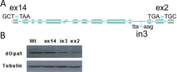
ThreeDrosophilalines with P-element/transposon insertions in thedOpa1gene were used in this study. The insertions in the coding sequence of 2ndexon, in the 3rdintron, and in the noncoding region of the 14th exon of the dOpa1 gene were designated as dOpa1þ/ex2, dOpa1þ/in3, and dOpa1ex14/ex14, re-spectively (Figure 1A; http://www.flybase.org). To determine if these insertions disrupted dOpa1 expression, we examined dOpa1 protein levels in these lines. Western blot analysis with a mouse polyclonal antibody against OPA1 showed that wild-type and dOpa1ex14/ex14 flies had similar levels of dOpa1 (Figure 1B), indicating that the insertion in exon 14 had no effect ondOpa1expression. In contrast, the protein levels in
dOpa1þ/ex2anddOpa1þ/in3were decreased. This indicated that the P-element insertion in exon 2 (dOpa1þ/ex2) and transposon insertion in intron 3 (dOpa1þ/in3) disrupteddOpa1expression. Since insertion in non-coding exon 14 had no effect on the dOpa1 protein level,dOpa1þ/ex14served as a control.
Somatic Homozygous Mutations indOpa1Resulted in a Rough and Glossy Eye Phenotype in AdultDrosophilaFlies
After we identified theDrosophilaortholog ofOPA1,dOpa1,
and confirmed that the insertions in the coding sequence of the 2ndexon and the 3rdintron disrupteddOpa1expression, we proceeded to test if loss ofdOpa1 would produce an eye phenotype. Eye phenotypes of dOpa1þ/ex2, dOpa1þ/in3, and
dOpa1þ/ex14 flies were examined by bright field microscopy (Figure 2A–2C). No gross eye phenotypes were observed, suggesting that haplo-insufficiency did not produce an observable phenotype in theDrosophilaeye.
We then crossed Drosophila dOpa1þ/ex2, dOpa1þ/in3, and
dOpa1þ/ex14. NodOpa1ex2/ex2ordOpa1in3/in3flies were obtained, whereas dOpa1ex14/ex14 flies were produced and appeared normal. Therefore, homozygous dOpa1ex2 and dOpa1in3 appear to be lethal, supporting the conclusion that the insertions in exon 2 (dOpa1þ/ex2) and intron 3 (dOpa1þ/in3) disrupted dOpa1 expression. To generate Drosophila with homozygous dOpa1in3/in3 mutant ommatidia without being impeded by the embryonic lethality of the homozygous mutant, we generated homozygous mutant ommatidia from heterozygous Drosophila by using somatic mutagenesis to eliminate thedOpa1þallele in certain eye cells.
The crosses with the dOpa1þ/Drosophila (Figure S2A) resulted in mosaic-eyed flies by the F2 cross (small clones). The mutant ommatidia contained three different cell types of eitherdOpa1þ/þ,dOpa1þ/, ordOpa1/. The different cell types could be identified by eye color (white, light red, and red, respectively), which corresponded to an increased copy number of the mini-white gene in the insertions. Small clone mosaics were generated for dOpa1ex2, dOpa1in3, and
dOpa1ex14, and later characterized by bright field and scanning electron microscopy (Figure 2). The homozygous mutant clones (dOpa1ex2/ex2 and dOpa1in3/in3) had a slightly rough eye phenotype with low penetrance (,5%–10%) compared with the respective parental (Figure 2D–2H) and controldOpa1ex14/ex14(Figure 2F and 2I) stocks.
To analyze the phenotype of the somatic homozygous
dOpa1 mutants with greater sensitivity, we generated‘‘large
Author Summary
Optic atrophies are a group of neurodegenerative disorders characterized by a gradual loss of vision, color vision defects, and temporal optic pallor. Autosomal dominant optic atrophy (DOA), a type of optic atrophy, contributes to a large portion of optic atrophy cases. Mutations of the optic atrophy 1 (OPA1) gene are responsible for this condition. Here we describe mutantDrosophilathat contain insertions in theDrosophila OPA1 ortholog, dOpa1. Heterozygous mutation causes no discernable eye phenotype, and homozygous mutation results in embryonic lethality. Using the powerful
Drosophila genetic techniques, we created eye-specific mutants, giving rise to cells with two mutant copies ofdOpa1only in the
somatic clones’’in the Drosophilaeye (Figure 2J–2R) using a mutation in theMinute gene located on the same chromo-somal arm asdOPA1. Homozygous mutations of theMinute
gene are lethal, therefore eliminatingMinute/dOpa1þ/þcells. As a result, generation of large somatic clones produced only two cell types:dOpa1þ/Minuteþ/anddOpa1/Minuteþ/þ[29]. We successfully generated large clone mosaics for insertions in exon 2 (Figure 2J, 2M, and 2P), intron 3 (Figure 2K, 2N, and 2Q), and exon 14 (Figure 2L, 2O, and 2R). IndOpa1ex2/ex2
and dOpa1in3/in3, but not the dOpa1ex14/ex14 clones, had a robust rough/glossy phenotype. In addition,dOpa1ex2/ex2and
dOpa1in3/in3, but not dOpa1ex14/ex14, clones exhibited tissue necrosis with variable onset and penetrance (Figure S3). The large clones of theMinutestock have a darker eye color. Thus, cells containing one copy of the Minute mutation were
characterized by a very deep red pigmentation. Light color cells represented dOpa1/ cells. The phenotypes of dOpa1
exon 2, intron 3, and exon 14 large clone mosaic eyes were scored (n.60; Figure S2B). Almost all of thedOpa1exon 2 and intron 3 large clones exhibited a severe rough/glossy phenotype, while all of the dOpa1 exon 14 large clones appeared normal.
ThedOpa1Mutation Is Genetically Reversible Via
P-Element Excision
To confirm that the phenotype of the somatic clones was the result of disruptingdOpa1 expression by the P-element insertion, we excised the P-element by crossing the stocks with KiD2–3 stocks. KiD2–3 stocks express a transposase that excises the P-elements (Figure S4). Excision of the P-element indOpa1ex2/ex2resulted in a complete reversal of the rough/ glossy phenotype in the large clones (Figure 3A and 3B). The P-element excision was confirmed by PCR using primers flanking the P-element insertion site. As expected after complete excision, the PCR amplification produced a 413 base pair fragment (Figure 3C, Lane 6). The precision of the excision was verified by DNA sequencing (Figure 3D). These results show that the rough glossy eye phenotype observed in ourdOpa1somatic clones was indeed caused by a P-element insertion in the dOpa1 gene, which can be genetically reversed.
Homozygous Mutation ofdopa1Causes Interommatidial Cell Death
All ommatidial units are uniformly patterned by the specific placement of interommatidial cells (IOC), also called lattice cells. It has been shown that apoptosis helps achieve the final pattern through removal of surplus IOC during development. The core of an ommatidium consists of four cone cells and two primary pigment cells, which are
Figure 2.Homozygous Mutation ofdOpa1Results in a Rough and Glossy Phenotype in the Somatic Clones of the AdultDrosophilaEye
(A–C) Bright field microscopy images of the adult eyes of the original heterozygous insertion lines are shown in (A), (B), and (C) fordOpa1þ/ex2,dOpa1þ/in3, anddOpa1þ/ex14, respectively. None of these stocks contained any gross eye phenotype. The mosaic-eyed flies produced by the F2 cross (small clones) containdOpa1þ/þ,dOpa1þ/, ordOpa1/cell types, which are white, light orange, and dark orange, respectively.
(D–I) (D), (E), and (F) are bright field images and (G), (H), and (I) are SEM micrographs of the small clonesdOpa1ex2,dOpa1in3
, anddOpa1ex14mutations, respectively. A weak rough phenotype with low penetrance was observed in the small clones of thedOPA1ex2anddOPA1in3mutations, but not the
dOPA1ex14mutation.
(J–R) (J), (K), and (L) are bright field images; (M), (N), and (O) are SEM micrographs; and (P,Q,R) are 103digital zooms of areas of interest in the large clones of thedOpa1ex2,dOpa1in3, anddOpa1ex14mutations, respectively. Glossy and rough phenotypes were observed with 100% penetrance in the large clones of thedOPA1ex2anddOPA1in3mutations, but not thedOPA1ex14mutation.
doi:10.1371/journal.pgen.0040006.g002
Figure 1.Insertion indOPA1Can Disrupt dOPA1 Expression
The location of each within dOpa1 and the nucleotide sequences flanking the insertion site in theDrosophilalines used in this study (A). Western blot analysis of severalDrosophilalines containing insertions near or withindOpa1. (B) showsdOpa1levels in adultDrosophila wild-typedOpa1(dOpa1þ/þ),Drosophilawith a transposon insertion in exon 14 (noncoding region) (dOpa1þ/ex14), intron 3 (dOpa1þ/in3), and exon
surrounded by IOC. The rough phenotypes observed in the adultDrosophila eye of the dOpa1/ somatic clones suggest that cells within the ommatidium fail to develop and pattern properly, presumably due to cells failing to regulate IOC apoptosis properly. The glossy phenotype is indicative of a failure to deposit lens and pigment material by the cone and pigment cells.
LargedOpa1somatic clones were generated with theMinute
gene mutation and GFP under control of a ubiquitous promoter (ubi), both of which are distal to the flippase recombination target sequence (FRT). As a result, the
dOpa1in3/in3 Minute/ ubi-GFP/ (GFP negative) clones can be distinguished from thedOpa1þ/in3Minuteþ/ubi-GFPþ/(GFP positive) clones [30]. To visualize the final, post-apoptotic ommatidial pattern during pupal development, pupae were isolated and dissected 42h after pupal formation (APF). Eye discs were stained using anti-armadillo antibody and Hoechst (Figures S5A and S5B and 4). GFP positive ommatidial units (dOpa1þ/in3Minuteþ/ubi-GFPþ/) had similar numbers of IOCs to those reported for wild-type eyes, but GFP-negative ommatidial units (dOpa1in3/in3 Minute/ ubi-GFP/) had a significantly reduced number of IOCs (p ¼ 5.05 3 108) associated with a high degree of mispatterning (Figure S5C). Cone and pigment cells of both genotypes were normal at this stage. The decreased number of IOCs in these clones indicates that mutation ofdOpa1may increase IOC cell death prior to 42h APF, and the reduction of IOCs results in the rough phenotype (mispatterning).
Previously, it was reported that mutations in OPA1 caused a decrease in ATP production [23] and an increase in cytochrome-c release [16,19,24,31–34]. Reduced ATP produc-tion caused by inhibited oxidative phosphorylaproduc-tion (OX-PHOS) would, in turn, be expected to increase mitochondrial reactive oxygen species (ROS). Chemically increased ROS can damage macromolecules and cellular membranes and in-crease apoptosis rates. To determine if inin-creased ROS production resulted in increased IOC cell death in thedOpa1
mutant, pupal retinae were stained for ROS production using dihydroethidium at different stages of development [35]. We did not observe dihydroethidium positive nuclei in any of the
dOpa1in3/in3clones (data not shown). This data suggests that the mutation ofdOpa1did not cause a significant increase in ROS production in the pupal retina prior to 42h APF.
Ommatidial Units of Adult Somatic HomozygousdOpa1
Mutant Flies Are Morphologically Perturbed
The glossy eye phenotype observed in the dOpa1in3 large clones suggests the cone and pigment cells did not secrete and deposit pigment properly [36]. A lens-depleted glossy eye phenotype was previously reported and had a defect in cone and pigment cell specification or proliferation [36,37]. At 42h APF, the pigmented cone cells appeared to be normal in morphology and number. To test if the glossy phenotype in our somatically generated dOpa1/ ommatidial units was caused by cone and pigment cell death later on, we performed confocal microscopy on the adultdOpa1in3large clones. This technique enabled the visualization of heterozygous (GFP-positive) and homozygous (GFP-negative) mutant ommatidial units, and the dissection of the ommatidial structures at a thickness of 2 microns. Using this approach, it was possible to reconstruct the three-dimensional structure of theDrosophila
eye. The ommatidial structure of thedOpa1/(GFP negative) eye was disorganized (Figure 5; Videos S1–S3), whereas the
dOpa1þ/ommatidia (GFP positive) were largely normal. The detailed structure of dOpa1þ/ ommatidia (GFP positive) revealed that they were structurally normal; the lattice cells were arranged in hexagonal shapes with cone and pigment cells centralized in the ommatidia (Figure 5; Videos S1–S3). In contrast,dOpa1/(GFP negative) ommatidia showed a loss of lattice cells (consistent with results in Figure 3), the cone cells were condensed, and exhibited severe cell surface defects. The space between cone and lattice cells was also dramatically increased, which is consistent with cone and/or pigment cell death. Our results suggest that the glossy phenotype in somatic mutant eyes was caused by damage or death of cone and pigment cells occurring after 42 h APF, which results in a decreased secretion of lens deposition.
To test if increased ROS levels could cause cone and pigment cell death in adultdOpa1/large clone eye tissues,
we measured the ROS levels in thedOpa1in3/in3 clones using MitoSOX (Invitrogen, M36008). dOpa1in3/in3 large clones exhibited increased ROS levels in the adult Drosophila eyes (Figure 5). Thus, the increase in ROS may be correlated with an increase in pigment and cone cell death, which is consistent with the decrease in production of lens material and glossy eye phenotypes. Using MitoSOX staining, we also show that dOpa1in3/in3 large clones exhibit higher levels of ROS indOpa1/cells compared todOpa1þ/cells (Figure 5). In
Figure 3.Reversal of thedOpa1Large Clone Eye Phenotype by P-Element Excision
The phenotypes present in large clones of thedOpa1ex2mutation were reversed, as documented by bright field microscopy (A) and SEM (B), by excision of the P-element. The excision of the P-element insertion was verified by PCR (C). Lane 1 contains 1lg of the 100 base pair ladder (NEB) for reference. Lane 2 is a negative reagent control without template DNA. Lane 3 contains DNA fromdOpa1þ/ex2amplified with the F1 primer, which anneals in the exon 2 flanking the P-element insertion and the R1 primer annealing inside the P-element. Lane 4 contains DNA from excised flies amplified with the F1 and R1 primers. Lane 5 contains DNA fromdOpa1þ/ex2and is amplified using F1 and R2. Lane 6 contains DNA excised from flies amplified with F1 and R2. A schematic representation of the annealing sites of the primer sets used in (C). We further verified the P-element excision by sequencing of the PCR product generated in lane 6 (D) and comparing with the known wild-type (wt) sequence.
this experiment, adultdOpa1 mutant large clone eyes were quickly dissected and stained using MitoSOX (2.5mM) in HBSS. MitoSOX staining on live dOPAþ/large clones eyes were performed on adultDrosophilaas described recently [38]. As shown in Figure 5, a significantly higher level of MitoSOX fluorescence was detected in dOpa1/ cells compared to
dOpa1þ/cells. The specificity of MitoSOX for superoxide was tested by performing MitoSOX stains in the presence of both SOD-1 (1000 units/ml) and Vitamin E (200 lg/mL); both dramatically reduced MitoSOX signal levels (data not shown).
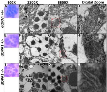
ThedOpa1Mutation Affects Mitochondrial Morphology
and Tissue Integrity
Previous studies have shown that OPA1 mutations induce mitochondrial fragmentation [13]. To determine if loss of
dOpa1 had similar effects on mitochondrial integrity in the
Drosophilaeye, we examined the morphology of mitochondria in wild-type and mutant large clone (dOPA1/) ommatidia. No difference was found in rhombomere structure (Figure 6A, 6E, and 6I), which suggests that the photoreceptor cells were not affected by thedOpa1mutation. In the homozygous mutants, mitochondria were scarce and significantly dysmor-phic (Figure 6J and 6K). These results suggest thatdOpa1 is important for mitochondrial morphology and that mutation ofdOpa1 may be associated with increased ROS production, leading to cone and pigment cell death.
The Rough Glossy Phenotype Can Be Partially Reduced by Antioxidant SOD-1, Vitamin E, and Genetically Expressed Human SOD1
Our observation that ROS production was increased in the homozygousdOpa1/mutant ommatidia and was associated with cone and pigment cell death suggested that antioxidants might ameliorate the glossy eye phenotype. In previous studies, feeding Drosophila with SOD-1 and vitamin E have been shown to inhibit cell death in the Parkinson’s disease
Drosophilamodel [39]. To test the effects of antioxidants on
Drosophilaeye phenotype, we added SOD-1 and vitamin E to the Drosophilafood. Ingestion of SOD-1 (1000 units/ml) and vitamin E (20 lg/ml) partially rescued the glossy eye phenotype in the dOpa1in3/in3 mutant ommatidum (Figure 7A–7E). Furthermore, MitoSOX (2.5 mM) staining of dis-sected Drosophila eyes showed treatment of antioxidants reduced the ROS level in dOpa1in3/in3 cells (Figure 7G and 7H) compared to those which were untreated (Figure 7F). A significant (p,0.05) reduction of ROS levels indOpa1in3large clone eye tissue was also found in the homogenates of antioxidant-treated samples (data not shown). In order to further demonstrate that the glossy eye phenotype ofdOPA1
mutant large clones arises as a result of excessive ROS generation by the mitochondria, we genetically tested if overexpression of human superoxide dismutase 1 (hSOD1, GenBank Accession Number NM_000454) is able to reverse the glossy eye phenotype of dOPA1mutant large clones. As shown in Figure S6, the overexpression of hSOD1 in the
Drosophila eye was achieved using a UAS/Gal4 system as previously described [39]. Figure 8A shows that expression of hSOD1 in the Drosophila eye resulted in a significant reduction of glossy eye phenotypes of dOPA1 mutant large clones (Right) in comparison to dOPA1 mutant large clone controls without the UAS-hSOD1 transgene (Left). The reversal of the rough eye phenotype ofdOPA1mutant large clones was not observed. In order to quantify the glossy eye phenotypes of our large clones, the phenotypes of dOPA1
mutant large clone eyes were scored based on the eye phenotype (n.40). As shown in Figure 8B, overexpression of hSOD1 in theDrosophilaeye resulted in dramatic reduction of the glossy eye phenotype ofdOPA1mutant large clones. The use of genetic methods to reverse the glossy eye phenotype of
dOPA1mutant large clones through the expression of hSOD1 indicates that this phenotype arises as a result of excessive ROS generation by the mitochondria and further supports a role for ROS in the pathogenesis of optic atrophy, suggesting that antioxidants may be beneficial in ameliorating these symptoms.
Discussion
Here, we report that the somatic mutation of theDrosophila
OPA1 ortholog, dOpa1, results in a rough/glossy eye pheno-type. It is very interesting that mutation ofOPA1in humans causes an autosomal dominant condition. When analyzing eye phenotype of the Drosophila eye we found that Drosophila
heterozygous for mutations indOPA1did not appear to have a gross phenotype. There are many possible explanations for this observation, some of which include the evolutionary differences between OPA1 in humans and its orthologs. However, the grossly normal eye structure in heterozygous
Figure 4. Homozygous Mutation of dOpa1 Causes a Loss of Inter-ommatidial Cells
Pupae were staged 42 h after pupae formation (APF), and large clone mosaics with GFP expression in the eye imaginal disk were collected and stained with Hoechst and for Armadillo. Eye discs were then analyzed and regions ofdOpa1in3/in3and/ordOpa1þ/in4ommatidial units were
photo-graphed and presented (A,C), using only channels with Armadillo signal. (A) illustrates a region of thedOpa1in3/in3ommatidial units. A red box marked with a b is used to indicate the ommatidial unit illustrated in (B). The different cell types are highlighted (B). The cone cells, c, are illustrated in yellow, the pigment cells, p, in blue, the IOCs in purple, and the bristle cells, b, in white. (C) shows the ommatidial units of adOpa1þ/in3Minuteþ/ ubi-GFPþ/ clonal region. A red box marked with a d indicates the ommatidial unit is shown in (D). No cell types are missing, and no disorganization is present. Cell types are represented as in (B).
mutantDrosophiladoes not imply normal function. It is still possible that the eyes are dysfunctional even though the eye structure is unaffected. We are in the process to analyze the heterozygous mutants for cardiac, skeletal muscle and photo-receptor function.
In the developingDrosophilaeye, the imaginal disc under-goes two phases of proliferation. First, all cells divide rapidly and asynchronously without regulation [40]. Then, the morphogenic furrow sweeps across the eye disc to pattern the cells. Subsequently, the cells differentiate into photo-receptor cells or undergo another round of division, which produces cone and pigment cells. During this process, the photoreceptor cells are specified first, they then retract and recruit four cone cells, which in turn recruit two pigment cells [41]. The cells remaining from the first phase of proliferation form ommatidial lattice cells [27]. These include the secondary, tertiary, and bristle cells that separate adjacent ommatidia, forming the precise hexagonal patterns of the adult Drosophila eye. After cell expansion, excess cells are eliminated by apoptosis [42]. Our results show that the rough/ glossy-eye phenotype of thedOpa1mutants is associated with lattice and cone/pigment cell death, and that death of lattice cells and cone/pigment cells occur at different developmental stages. In developingDrosophilaeye, the loss ofdOpa1causes a significant reduction in the number of lattice cells. However, the cone and pigment cells are structurally normal at 42 h APF stage, but later damage or death of cone and pigment cells results in decrease secretion of lens materials.
The underlying mechanism causing increased lattice cell death due to dOpa1 loss is still unknown. Previous studies showed that lattice cells respond to signals from cone and
pigment cells [43]. Our results show that loss ofdOpa1causes a significant reduction in the number of lattice cells in the presence of normal cone and pigment cells, indicating that death of lattice cells is not due to early defects in differ-entiation of retinal cell types. It has been shown that OPA1 reduces mitochondrial cytochrome-c release following cristae remodeling in the presence of activators of the mitochondrial apoptosis pathway [16]. Furthermore, electron microscopy and electron tomography studies suggest that OPA1 seques-ters cytochrome-c within mitochondrial cristae by narrowing cristae junctions, thereby inhibiting the redistribution of cytochrome-c to the inner membrane space [16]. However, the role of cytochrome-c in apoptosis inDrosophilahas only been recently established. Mendes and colleagues showed that
cyt c-dplays an important role inDrosophilaeye apoptosis; and mutation of cyt c-d causes an increase in the number of interommatidial cells and perimeter cells [44]. Thus we hypothesize that lattice cell death is the result of a cell autonomous death signal from mitochondria.
Our results show that cone and pigment cell death was correlated with increased ROS levels and mitochondrial fragmentation. This implies that the increased ROS produc-tion induces cell death, which is further supported by the reversal of these effects and the rescuing of the glossy eye phenotype by antioxidants. Increased ROS production can trigger apoptosis to eliminate deleterious cells [45], but the exact link between oxidative stress and specific apoptosis pathways remains elusive. Transient fluctuations in ROS levels may serve as an important regulator of mitochondrial respiration, but high and sustained ROS levels cause severe damage [46,47]. The rough/glossy phenotype is a result of the
Figure 5.HomozygousdOpa1Mutations Produce Morphologically Perturbed Ommatidial Units
Confocal microscopy analysis of the eyes of anesthetized adultdOpa1in3/in3large clones. A 3D reconstruction of a large clone withdOpa1þ/in3and
dOpa1in3/in3ommatidia was generated from reflectance (A), GFP fluorescence (B), and merged signals (C).dOpa1þ/in3anddOpa1in3/in3ommatidial units from (C) were magnified and are shown in (D) and (E), respectively. Note the abnormal morphological features of the cone cells in thedOpa1in3/in3
ommatidium. The red bar equals 2 microns. (F) shows thatdOpa1in3mutation causes higher ROS levels than control eyes. The MitoSOX fluorescent signals were measured in tissue homogenates from dissected control eyes (wild-type) anddOpa1in3large clone eyes. The data represent the mean6
standard deviation of three experiments, using 10-d-old flies and a total of 40 flies per genotype, *,0.05. ROS level indicated as MitoSOX fluorescence intensity normalized to microgram of eye tissue homogenate. dOpa1in3large clones exhibit relatively higher levels of ROS in dOPA1/cells than dOpa1þ/mutant cells. AdultdOpa1mutant large clone eyes were promptly dissected, stained, and imaged using MitoSOX in HBSS (G). MitoSOX fluorescent signal histogram plots (bottom row) of fluorescent images (middle row) were generated using jImage (values 0–256, left to right); corresponding light microscope images (top row) illustrate the eye after dissection and MitoSOX staining.
lack of cone and pigment cells. The consequence is insufficient secretion of lens material, which directly con-tributes to the glossy phenotype. This phenotype may occur because cone and pigment precursor cells undergo too few mitotic divisions, failing to enter the second mitotic wave due to insufficient ATP production. Alternatively, the phenotype could result from the misregulation of cone and or pigment cell differentiation. Our results suggest an additional possi-bility of cell damage and death from ROS after 42h APF. Importantly, antioxidants can partially rescue the glossy eye phenotype in thedOpa1intron 3 large clone ommatidia, but do not rescue the rough eye phenotype. This may further implicate the role of ROS in the pathogenesis of the eye phenotypes in thedOpa1mutants.
Our results show that loss of dOpa1causes a rough, glossy eye phenotype by two distinct pathogenic pathways. The lattice cell death observed in the developing eye is probably linked to increased cytochrome-c release. The glossy eye phenotype in the mutant flies is consistent with decreased lens material secretion from the cone/pigment cells, which are damaged by sustained high ROS levels. Since we were able to partially reverse the eye phenotype through antioxidant treatment and expression of human SOD, antioxidant may provide a fruitful approach for treating this condition.
Materials and Methods
Drosophila strains and genetics.The following stocks containing insertions inCG8479were used in this study as described inResults. y[d2] w[1118] Pi2 PfGMR-lacZ.C(38.1)g TPN1; Pfry[þt7.2]¼ neo-FRTg42D Pfw[þmC]¼lacWgl(2)s3475[s3475]/CyO y[þ] was a generous gift from the UCLA Undergraduate Research Consortium in Func-tional Genetics (URCFG) [30]. PBac[48]CG8479f02779 / CyO y[þ] and PBac[48] CG8479f03594 stocks were obtained from the Harvard
Drosophilastock center. The following stocks were used to generate
somatic clones. y[d2] w[1118] Pfry[þt7.2]¼ey-FLP.Ng2 Pf GMR-lacZ.C(38.1)gTPN1; Pfry[þt7.2]¼neoFRTg42D, y[d2] w[1118] Pfry[þt7.2]¼ey-FLP.Ng2 PfGMR-lacZ.C(38.1)gTPN1; Pfry[þt7.2]¼ neo-FRTg42D M(2)wþ, and y[d2] w[1118] Pfry[þt7.2]¼ey-FLP.Ng2 Pf GMR-lacZ.C(38.1)gTPN1; Pfry[þt7.2]¼neoFRTg42D M(2)wþUbi-GFP stocks
Figure 6.ThedOpa1Mutation Affects Mitochondrial Morphology and Tissue Integrity
Whole flies were sectioned and analyzed by hematoxylin and eosin (H&E) staining and standard microscopy (A), (E), (I) or TEM (B–D), (F–H), (J–L). (C), (G), and (H) contain red boxes with d, g, and i in them, respectively, indicating the region digitally zoomed in (D), (H), and (I). As shown in (A– C), there are no differences in the number of rhabdomeres among
dOpa1þ/þ(A),dOpa1þ/in3(E), anddOpa1in3/in3(I) ommatidial units (n.
40). Note: TEM analysis of similar sections revealed a significant difference in mitochondrial morphology along with abnormalities in the cells surrounding the rhabdomeres. dOpa1þ/þommatidial units contained many mitochondria (B–D). dOpa1þ/in3 ommatidial units contained
fragmented mitochondria (F–H).dOpa1in3/in3ommatidial units had few mitochondria (J–L), which all had very perturbed morphology. Severe tissue damage is visible in regions withindOpa1in3/in3ommatidial units.
dOpa1in3/in3ommatidial units were classified based on the morphology (number of sides) of the ommatidial units; we had previously observed that dOpa1in3/in3 ommatidial units adjacent to other dOpa1in3/in3
ommatidial units had four rather than normal six sides. doi:10.1371/journal.pgen.0040006.g006
Figure 7.Partial Reversal of the Rough, Glossy Phenotype ofdOpa1Mutants by the Antioxidants SOD-1 and Vitamin E
dOpa1in3large clones were either untreated, (A) and (C), or treated with 1,000 units/ml SOD-1 (B) or 20lg/ml vitamin E (D). Antioxidant treatment resulted in partial reversal of the rough eye phenotype.dOpa1mutant large clones that received no treatment (E, F), vitamin E (G), or SOD-1 (H) were dissected and stained with MitoSOX (F–H) to visualize ROS levels.dOpa1mutant large clones treated with antioxidants displayed lower levels of MitoSOX fluorescence indOpa1/
cells compared to untreated samples. MitoSOX fluorescent signal histogram plots (bottom row) of fluorescent images (middle row) were generated using JImage (Values 0–256, left to right); corresponding light microscope images (top row) illustrate the eye after dissection and MitoSOX staining.
were also the generous gifts of the UCLA URCFG. The y[1] w[1]; Ki[1] Pfry[þt7.2]¼Delta2–3g99B stock was used to generate excisions of P-Element used in this study; it was obtained from the Bloomington
Drosophila stock center. The following stocks were used to express
human superoxide dismutase 1 (hSOD1) in dOPA1in3somatic clones. UAS-hSOD1 transgenic flies were kindly provided by Dr. Bodmer [39], and Pfey3.5-GAL4.Exelgwere obtained from the Bloomington
Drosophilastock center.
Protein isolation.30 mg of adultDrosophilawere suspended in cold PBS and homogenized using a pestle. Cuticles were removed by centrifugation and the remaining tissue lysed in lysis buffer (Aviva), with protease inhibitors (Roche), on ice for 20 minute and used immediately for western blot analysis.
Western blot analysis of OPA1.Proteins were separated by 10%– 20% Tris-HCL polyacrylamide gel (Bio-Rad) electrophoreses, trans-ferred to PVDF membranes (Invitrogen), probed with a mouse polyclonal anti-OPA1 antibody (1:1,000) (Abnova) and isotype matched secondary antibodies conjugated to horseradish peroxidase (1:10,000). Signals were detected using the ECL Plus reagents (Amersham Biosciences).
Light and scanning electronic microscopy.Leica MZ95 dissecting scopes were used and images were acquired with a Qimaging MicroPublisher 5.0. For scanning electronic microscopy (SEM), anesthetized 1–5 days old Drosophila, were imaged using a FEI Company, Quanta 600 scanning electron microscope at a resolution of 3.5 nm at 30 kV.
DNA isolation.DNA was isolated from single adultDrosophilaflies by incubating the flies in DNA prep buffer (10mM Tris HCl [pH 8.2], 1mM EDTA [pH 8.0], 25mM NaCl, Proteinase K [200 ug/ml]) for 30
min at 558C, followed by inactivation of Proteinase K by 5 min at 958C.
Genotyping. DNA isolated from Drosophila was PCR amplified. Primers flanking the P-element insertion in exon 2 were used to verify the excision of the P-Element. The primer sequences are: F1 59 -GAGATTGAAAGGGCGATGG-39; R1 59-CCCACGGACATGC TAAGG-39. F2 59-TAAAATTCGGCAGTCCATCC-39; R2 59 -AATGTGTTTTGCCCACAGG-39The PCR products were sequenced, using standard Big Dye 3.1 procedures, in the UCI DNA Core sequencing facility. Primers flanking the junction between the transposon insertion and intron 3 were used to verify the presence of dOPA1in3. The primer sequences are: in3F 5
9-TCCAGACGACTGT CAAACCA-39; in3R 59-CCTCGATATACAGACCGATAAAAC-39. Pri-mers within the hSOD1 and Gal4 cDNA were used to genotype the presence of the UAS-hSOD1and eyeless-Gal4transgenes respectively. The primer sequences are: hSOD1F 59-TGCAGGTCCTCACTTTAAT CC-39; hSOD1R 59-CTTTGCCCAAGTCATCTGC-39; Gal4F 59-TCGA TTGACTCGGCAGCTCATCAT-39, Gal4R 59-GCGTCTTTGTTCCA GAATGCTGCT-39.
Dissection and staining ofDrosophilaretinae.Pupae were aged at 258C. Retinae were dissected into PBS and fixed in 4% Paraformal-dehyde/PBS and permeabilized in PBS/0.2% Triton X-100. Primary antibodies were: anti-armadillo N27A1 (1: 10, Developmental Studies Hybridoma Bank, DSHB). Secondary antibodies were Alexa-conju-gated (Molecular Probes).
Confocal microscopy analysis of the Drosophila eyes. Whole anaesthetized Drosophilawas placed in 35 mm FluoroDish (World Precision Instruments) for imaging with high numerical aperture objectives (403n.a. 0.8 and 1003n.a.1.0, both water immersion Zeiss Achroplan objectives) on Zeiss LSM 510 META NLO microscope. Eyes were placed in a droplet of water to prevent movement. Ommatidial units were observed using 488-line of argon laser in confocal reflectance and fluorescence (GFP, emission isolated through narrow 500–530 nm bandpass filter) channels simultane-ously. Stacks of images were acquired with 0.5lm or 1lm steps in Z-direction for 3-D reconstructions. Data were analyzed and processed using the LSM 510 3D software package.
Transmission electron microscopy. Whole Drosophila were fixed overnight in 4% paraformaldehyde at 48C and transferred to a post-fixation solution of 1% glutaraldehyde for at least 1 hour, then rinsed in PBS and placed in 1% osmium tetroxide for 20–60 min, and dehydrated by ethanol and propylene oxide immersion. A flat-embedding procedure was used after which the tissue block was trimmed using a single-edged razor blade under a dissecting microscope (Nikon). A short series of ultrathin (60–80 nm) sections containing ommatidium was cut from each block with an ultra-microtome (Reichert-Jung) and sequential sections were collected on mesh and formvar-coated slot grids. The sections were stained with uranylacetate and lead citrate to enhance contrast. Sections contain-ing granule cells and the hilus were examined with a Philips CM-10 transmission electron microscope and images of ommatidial units were captured with a Gatan digital camera.
Antioxidant treatment. Antioxidants were dissolved in solvents according to the manufacturer’s (Sigma) protocols and mixed in
Drosophilamedia. Solvents were mixed with instantDrosophilamedia as controls [29].
MitoSOX staining.MitoSOX staining on the large clone eyes were performed on adult Drosophila using a method based on the manufacturer recommendations and as described recently [48]. Briefly, Drosophila were dissected swiftly, removing eye tissue containing ommatidial units from the remainder of the head while keeping the eye intact. Eyes were then stained with MitoSOX (2.5mM) in HBBS (Gibco) for 30 minutes at 258C and then washed 4 times in HBBS and visualized using standard fluorescent microscopy techni-ques.
Supporting Information
Figure S1.DrosophilaCG8479 (dOpa1)Is an Ortholog of Human OPA1 An amino acid sequence alignment of human OPA1 (hOPA1), mouse Opa1 (mOpa1), Drosophila CG8479 (dOpa1), and yeast Mgm1 was performed using MUSCLE (http://phylogenomics.berkeley.edu/ cgi-bin/muscle/input_muscle.py). The three major domains of OPA1 and its homologs are highlighted in green (the basic domain), red (the GTPase domain), and yellow (the Dynamin Central Region) (http://lbbma.univ-angers.fr/lbbma.php?id¼9). Both National Center for Biotechnology Information (NCBI) BLAST searches and MUSCLE sequence alignments indicate thatCG8479is aDrosophilaortholog of human OPA1.
Figure 8.Overexpression ofhSOD1Reverses the Glossy Eye Phenotype ofdOPA1Mutant Large Clones
Bright field microscopy images of the adult eyes with (A) (left), and without (A) (right)hSOD1gene. AdultdOPA1mutant large clones were scored for the severity of a glossy eye phenotype (B). Eyes that contained more than 20% glossy ommatidial units indOPA1homozygous mutant (or wild-type chromosomal arm equivalent) were given a score of severe gloss. Eyes of an intermediate phenotype (less than 20% glossy ommatidial units) were given a score of slight gloss. dOPA1in3 UAS-hSOD1;ey-Gal4large clones were generated usingUAS-hSOD1transgenic flies, kindly provided by R. Bodmer (35) (Figure S6),eyeless-GAL4(ey-Gal4) transgenic flies (Bloomington). Presence of dOPA1in3,UAS-hSOD1, and ey-Gal4were all verified by PCR with primers as described in Materials and Methods.
Found at doi:10.1371/journal.pgen.0040006.sg001 (4.0 MB JPG).
Figure S2.Crossing Scheme for the Production of and Scoring of Resulting Clone Phenotypes
The generation of homozygous somatic mutants with the transposon insertions in both alleles by a series of crosses of the stocksdOpa1þ/in3 anddOpa1þ/ex14(A). The stocks with a transposon insertion in intron 3 (dOpa1þ/in3) and exon 14 (noncoding region) (dOpa1þ/ex14) (illustrated by the P and red triangle) were crossed with stocks containing flippase recombination target sequences at cytolocation 42D (FRT42D) (illustrated by FRT42D and the yellow triangle) of Chromosome 2 and flippase under the control of theeyelesspromoter on the X chromosome, and a white eye yellow body background (yw
ey-FLP). Males with yellow body and red eyes were selected. F1
progeny contained the expected numbers of all markers. The small clone mosaic males were then crossed to a stock that contained the mutant Minute gene (A) to produce large clone. This mutation is homozygous lethal and can be used to select againstdOpa1þ/þcells. The phenotypes produced by the large clone mosaic eyes of the
dOpa1ex2,dOpa1in3, and dOpa1ex14 mutations (n . 60) were scored based on the phenotype present (B).
Found at doi:10.1371/journal.pgen.0040006.sg002 (83 KB JPG).
Figure S3. Homozygous Mutation of dOpa1 Results in Necrosis of Cells in the Large Somatic Clones of the AdultDrosophilaEye Bright field microscopy images of the adult eyes of large clones of the
dOpa1ex2and dOpa1in3are shown in (A) and (B), respectively. The
onset and penetrance of this phenotype was variable, but was observed many times throughout the study.
Found at doi:10.1371/journal.pgen.0040006.sg003 (108 KB JPG).
Figure S4.DrosophilaHomozygous for the Excision ofdOpa1ex2Are Homozygous Viable and Do Not Exhibit Any Abnormal Phenotypes The crossing scheme for the excision of the P-element from exon 2 is illustrated in (A). (A)Drosophilahomozygous for the excision reversing the mutation is illustrated in (B) (eye) and (C) (body).
Found at doi:10.1371/journal.pgen.0040006.sg004 (452 KB JPG).
Figure S5. Homozygous Mutation of dOpa1 Results in Severe Disorganization of Ommatidial Units as a Result of Missing Interommatidial Cells
Pupae staged 42 h after pupae formation (APF) and mosaics with GFP expression in the eye imaginal disk were collected and stained with Hoechst and for Armadillo. A fluorescence micrograph of ommati-dial units containing GFP negative (dOpa1in3/in3Minuteþ/þubi-GFP/
clones) and GFP positive ommatidial units (dOpa1þ/in3Minuteþ/ ubi-GFP/þclones) is illustrated in (A). The white box marked with a d represents the area illustrated in Figure 4A in more detail. (B) illustratesdOpa1in3/in3Minuteþ/þubi-GFP/ommatidial units outlined in white. Note the severe disorganization of thedOpa1in3/in3Minuteþ/þ ubi-GFP/ommatidial units compared to thedOpa1þ/in3Minuteþ/ ubi-GFPþ/ommatidial units.dOpa1þ/in3Minuteþ/ubi-GFPþ/ommatidial units and dOpa1in3/in3 Minuteþ/þ ubi-GFP/ ommatidial units were scored for the number of different cell types based on morphology (C).
Found at doi:10.1371/journal.pgen.0040006.sg005 (376 KB JPG).
Figure S6.Crossing Scheme for the ProductiondOPA1Mutant Large Clone Phenotypes withUAS-hSOD1andey-Gal4
The stock with a P-element insertion in intron 3 (dOpa1þ/in3)
(illustrated by the P and red triangle) and FRT42D and yw ey-FLP
on the X chromosome were crossed with stocks containing
UAS-hSOD1transgene on Chromosome 2 [39] (A). Males without the CyO
balancer were selected and crossed to new females of the same genotype as in P. Females without CyO balancer were then selected and crossed to males with yw ey-FLP on the X chromosome and FRT42D on the second chromosome. Mosaic-eyed male progeny were then crossed to a stock that contained the CyO balancer to establish stocks. The genomic DNA of F3 males was isolated and genotyped for the presence of the hSOD1 transgene and transposon insertion in intron 3. Established stocks were genotyped after they had been established. The production of dOPA1 large clones with both UAS -hSOD1andey-Gal4were established by the crossing scheme illustrated in (B). Stocks containing ywey-FLPon the X chromosome as well as FRT42D, the dOPA1in3transposon insertion, and UAS-hSOD1 were crossed to stocks with theGal4transgene under the expression of the
eyelesspromoter (ey-Gal4) on the third chromosome. Straight-winged males were then crossed to a stock that contained the mutantMinute
gene to produce large clones. Large clones were scored and genotyped for the presence of theGal4transgene.
Found at doi:10.1371/journal.pgen.0040006.sg006 (566 KB TIF).
Video S1. dOPA1Large Clone Blue
Found at doi:10.1371/journal.pgen.0040006.sv001 (221 KB MPG).
Video S2.dOPA1Large Clone Green
Found at doi:10.1371/journal.pgen.0040006.sv002 (39 KB MPG).
Video S3.dOPA1Large Clone Reconstruction
Found at doi:10.1371/journal.pgen.0040006.sv003 (251 KB MPG).
Acknowledgments
We would like to thank John Olson and the UCLA Undergraduate Research Consortium in Functional Genomics sponsored by the Howard Hughes Medical Institute for providing theDrosophilastocks used in this study. We are very grateful to Tatiana B. Krasieva, the Beckman Laser Institute and Medical Clinic, University of California Irvine, for her assistance with the confocal microscopy analysis; and to Dr. Zhiyin Shan in Dr. Ribak’s Lab, University of California Irvine, for the TEM analysis; and to Danling Wang in Dr. Zhuohua Zhang’s Lab at the Burnham Institute for her assistance for the SEM analysis. We would like to thank Bruce J. Tromberg, Virginia Kimonis, Arnold Starr, and Swaraj Bose for stimulating discussions. We are also very grateful to Leslie Thompson for critically reading this manuscript.
Author contributions. WY and TH conceived and designed the experiments and analyzed data. WY, JM, JJT, ST, PKL, and KN performed the experiments. WY, CBB, DCW, and TH wrote the manuscript.
Funding. This project was partially supported by a National Institutes of Health (NIH) Clinical Associate Physician Award, the Howard Hughes Biomedical Science Program, and the UCI Junior Physician Scientists Award to TH. Additional funding was derived from NIH grants NS213228 and AG 243773 awarded to DCW and the Ellison New Opportunity Award, by NIH multidisciplinary exercise fellowship AR 47752 awarded to JT. TH is also partially supported by an NCH R03, the Susan Komen Breast Cancer Foundation, and the Helen & Larry Hoag Foundation.
Competing interests.The authors have declared that no competing interests exist.
References
1. Bette S, Schlaszus H, Wissinger B, Meyermann R, Mittelbronn M (2005) OPA1, associated with autosomal dominant optic atrophy, is widely expressed in the human brain. Acta Neuropathol (Berl) 109: 393–399. 2. Kjer B, Eiberg H, Kjer P, Rosenberg T (1996) Dominant optic atrophy
mapped to chromosome 3q region. II. Clinical and epidemiological aspects. Acta Ophthalmol Scand 74: 3–7.
3. Votruba M, Aijaz S, Moore AT (2003) A review of primary hereditary optic neuropathies. J Inherit Metab Dis 26: 209–227.
4. Carelli V, Ross-Cisneros FN, Sadun AA (2004) Mitochondrial dysfunction as a cause of optic neuropathies. Prog Retin Eye Res 23: 53–89.
5. Votruba M, Moore AT, Bhattacharya SS (1998) Clinical features, molecular genetics, and pathophysiology of dominant optic atrophy. J Med Genet 35: 793–800.
6. Kjer P, Jensen OA, Klinken L (1983) Histopathology of eye, optic nerve and brain in a case of dominant optic atrophy. Acta Ophthalmol (Copenh) 61: 300–312.
7. Toomes C, Marchbank NJ, Mackey DA, Craig JE, Newbury-Ecob RA, et al. (2001) Spectrum, frequency and penetrance of OPA1 mutations in dominant optic atrophy. Hum Mol Genet 10: 1369–1378.
8. Ozden S, Duzcan F, Wollnik B, Cetin OG, Sahiner T, et al. (2002) Progressive autosomal dominant optic atrophy and sensorineural hearing loss in a Turkish family. Ophthalmic Genet 23: 29–36.
9. Chen AS, Kovach MJ, Herman K, Avakian A, Frank W, et al. (2000) Clinical heterogeneity in autosomal dominant optic atrophy in two 3q28-qter linked central Illinois families. Genet Med 2: 283–289.
10. Gorgone G, Li Volti S, Cavallaro N, Conti L, Profeta GM, et al. (1986) Familial optic atrophy with sex-influenced severity. A new variety of autosomal-dominant optic atrophy? Ophthalmologica 192: 28–33. 11. Huang T, Whang JD, Kimonis V (2006) Sex-influenced autosomal dominant
optic atrophy is caused by mutations of IVS9þ2A.G in the OPA1 gene. Genet Med 8: 59.
Nuclear gene OPA1, encoding a mitochondrial dynamin-related protein, is mutated in dominant optic atrophy. Nat Genet 26: 207–210.
13. Alexander C, Votruba M, Pesch UE, Thiselton DL, Mayer S, et al. (2000) OPA1, encoding a dynamin-related GTPase, is mutated in autosomal dominant optic atrophy linked to chromosome 3q28. Nat Genet 26: 211–215. 14. Pesch UE, Leo-Kottler B, Mayer S, Jurklies B, Kellner U, et al. (2001) OPA1 mutations in patients with autosomal dominant optic atrophy and evidence for semi-dominant inheritance. Hum Mol Genet 10: 1359–1368. 15. Misaka T, Miyashita T, Kubo Y (2002) Primary structure of a
dynamin-related mouse mitochondrial GTPase and its distribution in brain, subcellular localization, and effect on mitochondrial morphology. J Biol Chem 277: 15834–15842.
16. Frezza C, Cipolat S, Martins de Brito O, Micaroni M, Beznoussenko GV, et al. (2006) OPA1 controls apoptotic cristae remodeling independently from mitochondrial fusion. Cell 126: 177–189.
17. Chan DC (2006) Dissecting mitochondrial fusion. Dev Cell 11: 592–594. 18. Duvezin-Caubet S, Jagasia R, Wagener J, Hofmann S, Trifunovic A, et al.
(2006) Proteolytic processing of OPA1 links mitochondrial dysfunction to alterations in mitochondrial morphology. J Biol Chem 281: 37972–37979. 19. Cipolat S, Rudka T, Hartmann D, Costa V, Serneels L, et al. (2006)
Mitochondrial rhomboid PARL regulates cytochrome c release during apoptosis via OPA1-dependent cristae remodeling. Cell 126: 163–175. 20. Cipolat S, Martins de Brito O, Dal Zilio B, Scorrano L (2004) OPA1 requires
mitofusin 1 to promote mitochondrial fusion. Proc Natl Acad Sci U S A 101: 15927–15932.
21. Lee YJ, Jeong SY, Karbowski M, Smith CL, Youle RJ (2004) Roles of the mammalian mitochondrial fission and fusion mediators Fis1, Drp1, and Opa1 in apoptosis. Mol Biol Cell 15: 5001–5011.
22. Zhang Y, Chan DC (2007) New insights into mitochondrial fusion. FEBS Lett 581: 2168–2173.
23. Lodi R, Tonon C, Valentino ML, Iotti S, Clementi V, et al. (2004) Deficit of in vivo mitochondrial ATP production in OPA1-related dominant optic atrophy. Ann Neurol 56: 719–723.
24. Arnoult D, Grodet A, Lee YJ, Estaquier J, Blackstone C (2005) Release of OPA1 during apoptosis participates in the rapid and complete release of cytochrome c and subsequent mitochondrial fragmentation. J Biol Chem 280: 35742–35750.
25. Alavi MV, Bette S, Schimpf S, Schuettauf F, Schraermeyer U, et al. (2007) A splice site mutation in the murine Opa1 gene features pathology of autosomal dominant optic atrophy. Brain 130: 1029–1042.
26. Davies VJ, Hollins AJ, Piechota MJ, Yip W, Davies JR, et al. (2007) Opa1 deficiency in a mouse model of Autosomal Dominant Optic Atrophy impairs mitochondrial morphology, optic nerve structure and visual function. Hum Mol Genet 16: 1307–1318.
27. Brachmann CB, Cagan RL (2003) Patterning the fly eye: the role of apoptosis. Trends Genet 19: 91–96.
28. McQuibban GA, Lee JR, Zheng L, Juusola M, Freeman M (2006) Normal mitochondrial dynamics requires rhomboid-7 and affects Drosophila lifespan and neuronal function. Curr Biol 16: 982–989.
29. Liao TS, Call GB, Guptan P, Cespedes A, Marshall J, et al. (2006) An efficient genetic screen in Drosophila to identify nuclear-encoded genes with mitochondrial function. Genetics 174: 525–533.
30. Chen J, Call GB, Beyer E, Bui C, Cespedes A, et al. (2005) Discovery-based science education: functional genomic dissection in Drosophila by under-graduate researchers. PLoS Biol 3: e59. doi: 10.1371/journal.pbio.0030059
31. Gottlieb E (2006) OPA1 and PARL keep a lid on apoptosis. Cell 126: 27–29. 32. Kamei S, Chen-Kuo-Chang M, Cazevieille C, Lenaers G, Olichon A, et al. (2005) Expression of the Opa1 mitochondrial protein in retinal ganglion cells: its downregulation causes aggregation of the mitochondrial network. Invest Ophthalmol Vis Sci 46: 4288–4294.
33. Misaka T, Murate M, Fujimoto K, Kubo Y (2006) The dynamin-related mouse mitochondrial GTPase OPA1 alters the structure of the mitochon-drial inner membrane when exogenously introduced into COS-7 cells. Neurosci Res 55: 123–133.
34. Olichon A, Elachouri G, Baricault L, Delettre C, Belenguer P, et al. (2006) OPA1 alternate splicing uncouples an evolutionary conserved function in mitochondrial fusion from a vertebrate restricted function in apoptosis. Cell Death Differ 14: 682–692.
35. Ali SS, Xiong C, Lucero J, Behrens MM, Dugan LL, et al. (2006) Gender differences in free radical homeostasis during aging: shorter-lived female C57BL6 mice have increased oxidative stress. Aging Cell 5: 565–574. 36. Flores GV, Daga A, Kalhor HR, Banerjee U (1998) Lozenge is expressed in
pluripotent precursor cells and patterns multiple cell types in the Drosophila eye through the control of cell-specific transcription factors. Development 125: 3681–3687.
37. Mandal S, Guptan P, Owusu-Ansah E, Banerjee U (2005) Mitochondrial regulation of cell cycle progression during development as revealed by the tenured mutation in Drosophila. Dev Cell 9: 843–854.
38. Tong JJ, Schriner SE, McCleary D, Day BJ, Wallace DC (2007) Life extension through neurofibromin mitochondrial regulation and antioxidant therapy for neurofibromatosis-1 in Drosophila melanogaster. Nat Genet 39: 476– 485.
39. Wang D, Qian L, Xiong H, Liu J, Neckameyer WS, et al. (2006) Antioxidants protect PINK1-dependent dopaminergic neurons in Drosophila. Proc Natl Acad Sci U S A 103: 13520–13525.
40. Ready DF, Hanson TE, Benzer S (1976) Development of the Drosophila retina, a neurocrystalline lattice. Dev Biol 53: 217–240.
41. Wolff T, Ready DF (1991) The beginning of pattern formation in the Drosophila compound eye: the morphogenetic furrow and the second mitotic wave. Development 113: 841–850.
42. Monserrate JP, Baker Brachmann C (2007) Identification of the death zone: a spatially restricted region for programmed cell death that sculpts the fly eye. Cell Death Differ 14: 209–217.
43. Miller DT, Cagan RL (1998) Local induction of patterning and pro-grammed cell death in the developing Drosophila retina. Development 125: 2327–2335.
44. Mendes CS, Arama E, Brown S, Scherr H, Srivastava M, et al. (2006) Cytochrome c-d regulates developmental apoptosis in the Drosophila retina. EMBO J Rep 7: 933–939.
45. Morey M, Corominas M, Serras F (2003) DIAP1 suppresses ROS-induced apoptosis caused by impairment of the selD/sps1 homolog in Drosophila. J Cell Sci 116: 4597–4604.
46. Simon HU, Haj-Yehia A, Levi-Schaffer F (2000) Role of reactive oxygen species (ROS) in apoptosis induction. Apoptosis 5: 415–418.
47. Curtin JF, Donovan M, Cotter TG (2002) Regulation and measurement of oxidative stress in apoptosis. J Immunol Methods 265: 49–72.