Distribution of AMPA-type
glutamate receptor subunits
in the chick visual system
Departamento de Fisiologia e Biofísica, Instituto de Ciências Biomédicas, Universidade de São Paulo, 05508-900 São Paulo, SP, Brasil
R.S. Pires and L.R.G. Britto
Abstract
Several glutamate receptor (GluR) subunits have been characterized during the past few years. In the present study, subunit-specific antisera were used to determine the distribution of the AMPA-type glutamate receptor subunits GluR1-4 in retinorecipient areas of the chick brain. Six white leghorn chicks (Gallus gallus, 7-15 days old, unknown sex) were deeply anesthetized and perfused with 4% buff-ered paraformaldehyde and brain sections were stained using immunoperoxidase techniques. The AMPA-type glutamate receptor subunits GluR1, GluR2/3 and GluR4 were present in several retinorecipient areas, with varying degrees of colocalization. For example, perikarya in layers 2, 3, and 5 of the optic tectum contained GluR1, whereas GluR2/3 subunits appeared mainly in neurons of layer 13. The GluR4 subunit was only detected in a few cells of the tectal layer 13. GluR1 and GluR2/3 were observed in neurons of the nucleus geniculatus lateralis ventralis, whereas GluR4 was only pres-ent in its neuropil. Somata in the accessory optic nucleus appeared to contain GluR2/3 and GluR4, whereas GluR1 was the dominant sub-unit in the neuropil of this nucleus. These results suggest that different subpopulations of visual neurons might express different combina-tions of AMPA-type GluR subunits, which in turn might generate different synaptic responses to glutamate derived from retinal gan-glion cell axons.
Correspondence
R.S. Pires
Departamento de Fisiologia e Biofísica
Instituto de Ciências Biomédicas Universidade de São Paulo Av. Prof. Lineu Prestes, 1524 05508-900 São Paulo, SP Brasil
Fax: 55 (011) 818-7426 E-mail: [email protected] Presented at the XI Annual Meeting of the Federação de Sociedades de Biologia Experimental, Caxambu, MG, Brasil, August 21-24, 1996. Research supported by FAPESP, CNPq, FINEP and CAPES.
Received April 3, 1996 Accepted November 6, 1996
Key words •Glutamate
•Receptors
•Neurotransmitters
•Visual system
•Visual pathways
Glutamate receptors (GluR) have been categorized into two groups called ionotropic (iGluRs) and metabotropic (mGluRs) recep-tors. The iGluRs comprise cation-specific ion channels and are traditionally classified into three types, mainly based on pharmaco-logical and biophysical properties: α -amino-3-hydroxy-5-methyl-4-isoxazolepropionate (AMPA)-type receptors (composed of GluR1-4 subunits), kainate-type receptors (GluR5-7, KA1-2 subunits), and N-methyl-D-aspartate (NMDA)-type receptors (NMDAR1, NMDA R2A-D subunits) (1). Recent molecular
anti-sera against the AMPA-type glutamate re-ceptor subunits (GluR1-4) were used to de-termine the distribution of subunits in the visual system of the chick.
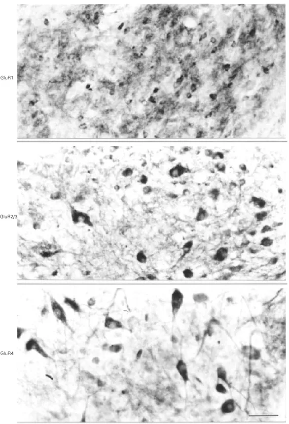
Six 7-15-day old white leghorn chicks (Gallus gallus) were used. The animals were anesthetized with ketamine and xylazine and perfused through the heart with phosphate buffered saline and 4% paraformaldehyde in 0.1 M sodium phosphate buffer (PB), pH 7.4. After 3-5 h of postfixation, the brains were transferred to 30% (w/v) sucrose in PB to ensure cryoprotection. Coronal sections (30 µm) of the frozen brains were cut with a sliding microtome. The free-floating brain sections were incubated with rabbit antisera against GluR1, GluR2/3 and GluR4 subunits (Chemicon, Temecula, CA) diluted 1:100-1:250 in PB containing 0.3% Triton X-100 for 14-18 h at 4oC. After washing 3 times (15 min each) with PB, the sections were incu-bated with a biotinylated goat anti-rabbit antiserum diluted 1:200 in PB for 1 h at room temperature, and then washed with PB and incubated with an avidin-biotin-peroxidase complex (ABC Elite, Vector Labs., Burlingame, CA). Following the reaction with 0.05% 3-3’-diaminobenzidine and a 0.01% hydrogen peroxide in PB and intensi-fication with 0.05% osmium tetroxide in water, the sections were mounted on gelatin and chromoalumen-coated slides, dehy-drated, cleared, and coverslipped with Permount. The intensity of AMPA-type sub-unit immunoreactivity in the neuropil of retinorecipient areas of the chick brain was subjectively rated from absent to intense. A similar scale was used to rate the number of stained neurons. Figure 1 illustrates examples of immunohistochemical staining that were rated according to this subjective scaling. No attempt was made to quantify the inten-sity of staining, but only to determine the number of somata that were stained well above the background.
Moderate to intense perikaryon staining for GluR1-like immunoreactivity was
ob-served in the nucleus geniculatus lateralis ventralis, nucleus lateralis anterior, nucleus lentiformis mesencephali, layers 2, 3, and 5 of the optic tectum, and the nucleus supra-chiasmaticus. On the other hand, neuropil staining for the same subunit was moderate to intense in the ventral geniculate complex, griseum tecti, nucleus lentiformis mesen-cephali, nucleus of the basal optic root, lay-ers 2, 3, and 5 of the optic tectum, and nucleus suprachiasmaticus. Perikarya exhib-iting GluR2/3 immunoreactivity were mod-erate to intense in the nucleus geniculatus lateralis ventralis, nucleus of the basal optic root, layers 10 and 13 of the optic tectum, nucleus opticus principalis thalami, and nucleus suprachiasmaticus. Moderate to in-tense neuropil staining for GluR2/3 was ob-served in the griseum tecti, layers 3 and 13 of the optic tectum, nucleus opticus principalis thalami, and nucleus suprachiasmaticus. GluR4 immunoreactivity was much less fre-quent in perikarya than GluR1 and GluR2/3 immunoreactivities. Indeed, moderate GluR4 immunoreactivity was only observed in perikarya of the nucleus of the basal optic root. Neuropil staining for GluR4 was mod-erate to intense in the ventral geniculate complex, griseum tecti and the nucleus lentiformis mesencephali.
Figure 1 - Photomicrographs of coronal sections of the chick brain at the level of the nucleus of the basal optic root, pro-cessed for GluR1, GluR2/3, and GluR4 immunoreactivity. Neu-ropil staining is intense for GluR1, and slight for GluR2/3 and GluR4. Perikarya staining is slight for GluR1, and moder-ate for GluR2/3 and GluR4. Scale bar = 40 µm.
GluR1
GluR2/3
GluR1 GluR2/3 GluR4
Perikarya Neuropil Perikarya Neuropil Perikarya Neuropil
Absent Slight Moderate Intense
detected in layers 1, 14, and 15 of the optic tectum, and in the nucleus pretectalis diffusus. Table 1 shows a detailed account of the distribution of GluR subunits in the perikarya and neuropil of retinorecipient areas of the chick brain.
Previous immunohistochemical studies have shown the distribution of AMPA-type glutamate receptor subunits throughout the rat CNS. These studies did not focus on the localization of these subunits in the visual system. Indeed, there is little information concerning the distribution of GluRs in the visual system of other species. We found a moderate to intense staining for all AMPA subunits in the nucleus geniculatus lateralis ventralis. Petralia and Wenthold (3) noted a
moderate immunoreactivity with antisera against the same AMPA-type subunits in this structure of the rat brain. Mappings of the distribution of these subunits have also revealed moderate immunoreactivity of GluR1 in the neuropil of the zonal and super-ficial layers of the superior colliculus, and moderate to intense perikarya staining for GluR2/3 immunoreactivity in the deep lay-ers of the same structure (3,4). Several lines of evidence indicate that the rodent superior colliculus is equivalent to the avian optic tectum (5). Indeed, in the present study we found populations of neurons in layers 2, 3, and 5 of the chick optic tectum that stained moderately to intensely with antisera against GluR1. Neurons in layers 10 and 13 of the
Table 1 - Distribution of GluR subunits in the retinorecipient areas of the chick brain.
AP, Area pretectalis; GLdp, nucleus geniculatus lateralis, pars dorsalis principalis; GLv, nucleus geniculatus lateralis, pars ventralis; GT, griseum tecti; IGL, intergeniculate leaflet; LA, nucleus lateralis anterior thalami; LMmc, nucleus lentiformis mesencephali, pars magnocellularis; nBOR, nucleus of the basal optic root; TeO, optic tectum; OPT, nucleus opticus principalis thalami; SCNl, nucleus suprachiasmaticus, pars lateralis.
AP GLdp
GLv GT IGL LA LMmc
nBOR TeO Layer 2
Layer 3
Layer 4
Layer 5
Layer 6
Layer 7
Layer 8
Layer 9
Layer 10
Layer 11
Layer 12
Layer 13 Structures
optic tectum showed moderate to intense immunoreactivity for GluR2/3. In the rat, GluR1 was also found in the most superficial layers of the superior colliculus, whereas GluR2/3 was predominant in its deep layers (3,4). This pattern is similar to that found in the chick and described above.
Recent in situ hybridization studies re-vealed abundant expression of GluR1-4 mRNAs in the nucleus geniculatus lateralis ventralis, and moderate expression of GluR1 and GluR2 mRNAs in the rat superior col-liculus (6,7). The immunolabeling pattern of AMPA receptor subunit distribution in our study resembles that seen in these in situ
hybridization mappings. A notable differ-ence between our results and [3H]-AMPA binding data obtained in rat brain is the low binding in the nucleus geniculatus lateralis ventralis (8,9), as compared to the moderate to intense staining observed here and the strong hybridization signal seen in the in situ hy-bridization experiments (7). Perhaps this dif-ference reflects multiple receptor popula-tions labeled by AMPA with different affini-ties (10), or simply a methodological artifact. Induced oocyte expression of AMPA-type subunits showed that each subunit has its own pharmacological and physiological properties, and that in vivo responses to
glutamate may reflect the existence of mul-tiple receptors with different combinations of shared subunits (1). For example, the Ca2+ permeability and current-voltage (I-V) rela-tion of a channel varies with the subunit composition of the receptor. Receptors con-taining the GluR2 subunit showed low Ca2+ permeability, a major feature of AMPA-type receptors, whereas receptors lacking the GluR2 subunit showed high Ca2+ permeabil-ity. The present study revealed that GluR2/3 subunits were present in most visual areas, although with varying expression levels. In these areas, the GluR2/3 subunits are colocalized with GluR1 and/or GluR4 sub-units. If the GluR2/3 subunits comprise func-tional receptors with GluR1 and GluR4 in the visual system, one could expect that most of the AMPA-type GluRs in these areas would exhibit a low Ca2+ permeability. Our experiments do not permit conclusions about this question, which could be obtained by a combination of immunoprecipitation and patch-clamp experiments.
In summary, the present study revealed an extensive distribution of AMPA-type subunits in various retinorecipient areas of the chick brain, and suggests that several patterns of response to glutamate derived from retinal ganglion cells may exist in the visual system.
References
1. Sprengel R & Seeburg PH (1995). Ionotropic glutamate receptors. In: North RA (Editor), Handbook of Receptors and Channels, Ligand- and Voltage-Gated Ion Channels. CRC Press, Boca Raton, Florida. 2. Massey SC (1990). Cell types using
glutamate as neurotransmitter in the ver-tebrate retina. Progress in Retinal Re-search, 9: 399-425.
3. Petralia RS & Wenthold RJ (1992). Light and electron immunocytochemical local-ization of AMPA-selective glutamate re-ceptors in the rat brain. Journal of Com-parative Neurology, 318: 329-354.
4. Martin LJ, Blackstone CD, Levey AI, Huganir RL & Price DL (1993). AMPA glutamate receptor subunits are differen-tially distributed in rat brain. Neurosci-ence, 53: 327-358.
5. Vanegas H (1984). Comparative Neurol-ogy of the Optic Tectum. Plenum Press, New York.
6. Matute C, Nguyen Q-T & Miledi R (1993). mRNAs coding for neurotransmitter recep-tors in rabbit and rat visual areas. Journal of Neuroscience Research, 35: 652-663. 7. Sato K, Kiyama H & Tohyama M (1993).
The differential expression patterns of messenger RNAs encoding non-N -meth-yl-D-aspartate glutamate receptor subunits (GluR1-4) in the rat brain. Neuroscience, 52: 515-539.
8. Monaghan DT, Yao D & Cotman CW (1984). Distribution of [3H]AMPA binding sites in rat brain as determined by quanti-tative autoradiography. Brain Research, 324: 160-164.
9. Rainbow TC, Wieczorek CM & Halpain S (1984). Quantitative autoradiography of binding sites for [3H]AMPA, a structural analogue of glutamic acid. Brain Re-search, 309: 173-177.