Meis regulation and function
during eye and nervous system development
Joana Sofia Soares Santos
Tese de doutoramento em Ciências Biomédicas
Porto 2011
Joana Sofia Soares Santos
Meis regulation and function
during eye and nervous system development
Tese de Candidatura ao grau de Doutor em Ciências Biomédicas submetida ao
Instituto de Ciências Biomédicas Abel Salazar da Universidade do Porto
Orientador – Professor Doutor Fernando Casares
Categoria – Investigador Principal
Afiliação – Centro Andaluz de Biología del Desarrollo,
CSIC-Universidad Pablo de Olavide, Espanha
Co-orientador – Professor Doutor Pedro Rodrigues
Categoria – Professor Associado
Este trabalho foi financiado pela
O autor desta tese declara que interveio na concepção e na execução do trabalho
experimental, na interpretação dos resultados e na redacção dos artigos publicados:
Bessa, J., Tavares M.J., Santos J., Kikuta, H., Laplante M., Becker T.S., Gómez-Skarmeta J.L. and Casares, F. (2008) meis1 regulates cyclinD1 and c-myc expression and controls the proliferation of the multipotent cells in the developing zebrafish eye.
Development 135, 799-803.
Santos J.S., Fonsenca N.A., Vieira C.P., Vieira J., Casares F. (2010) Phylogeny of the Teashirt-related zinc finger (Tshz) gene family and analysis of the developmental
expression of tshz2 and tshz3b in the zebrafish. Developmental Dynamics 239 (3),
Summary
Resumo
i
General introduction
III
1
1.1. Danio rerio as an animal model to study vertebrate development
1.2. Analogies between fly and vertebrate eye development
1.3. The meis gene family of transcription factors
1.4. teashirt gene family, general importance
1.5. References
ii
1
2
Chapter I
Chapter II
Supplementary Information
Conclusions
Abbreviations
Acknowledgments
3
4
5
6
7
2
7
7
12
18
34
47
64
68
75
Meis regulation and function during eye and nervous system development i
Summary
The development of organs is a complex process controlled by gene regulatory
networks. In these networks, transcription factors control the expression of target genes
which, in turn, determine the specific properties of developing cells. In this thesis, we have
focused on two families of transcription factors that have widespread roles in normal
development and disease: the meis and teashirt gene families. For this, we have resorted to the zebrafish, as a vertebrate animal model, with special focus on the formation of the
retina.
In Chapter I, we analysed the requirement of meis genes during the early retinal
development. Previous work had shown that, in Drosophila, the meis gene homologue
homothorax (hth) was required for the maintenance of the undifferentiated and
proliferative state of retinal progenitors. Due to the partial conservation of the gene
network controlling early eye specification between vertebrates and invertebrates, we
asked whether meis genes were expressed in retinal progenitors and if they did, what their role was. We found that of the four meis paralogous genes, meis1 was expressed in retinal progenitors. In addition, using a combination of strategies, we were able to show
that meis1 was required in retinal progenitors to sustain their proliferative growth. These results indicated that Meis/Hth shared a conserved function.
Since the eye regulatory gene networks in vertebrates and Drosophila show significant similarities, we also decided to study other class of genes, which encode the
Teashirtzinc-finger transcription factors (Tsh). In Drosophila, the two tsh paralogues, tsh
and tiptop (tio) were known to collaborate with hth and eyeless (the Drosophila pax6
homologue) during fruit fly eye development. However, very little was known, even at the
level of expression, about this gene family in vertebrates. In zebrafish only tshz1 had been
described so far. We found new tshz genes in zebrafish and placed them in a comprehensive phylogeny. We also described the expression pattern of two of them,
tshz2 and tshz3b, and compared it with that of meis and pax6 genes. We concluded that
tshz, meis and pax6 gene families have coexpression domains in the brain that open the possibility of interaction between their gene products. This work is addressed in Chapter
Meis regulation and function during eye and nervous system development ii
Resumo
O desenvolvimento dos orgãos é um processo complexo, controlado por redes de
regulação genética. Nestas redes, os factores de transcrição controlam a expressão de
genes-alvo que, por sua vez, determinam as propriedades específicas das células em
desenvolvimento. Nesta tese, centramo-nos em duas famílias de factores de transcrição
que têm funções generalizadas no desenvolvimento normal e em doenças: as famílias de
genes meis e teashirt. Para isso, recorremos ao peixe-zebra como um modelo de animal vertebrado, com especial ênfase na formação da retina.
No Capítulo I, analisamos a exigência dos genes meis durante o início do desenvolvimento da retina. Trabalhos anteriores mostraram que, em Drosophila, o gene
homólogo de meis, homothorax (hth), é necessário para a manutenção do estado indiferenciado e proliferativo das células progenitoras da retina. Devido à conservação
parcial da rede de genes que controlam o início da especificação do olho, entre animais
vertebrados e invertebrados, perguntamo-nos se os genes meis eram expressos nas
células progenitoras da retina e, se assim fosse, qual o seu papel. Nós descobrimos que,
dos quatro genes parálogos meis, meis1 era expresso nas células progenitoras da retina.
Além disso, usando uma combinação de estratégias, fomos capazes de mostrar que o
meis1 era necessário para manter o crescimento proliferativo das células progenitoras da
retina. Estes resultados indicaram que Meis/Hth compartilham uma função conservada.
Uma vez que as redes de regulação genética do olho em vertebrados e em
Drosophila apresentam semelhanças significativas, nós decidimos estudar também outra
classe de genes, os quais codificam os factores de transcrição Teashirt “zinc-finger”
(Tsh). Em Drosophila, os dois genes tsh parálogos, tsh e tiptop (tio) eram conhecidos por
colaborarem com hth e eyeless (o homólogo pax6 de Drosophila) durante o desenvolvimento do olho da mosca da fruta. No entanto, muito pouco era conhecido,
mesmo a nível de expressão, sobre esta família de genes em vertebrados. No
peixe-zebra apenas tshz1 tinha sido descrito até agora. Nós encontramos novos genes tshz do
peixe-zebra e colocamo-los numa filogenia. Nós também descrevemos os padrões de
expressão de dois deles, tshz2 e tshz3b, e comparamo-los com os dos genes meis e
pax6. Nós concluímos que as famílias de genes meis, tshz e pax6 têm domínios de coexpressão no cérebro que indicam a possibilidade de interacção entre os seus
Meis regulation and function during eye and nervous system development 1
General introduction
Meis regulation and function during eye and nervous system development 2 1.1. Danio rerio as an animal model to study vertebrate development
Zebrafish (Danio rerio) has become an important animal model to study the mechanism of vertebrate development and disease, since its introduction by George Streisinger.
Zebrafish presents several advantages when used as a model system, which have often
been cited in literature. For instance, it is easy to maintain in small spaces, it reaches
sexual maturity in only 3-4 months and a pair of fish can produce more than 200 eggs per
mating. Additionally, eggs and embryos are transparent and, therefore, it is easy to follow
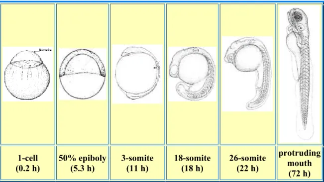
the development with just a standard dissection microscope. Development is so quick that
merely 24 hours post-fertilization (hpf) all the major organs are formed (Fadool and
Dowling, 2008) (Figure 1).
1-cell
(0.2 h)
50% epiboly
(5.3 h)
3-somite
(11 h)
18-somite
(18 h)
26-somite
(22 h)
protruding
mouth
(72 h)
Figure 1. Diagram representing some stages of the zebrafish development. At 1-cell stage, after
fertilization, the embryo is divided in two structures, the yolk and on the top of the yolk, the cell. At
11 hours post-fertilization (hpf) stage, the first 5 to 6 somites appear at the rate of about 3 per
hour. The brain primordium has now distinctively thickened into the neural keel and one can first
distinguish the optic vesicle. At 18 hpf, the otic placode (the precursor of the fish ear) is present
beside the hindbrain rudiment that is midway between the optic vesicle and the first somite. At 22
hpf stage of development, the lens and the midbrain-hindbrain boundary are visible. (Adapted
from http://www.neuro.uoregon.edu/k12/Development%20Stages.html).
Zebrafish is a particularly good model to study eye development and morphology,
Meis regulation and function during eye and nervous system development 3 vertebrate lineage. The eye develops from three distinct embryological tissues called
neuroectoderm, skin ectoderm and head mesenchyme. Neural retina arises from the
neuroectoderm. The zebrafish neural retina, as happens in many classes of vertebrates,
is composed of seven cell types, six neurons (interneurons, amacrine, horizontal, bipolar,
ganglion and photoreceptors cells), and a single glial cell, called Muller cell (Fadool and
Dowling, 2008).
The retina has a well-characterized laminar organization. The ganglion cells are
present innermost in the retina, closest to the lens, and the photoreceptors (rods and
cones) lie outermost against the pigment epithelium. Thus, light must pass through all the
retina layers before activating the photoreceptors. The light is then translated into an
electrical message that stimulates the succeeding neurons of the retina, it is transmitted to
the brain through the optic nerve, and it is processed to form an image
(http://webvision.med.utah.edu/sretina.html). All vertebrates have a retina with three
layers of nerve cell bodies and two layers of synapses. The outer nuclear layer (ONL)
contains the cell bodies of the rods and cones, the inner nuclear layer (INL) contains the
cell bodies of the bipolar, horizontal and amacrine cells, and, lastly, the ganglion cell layer
(GCL) is composed of the bodies of ganglion cells and displaced amacrine cells. Dividing
these nerve cell layers one finds the outer plexiform layer (OPL) and the inner plexiform
layer (IPL), where synaptic contacts occur (http://webvision.med.utah.edu/sretina.html)
(Figure 2).
Figure 2. Diagram of a vertebrate
differentiated retina´s layers. Retina is up
and lens is down. R – rod photoreceptors;
C – cone photoreceptors; H – horizontal
cells; B – bipolar cells; I – interplexiform
cells; M – Muller cells; Am – amacrine cells;
G – ganglion cells. (Adapted from:
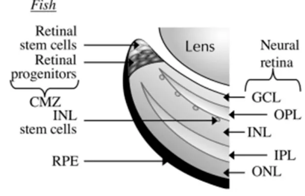
Meis regulation and function during eye and nervous system development 4 In zebrafish and in larval amphibians, but not in other vertebrates, retinal
neurogenesis also occurs postembryonically, throughout the animal’s life, in a stem cell
containing zone, a region called cilliary marginal zone (CMZ) or circumferential germinal
zone (Raymond et al., 2006; Amato et al., 2004). The CMZ lies between the neural retina
and the iris and contains multipotent, self-renewing retinal stem cells, which gene
expression recapitulates the expression that occurs in retinoblasts during embryonic
development (Raymond et al., 2006; Amato et al., 2004). In fact, in fish and amphibians only the most central region of the retina is formed during embryogenesis and most part of
the retina develops from the addition of new cells from the CMZ (Moshiri et al., 2004).
CMZ can also regenerate retina after injury (Moshiri et al., 2004) (Figure 3).
As mentioned before, zebrafish neural retina development is very similar to that of
other vertebrates. During neurulation, six3 and pax6 expression in the anterior neural plate specify the eye tissues (Kobayashi et al., 2001; Puschel et al., 1992). Through
subsequent morphogenetic mechanisms, eyes develop from optic vesicles outpouching Figure 3. Localisation of the cilliary marginal zone in fish. Multipotent retinal stem cells are
found in the most peripheral zone of the cilliary marginal zone of zebrafish. CMZ – cilliary
marginal zone; GCL – ganglion cell layer; INL – inner nuclear layer; IPL – inner plexiform
layer; ONL – outer nuclear layer; OPL – outer plexiform layer; RPE – retinal pigment
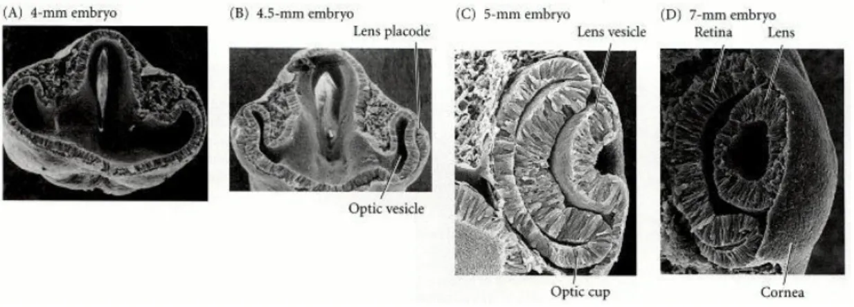
Meis regulation and function during eye and nervous system development 5 from two sides of the developing neural tube (forebrain) (Fadool and Dowling, 2008). At
24 hpf, the optic cup develops after the invagination of these masses and formation of the
optic lumen. The inner layer of the optic lumen gives rise to the neural retina while the
outer layer produces the retinal pigment epithelium (RPE) (Fadool and Dowling, 2008)
(Figure 4).
Retinal neurogenesis is an orderly process. The first cells to differentiate are
ganglion cells. Neural differentiation then follows an inner to outer retinal order.
Differentiated ganglion cells appear around 28 and 32 hpf in the ventralnasal retina.
Neurogenesis then spreads dorsally around to the ventral temporal retina in a wave-like
manner reminiscent of the movement of the morphogenetic furrow in Drosophila (Fadool
and Dowling, 2008; Hu and Easter, 1999). There are other similarities to Drosophila. The wave of differentiation is associated with sonic hedgehog expression and the specification of ganglion cells needs atonal5 (ath5) expression, a homologue of the Drosophila gene
atonal (Fadool and Dowling, 2008) (Figure 5). After the differentiation of the ganglion cells, the differentiation of amacrine cells, interneurons and retinal lamination takes place. At 48
hpf, lamination is across most of the retina. Muller glial cells are among the last to be Figure 4. Vertebrate eye development (adapted from Gilbert, S.F., Part 3. Later embryonic development – Chapter 12. The central nervous system and the epidermis – Development of the Vertebrate Eye In: Developmental Biology, 6th Ed; Sinauer Associates, Inc: Massachusetts, 2000 (web version))
(A, B) The optic vesicle evaginates from the brain and interacts with the overlying ectoderm,
inducing a lens placode. (B, C) The overlying ectoderm differentiates into lens cells as the
optic vesicle folds in on itself, and the lens placode becomes the lens vesicle. (C) The optic
vesicle becomes the neural and pigmented retina as the lens is internalized. (D) The lens
Meis regulation and function during eye and nervous system development 6 formed (Fadool and Dowling, 2008).
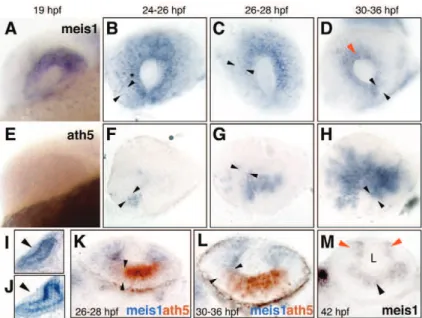
Figure 5. The spatiotemporal pattern of retinal ganglion cells (RGC) neurogenesis in zebrafish.
Spread of aht5 mRNA (A-C), ath5:GFP (D-F) and RGC differentiation (G-I) across the retina. (A-C) Whole-mount embryos stained with an antisense ath5 riboprobe. (D-I) Whole-mount embryos double-stained with anti-GFP (D-F) and zn5 (G-I) antibodies. The ath5 wave leads the RGC differentiation wave by several hours. Arrows in A, D, H and I indicate the
ventralnasal patch. Arrowheads in H and I indicate zn5+ RGCs. Asterisks mark the location of
choroid fissure, which delineates the boundary between nasal and temporal retina. Anterior
Meis regulation and function during eye and nervous system development 7 1.2. Analogies between fly and vertebrate eye development
Morphologically, the eyes of insects and vertebrates are completely different.
Nevertheless, the mechanism of development of this sensory organ is similar in some
core aspects. The most remarkable is the role of Pax6. Mammal’s pax6 and its fly homologue, eyeless (ey), when mutated lead to the abolishment of eye development, and
when under targeted expression both are capable of inducing ectopic eye (Gehring,
2002). Fly and vertebrate eye development is similar in other remarkable aspects.
Drosophila retina neuronal differentiation spreads like a wave across the eye imaginal
disc. Neurogenesis in zebrafish retina also spreads through several sequential waves. In
both cases, the progression of these waves requires the fly gene atonal and its vertebrate
homologue ath5. This suggests that the molecular mechanisms driving the retina neurogenic wave have been conserved from flies to fish (Kay et al., 2001; Brown et al.,
2001; Masai et al., 2000) (Figure 5).
The parallelisms found in these previous works in Drosophila and zebrafish eye
development led us to study the role of other genes, known to be involved in Drosophila
eye development, in vertebrates. In the next chapters, we will describe the studies on the
role played by homothorax (hth) and teashirt (tsh) homologues during zebrafish development.
1.3.
The
meis
gene family of transcription factors
Hox proteins are transcriptional regulators essential for cell fate specification in early
embryonic development. However, these proteins have poor specificity and affinity with
their DNA sequence targets. Consequently, they need cofactors to enhance their DNA
specificity and affinity (Mann and Affolter, 1998). There are two vertebrate families of Hox
cofactors: Pbx and Meis, which belong to the TALE (Three Amino acid Loop Extension)
homeodomain superfamily (Choe et al., 2002). The Meis class comprises fly Hth and vertebrate Prep and Meis homeoproteins. Hth and mouse Meis1 protein are highly
homologous in two regions called homeodomain (HD) and MH domain (for Meis and
Homothorax). The MH domain is found only in Hth and in mouse, human and Xenopus
Meis1 homologues (Pai et al., 1998); Prep1 and Meis are able to form complexes with Pbx in the absence of a DNA target and there is a strong correlation between meis gene
Meis regulation and function during eye and nervous system development 8 et al., 1997; Saleh et al., 2000). In several developmental pathways, Pbx1 binds with Meis family members in the cytoplasm to be imported to the nucleus, but during mammalian
female genital tract development the control of Pbx1 intracellular distribution is
independent of Meis proteins (Dintilhac et al., 2005). While Meis proteins are present in a
specific pattern of expression during development, Prep1 is expressed ubiquitously in the
embryo (Ferretti et al., 1999). In Drosophila, Hth is required for the nuclear localization of
the PBX homologue, Extradenticle (Pai et al., 1998; Rieckhof et al., 1997), suggesting that interactions between Meis and Pbx molecules have been conserved from Drosophila to mouse (Toresson et al., 2000). meis1 (myeloid ecotropic viral integration site 1) was first
identified as a new site of viral integration in myeloid leukemic tumours arising in BXH-2
mice (Moskow et al., 1995). It was mapped on mouse chromosome 11 and on human
chromosome 2p23-p12 (Moskow et al., 1995; Smith et al., 1997). Meis homeobox proteins have important roles in vertebrate development and disease as one of the Hox cofactors,
but they also have Hox-independent functions (Moens and Selleri, 2006; Choe et al., 2002; Shen et al., 1997). Choe and colleagues (2009) proposed that Meis proteins act as
Hox cofactors through the modulation of the accessibility of histone deacetylases and
CBP histone modification enzymes to Hox-regulated promoters, during zebrafish
development. Meis proteins are not just involved in DNA binding and complex stabilization
on the target DNA but also recruit TALE partner proteins into the nucleus (Geerts et al.,
2005). Meis proteins also form complexes with other homeodomain transcription factors,
like Engrailed and Pdx, and also with bHLH proteins, like MyoD (Sagerstrom, 2004; Heidt
et al., 2007). Meis genes have been reported to have several different full-length splice
variants, which probably recruit different proteins, resulting in varying transcriptional
activity of the target promoter sequence with functional and tissue distribution differences
(Geerts et al., 2005). Noro and colleagues (2006) reported a homeodomain-less isoform encoded by gene meis1 and hth, suggesting that alternative splicing is an evolutionary
conserved mechanism to expand and diversify these transcription factors’ functions.
Nevertheless, Drosophila Meis protein Hth in vivo function has been much better studied
(Mann and Affolter 1998 and Ryoo et al., 1999). Hth is required early in development for a normal eye formation and is an important suppressor of eye differentiation (Pichaud and
Casares, 2000, Pai et al., 1998). Data from Drosophila studies suggested that other proteins may function as Hox cofactors, such as Teashirt (Tsh). Vertebrate genomes
contain tsh homologs that, when expressed in flies, can rescue tsh mutant phenotypes
(Manfroid et al., 2004). However, it is not yet known if these proteins also function as Hox cofactors in vertebrates.
Meis regulation and function during eye and nervous system development 9 mentioned above, the meis1 gene was first identified as a major integration site for leukemogenic virus in a murine leukemia model. Similar oncogenic functions were also
proposed for its paralogues meis2 and meis3 genes (Geerts et al., 2005; Fujino et al., 2001; Hara et al., 2008). Several studies show that meis genes enhance early cell
proliferation and suppress differentiation during development (Geerts et al., 2005). meis
genes also play a role in oncogenesis, being best defined in leukemia (Argiropoulos et al.,
2008; Wong et al., 2007; Lawrence et al., 1999; Geerts et al., 2005; Esparza et al., 2008; Kumar et al., 2009; Li et al., 2009, Rozovskaia et al., 2001; Sitwala et al., 2009). Indeed, in human leukemia, high expression of meis1 was found in bone marrow cells of acute
myeloid leukemia (AML) patients (Geerts et al., 2005; Afonja et al., 2000). It is reported that meis1 cooperates with Hox genes to induce AML in mice and these genes are
coexpressed in human AML (Calvo et al., 2001; Kroon et al., 1998; Thorsteinsdottir et al., 2001). Recent work offers some inklings on the molecular mechanisms involved: GSK-3
maintains the MLL (Mixed lineage leukemia) leukemia stem cell transcriptional program by
promoting the conditional association of CREB, and its coactivators TORC and CBP
(CREB binding protein) with homedomain protein Meis1, a critical component of the
MLL-subordinate program, which in turn facilitates Hox-mediated transcription and oncogenesis
(Wang et al., 2010). There is some evidence promoting a linkage of the leukemogenic activities of Meis1 to the cyclin cell cycle control pathway (Argiropoulos et al., 2010).The
role of meis genes in other forms of cancers is still mostly unknown, but upregulation was found in pancreatic endocrine neoplasms, in lung adenocarcinoma tumours (Fernandez et al., 2004), in neuroblastomas (Geerts et al., 2005, 2003; Jones et al., 2000; Spieker et al.,
2001), in ovarian carcinomas (Crijns et al., 2007) and also in nephroblastomas (Dekel et al., 2006).
In mice, Meis1 is also required for normal hematopoiesis and angiogenesis (Azcoitia
et al., 2005; Hisa et al., 2004; Pineault et al., 2002). Hu and colleagues (2009) reported
that HoxA9 indirectly modulates Meis1 and that this process is biologically important
during normal hematopoiesis, and Simsek and colleagues (2010) found that Meis1
regulates hematopoietic stem cells metabolism through transcriptional activation of Hif-1a
(Hypoxia Inducible Factor 1). In zebrafish, Meis1 and Pbx are involved in erythropoietic
cell lineage specification and in myelopoiesis inhibition (Pillay et al., 2010). Meis1 was also recently implicated as a regulator of endothelial cell development in zebrafish
(Minehata et al., 2008). meis1 knock-down has a profound effect on primitive
erythropoiesis and erythromyeloid progenitor cells and also severely disrupts the proper
development of the vasculature (Cvejic et al., 2011).
Meis regulation and function during eye and nervous system development 10 system (CNS) of vertebrates. Meis2 (and Pax6) is expressed during early fetal forebrain
development in humans (Larsen et al., 2010). In mice, Meis1 and Meis2 are present and
define distinct territories in the developing telencephalon (Toresson et al., 2000). Previous studies suggest that microRNA-9 regulates neurogenesis in mice telencephalon through
regulations of several transcription factors, including Meis2 (and Pax6) (Shibata et al., 2011). Elkouby and colleagues (2010) reported that earliest expression of Wnt3a protein
in paraxial mesoderm directly activates Meis3 in the overlying neuroectoderm to induce
posterior cell fates, suggesting a new model for neural anteroposterior patterning. In
zebrafish, Meis2 is expressed in distinct domains of the central nervous system during
development, with the strongest expression in the hindbrain (Biemar et al., 2001; Cecconi
et al., 1997). In chicken, Meis2 is necessary and sufficient for tectal development through
direct interaction with Otx2, a transcription factor required for the formation of all forebrain-
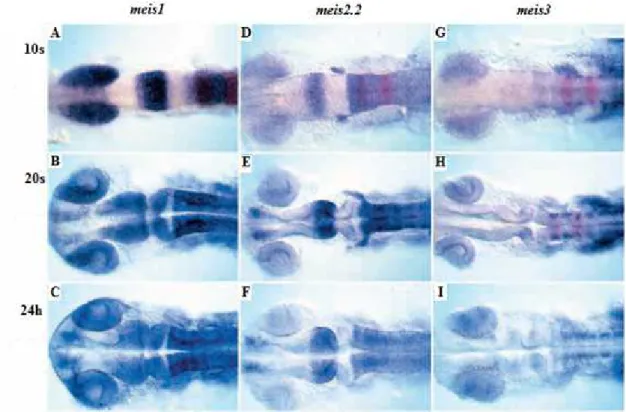
and midbrain-derived structures (Agoston and Schulte, 2009). In zebrafish, all Meis
proteins have been suggested to promote hindbrain fates (Choe et al., 2002; Waskiewicz
et al., 2001) (Figure 6). It was defined that Meis proteins act in the same pathway as Pbx
in zebrafish hindbrain development and may function as a DNA-binding partner of Pbx
proteins and as a post-transcriptional regulator of Pbx protein levels (Waskiewicz et al.,
2001). Zebrafish Meis3 act synergistically with Pbx4 and Hoxb1b to promote hindbrain
fates during development (Vlachakis et al., 2001), and Meis1 interacts functionally with
Irx7 to activate anterior hindbrain markers, such as hoxb1a, hoxa2 and krox20 (Stedman
et al., 2009). In rhombomere 3, krox20 transcription is controlled directly by Meis, probably by Meis2 (Wassef et al., 2008). Through morpholino-mediated knockdown of Meis1
protein in zebrafish, Erickson and colleagues (2010) concluded that Meis1 contributes to
retinotectal map formation by specifying positional information in both the retina and
tectum.
In Xenopus, Meis1 and Pbx1 form a transcriptional activation complex and regulate
hindbrain and neural crest development (Maeda et al., 2002, 2001). Xenopus Meis3 is required for hindbrain patterning during development (Dibner et al., 2001, 2004) and is
also essential for primary neuron and neural crest cells fates (Gutkovich et al., 2010). In
Xenopus, Meis1 and Pbx1 are important direct or indirect regulators of Zic3 expression,
which is involved in the control of left-right asymmetry and neural tube closure in
vertebrates (Kelly et al., 2006).
Recent studies revealed linkage of the meis1 locus to the human neurologic
disorder Restless Legs Syndrome (Trenkwalder et al., 2009; Xiong et al., 2009).
In the olfactory epithelium (OE) of mice, slowly dividing self-renewing precursors
Meis regulation and function during eye and nervous system development 11 OE (Tucker et al., 2010).
In mice and chicken limbs, meis1 and meis2 are expressed in the proximal domain, and the ectopic expression of meis1 in the distal domain disrupts its development and induces distal-to-proximal transformations in a way similar to Drosophila’s homologue hth
(Mercader et al., 1999, 2000, 2009; Capdevila et al., 1999).
Sánchez-Guardado and colleagues (2011)reported meis1 and meis2 expression in
the developing chicken inner ear, suggesting a possible role of Meis assigning regional
identity in the morphogenesis, patterning, and specification of the developing inner ear.
meis3 is expressed in pancreatic islets and cultured β-cells and is required for β-cell survival, and has PDK1 (3-phosphoinositide-dependent protein kinase 1) as a direct target
that mediates Meis3 role in β-cell survival (Liu et al., 2010). Meis1 may also be involved in pancreas organogenesis through its downregulation by the homeobox transcription factor
PDX-1 (von Burstin et al., 2010).
Meis regulation and function during eye and nervous system development 12 As mentioned before, eye development and structure are very different in
invertebrates and vertebrates but some components of the regulatory network involved in
eye development are conserved (Heine et al., 2008). Postmitotic neurons in the compound eye of Drosophila melanogaster are generated in the wake of a single
neurogenic wave, called morphogenetic furrow (MF), that goes across the eye imaginal
disc (Heine et al., 2008). Proliferating progenitor cells ahead of the MF express the protein
Hth, which together with Ey and Tsh promotes rapid, asynchronous proliferation of retinal
progenitor cells and prevents their premature differentiation (Bessa et al., 2002). hth
homologs, meis1 and meis2, are expressed in the eye during development (Hisa et al.,
2004; Heine et al., 2008). Heine and colleagues (2008) showed that Meis have an evolutionary conserved role in maintaining the proliferating state of early retinal progenitor
cells and their results also place these proteins upstream of CyclinD1 and Pax6 in retinal
progenitor cells. pax6 is an essential gene for eye development in numerous organisms,
including humans, but the direct upstream molecular regulators of pax6 expression in the developing eye are poorly known (Zhang et al., 2002). In mice, pax6 lens placode
enhancer is bound by Meis1 and Meis2 directly, controlling pax6 expression during early lens ectoderm induction (Zhang et al., 2002). The role of the transcription factors Meis
during zebrafish eye development is addressed in Chapter I.
In the medaka fish (Oryzias latipes), Meis2 is one of the main targets of the miRNA
miR-204 function and together with altered regulation of the Pax6 pathway, the
abnormally elevated levels of Meis2 resulting from miR-204 inactivation are largely
responsible for microphthalmia, abnormal lens formation, and altered dorsoventral
patterning of the retina, which is associated with optic fissure coloboma (Conte et al., 2010).
1.4.teashirt gene family, general importance
tsh is the founder member of a family of evolutionary conserved zinc-finger genes, which encode proteins that regulate development, and was first described in Drosophila
melanogaster (Fasano et al., 1991). Drosophila Tsh protein has three distantly spaced zinc-finger motifs (Fasano et al., 1991). In the Drosophila embryo, tsh expression is present in parasegments 3-13, the central nervous system, first and second midgut
constrictions, proximal part of leg imaginal discs and dorsal vessel during hearth
morphogenesis (Alexandre, 1996).
Meis regulation and function during eye and nervous system development 13 (AP) determination of the trunk segments and repression of the anterior head
development through its function has a Hox cofactor (Fasano et al., 1991; Roder et al.,
1992; Bhojwani et al., 1997; Robertson, 2004). In different combinations with Cubitus interruptus (Ci) and Armadillo, Tsh takes part in the specification of naked cuticle along
the AP axis and within each trunk segment in the larval epidermis (Angelats, 2002). Tsh
mediates trunk-specific Wingless (Wg) signalling activity (naked cell-fate) by binding to
Armadillo, and Tsh, in addition to Ci, is required for normal Hedgehog (Hh) maintenance
of wg expression in the trunk (Gallet et al., 1998). Tsh is also required positively for the expression of the Hh target gene rhomboid (rho) (Gallet et al., 2000). In trunk
parasegments 3-13, Tsh directly interacts with and limits sex combs reduced (scr) transcription and salivary gland induction (Andrew, 1994, 1998; Taghli-Lamallem, 2007).
Tsh is necessary for normal morphogenesis of the anterior and central midgut structures.
tsh expression in the central midgut mesoderm is regulated by Ultrabithorax,
Abdominal-A, Decapentaplegic (Dpp) and Wg (Mathies, 1994). The Wg pathway regulates Tsh
post-translationally through its phosphorylation and nuclear accumulation, resulting in the
recruitment of Tsh to this pathway (Gallet et al., 1999). Tsh, in cooperation with Brinker, is required for the wingless-mediated repression of ultrabithorax and labial in the midgut
(Waltzer, 2001; Saller, 2002). Embryonic midgut tsh expression is dependent not only upon hox genes, like antennapedia (antp), but also upon extradenticle (exd), and normal
morphogenesis of the gut relies on the normal expression of these genes (Rauskolb et al.,1994).
In the Drosophila wing imaginal disc, tsh expression marks the presumptive notum
(Ng et al., 1996), and Tsh collaborates with Hth to block wing blade development, probably by repressing some of the activities of the Notch pathway at the dorsoventral
compartment boundary (Casares and Mann, 2000). Tsh has also been identified as a
positive regulator of hth during hinge specification (Azpiazu and Morata, 2000; Soanes et
al., 2001). Repression of tsh through the combined action of Wg and Dpp, and subsequent repression of hth are necessary events for the specification of the wing field
(Wu and Cohen, 2002; Zirin and Mann, 2004).
Tsh is also involved in proximal leg morphogenesis, where it is required for proper
growth and cell differentiation (Wu and Cohen, 2000; Erkner et al., 1999). Grunge (Gug), an Atrophin-like protein, is a positive regulator of tsh, specifically in ventroproximal cells of the proximal leg (Erkner et al., 2002).
tsh is expressed in the eye imaginal disc, anterior to the morphogenetic furrow, and is part of the gene network responsible for the specification of eye identity (Pan and
later-Meis regulation and function during eye and nervous system development 14 acting transcription factors (Sine oculis (So), Eyes absent (Eya) and Dachshund (Dac)),
and to promote cell proliferation, proposing that regulation of hth, ey and tsh is critical for
the transition from an uncommitted proliferative state to a mature differentiated state
during Drosophila eye development (Bessa et al., 2002). Recently, it has been shown that
Hth and Tsh require Yorki (Yki), a downstream component of the Hippo tumour
suppressor pathway, to induce proliferation and survival of the undifferentiated cells in the
eye imaginal disc (Peng et al., 2009). tsh has to be first expressed in undifferentiated cells and later has to be turned off to allow retinal cells differentiation, i.e. its expression must
be transient. Tsh role as a regulator of eye specification has been proposed to be due to
its ability to make eye disc cells responsive to Wg and Dpp signalling in an eye-specific
manner (Bessa et al., 2005). Tsh has also functions along the eye disc dorsoventral (DV)
axis, as it suppresses eye development close to the ventral margin and promotes eye
development near the dorsal margin (Singh et al., 2002). These Tsh functions, require
expression of early DV patterning genes; Iroquois-Complex (Iro-C) and Delta (Dl) in the
dorsal part and Serrate (Ser) in the ventral (Singh et al., 2004).
tsh has a paralogue gene called tiptop (tio) (Laugier et al., 2005). Drosophila’s Tio and Tsh proteins show the largest similarity in their three shared zinc-finger motifs
(Laugier et al., 2005). Curiously, both Drosophila melanogaster’s tio and the single
Anopheles gambiae’s tiotsh gene have a conserved fourth zinc-finger motif in the
C-terminal part, which is also present in vertebrate Tsh proteins (Laugier et al., 2005; Caubit
et al., 2000). During Drosophila’s early embryogenesis, tsh and tio expressions are present in distinct domains: tsh in the trunk and tio in parts of the head and tail. But later in
embryo development, they have both common and tissue-specific expression patterns
(Laugier et al., 2005). In contrast, tsh and tio are coexpressed in the imaginal discs
(Bessa et al., 2009). In both embryonic and larval development, Tio and Tsh are functionally similar, since Tio can partially rescue Tsh loss. Tio and Tsh can repress each
other’s expression and Tsh also auto-regulates its own expression through a negative
feedback loop (Laugier et al., 2005; Bessa et al., 2009). These authors suggest that these
mechanisms could be a way to ensure control of total Tio/Tsh levels.
tsh has also been described in other insects. A single tiotsh gene was described in
Tribolium castaneum (Shippy et al., 2008). In this same work, it is suggested that
Drosphilatsh and tio paralogues resulted from a duplication event after divergence from the mosquito lineage. Thermobia domestica has one tsh gene that, curiously, is not
expressed strongly in the abdomen, unlike Drosophila’s tsh (Peterson et al., 1999).
tsh genes have also been described in vertebrates but not so extensively.
Meis regulation and function during eye and nervous system development 15 three first and widely spaced zinc-finger motifs. However, vertebrates have additional
zinc-fingers and a vertebrate-specific homeodomain (Koebernick et al., 2006; Onai et al.,
2007; Caubit et al., 2000) (Figure 7). Despite these differences, the three mice tshz genes can rescue Tsh loss-of-function in flies, suggesting that the molecular function of Tshz is
phylogenetically conserved (Manfroid et al., 2004). In mammals, three tshz genes have been described so far (tshz1-3) and the presence of tshz2 and tshz3 was recently
detected in mice ureters and tshz3 in human embryonic renal pelvis (Jenkins et al., 2009). These authors and others (Caubit et al., 2008) also suggest that Tshz3 may have a function in ureter morphogenesis in mice and mutations of the gene in humans may be
one of the causes for congenital pelvi-ureteric junction obstruction (PUJO) (Jenkins et al., 2009). In mice, Tshz3 is required for proximal ureteric smooth muscle cell differentiation,
downstream of Sonic hedgehog (Shh) and Bone morphogenetic protein 4 (Bmp4) (Caubit
et al., 2008). Mice Tshz proteins were also suggested to have a role in the establishment
of regional identity and specification in the forebrain (Caubit et al., 2005). In mice tshz3 is expressed in multiple areas of the brainstem involved in respiration and its deficiency
leads to breathe failing and death at birth (Caubit et al., 2010). Mice tshz1 and tshz2 are detected in neural tube, somites, in developing limbs, branchial arches and gut,
suggesting that Tshz and fly Tsh may share conserved functions in axis patterning and
limbs specification (Caubit et al., 2000; Long et al., 2001). In humans, reduced expression
of tshz3 may be involved in development of Alzheimer’s disease (Kajiwara et al., 2009) and human tshz2 and tshz3 are possible candidates for tumour suppressor genes and expression of both is downregulated in breast and prostate cancer (Yamamoto et al.,
Meis regulation and function during eye and nervous system development 16 Like mammals, the genome of the amphibian Xenopus tropicalis also has three
tshz-related genes (tshz1-3) (Onai et al., 2007). In frogs, at tailbud and larval stages of
development, tshz3 is expressed mainly in the caudal central nervous system (hindbrain and spinal cord) and in the caudal branchial arch (Onai et al., 2007). These authors
suggest that Xtshz3 is an important promoting factor for the dorsal axis determination in
Xenopus embryo and this function is exerted by facilitating canonical Wnt signalling.
These authors also propose that Xtshz3 play a role in AP axis patterning. In fact, Xtshz1
has also been demonstrated to be essential for the AP patterning of the central nervous
system (CNS) (Koebernick et al., 2006). Xtshz1 also controls segmental migration of cranial neural crest cells. Besides expression in the caudal CNS and migrating cranial
neural crest cells, Xtshz1 is also detected in diencephalon, pronephros and olfactory
placodes (Koebernick et al., 2006).
In chicken, tshz3 is expressed in the central and in the peripheral nervous system and in
myotendinous junctions, muscles and connective tissues. In somites, chicken tshz3 is activated by FGF8 (Manfroid et al., 2006).
Until now only tshz1 had been described in zebrafish (Wang et al., 2007). From 2 to 25-somite stages of development, tshz1 is expressed in the posterior neural tube. From
prim-5 stage onwards, its expression in this domain gradually diminishes and shifts to the
anterior neural tissues including the telencephalon, the tectum opticum, the
midbrain-hindbrain boundary, the midbrain-hindbrain and the eyes (Figure 8). In Chapter II, we report the
finding and description of other tshz genes during zebrafish development.
Figure 7. Structural organization of Human (TSH1 and TSH2), Mouse (mTsh1 and mTsh2)
and Drosophila (DTsh) Tsh proteins. Proteins are aligned according to the position of conserved motifs. Znf1, Znf2 and Znf3 boxes represent typical Teashirt zinc-finger motifs
(Cx2Cx12HMx4H). Spacing between these motifs is variable and is indicated by the number
above the line. Triangles indicate additional potential zinc-finger motifs (Cx2Cx12Hx3-4H)
which are not Tsh-like. AD: indicates a domain rich in glutamic and aspartic residues. Grey
Meis regulation and function during eye and nervous system development 17 Figure 8. Spatial and temporal expression patterns of tshz1 revealed by RNA in situ
hybridization. tshz1 expression is in blue and krox-20 is in red. (A, B, E, G, I and K) are lateral views and (C, D, F, H, J and L) are dorsal views. The inset in (D) is an anterior view (dorsal to
the top) of the forebrain of the hybridized embryo. (A) At the 2-somite stage (10 hpf), tshz1
expression is initiated in the hindbrain and anterior spinal cord. (B) At the 14-somite stage (16
hpf), tshz1 is expressed throughout the spinal cord. (C) Dorsal view of the hindbrain region (red box in B). The expression domain of tshz1 has a clear anterior boundary at the rostral margin of rhombomere 7 (r7). (D) At the prim-5 stage (24 hpf), tshz1 is expressed in the spinal cord and pectoral fin buds (black arrows) and in the dorsal forebrain (white arrow in the inset).
(E-F) At the long-pec stage (48 hpf), tshz1 is expressed in the olfactory bulb (ob), tectum opticum (tec), mid-hindbrain boundary (mhb), hindbrain (h), first pharyngeal arch (1pa) and in
the eye (dash line-circled), with very weak expression in the olfactory placodes (arrows in F).
(G-H) At the protruding mouth stage (72 hpf), lower levels of tshz1 transcripts are observed in the olfactory bulb, pharyngeal arch and midbrain-hindbrain boundary. (I-L) At the early larvae
stages (96-120 hpf), reduced expression of tshz1 is seen in the tectum and first arch. By 120 hpf, no tshz1 expression is detectable in the pharyngeal arches, but is still observed in restricted areas of the telecephalon, tectum, mid-hindbrain boundary and hindbrain. (Adapted
Meis regulation and function during eye and nervous system development 18 1.5.References
Afonja, O., Smith, J.E., Cheng, D.M., Goldenberg, A.S. (2000) MEIS1 and HOXA7 genes in human acute myeloid leukemia Leukemia Research. 24, 849-855.
Agoston, Z. and Schulte, D. (2009) Meis2 competes with the Groucho co-repressor Tle4 for binding to Otx2 and specifies tectal fate without induction of a secondary
midbrain-hindbrain boundary organizer. Development 136, 3311-3322.
Alexandre, E., Graba, Y., Fasano, L., Gallet, A., Perrin, L., De Zulueta, P., Pradel, J., Kerridge, S., Jacq, B. (1996) The Drosophila Teashirt homeotic protein is a DNA-binding protein and module, a HOM-C regulated modifier of variegation, is a likely candidate for being a direct target gene. Mechanisms of Development 59, 191-204.
Amato, M.A., Arnault, E., Perron, M. (2004) Retinal stem cells in vertebrates: parallels and divergences. Int. J. Dev. Biol. 48, 993-1001.
Andrew, D.J. (1998) Regulation and Formation of the Drosophila Salivary Glands. Annals New York Academy of Sciences,55-69.
Andrew, D.J., Horner, M.A, Petitt, M.G., Smolik, S.M., Scott, M.P. (1994) Setting limits on homeotic gene function: restraint of Sex combs reduced activity by teashirt and other
homeotic genes. The EMBO Journal 13 (5), 1132-1144.
Angelats, C., Gallet, A., Therond, P., Fasano, L., Kerridge, S. (2002) Cubitus Interruptus Acts to Specify Naked Cuticle in the Trunk of Drosophila Embryos
Developmental Biology. 241, 132-144.
Argiropoulos, B., Palmqvist, L., Yung, E., Kuchenbauer, F., Heuser, M., Sly, L.M., Wan, A., Krystal, G., Humphries, R.K. (2008) Linkage of Meis1 leukemogenic activity to multiple downstream effectors including Trib2 and Ccl3. Experimental Hematology 36, 845-859.
Meis regulation and function during eye and nervous system development 19 of Meis1 to cell-cycle entry and transcriptional regulation of cyclin D3. Blood 115 (20), 4071-4082.
Azcoitia, V., Aracil, M., Martınez-A, C., Torres, M. (2005) The homeodomain protein Meis1 is essential for definitive hematopoiesis and vascular patterning in the mouse
embryo. Developmental Biology 280, 307-320.
Azpiazu, N. and Morata, G. (2000) Function and regulation of homothorax in the wing imaginal disc of Drosophila. Development 127, 2685-2693.
Bessa, J., and Casares, F. (2005) Restricted teashirt expression confers eye-specific responsiveness to Dpp and Wg signals during eye specification in Drosophila.
Development 132, 5011-5020.
Bessa, J., Carmona, L., Casares, F. (2009) Zinc-finger Paralogues tsh and tio Are Functionally Equivalent During Imaginal Development in Drosophila and Maintain Their Expression Levels Through Auto- and Cross-Negative Feedback Loops. Developmental
Dynamics 238, 19–28.
Bessa, J., Gebelein, B., Pichaud, F., Casares, F., Mann, R.S. (2002) Combinatorial control of Drosophila eye development by Eyeless, Homothorax, and Teashirt. Genes & Development 16, 2415-2427.
Biemar, F., Devosa, N., Martial, J.A., Driever, W., Peers, B. (2001) Cloning and expression of the TALE superclass homeobox Meis2 gene during zebrafish embryonic
development. Mechanisms of Development 109, 427-431.
Bhojwani, J., Shashidhara, L.S., Sinha, P. (1997) Requirement of teashirt (tsh) function during cell fate specification in developing head structures in Dev Genes Evol 207,
137-146.
Brown, N.L., Patel, S., Brzezinski, J., Glaser, T. (2001) Math5 is required for retinal ganglion cell and optic nerve formation. Development 128, 2497-2508.
Meis regulation and function during eye and nervous system development 20 mechanisms of cooperativity with Hoxa9 in myeloid leukemia. PNAS 98 (23), 13120-13125.
Capdevila, J., Tsukui, T., Esteban, C.R., Zappavigna, V., Izpisu, J.C., Belmonte, I.
(1999) Control of Vertebrate Limb Outgrowth by the Proximal Factor Meis2 and Distal Antagonism of BMPs by Gremlin. Molecular Cell. 4, 839-849.
Casares, F. and Mann, R.S. (2000) A dual role for homothorax in inhibiting wing blade development and specifying proximal wing identities in Drosophila. Development. 127,
1499-1508.
Caubit, X., Coré, N., Boned, A., Kerridge, S., Djabali, M., Fasano, L. (2000) Vertebrate orthologues of the Drosophila region-specific patterning gene teashirt. Mechanisms of
Development 91, 445-448.
Caubit, X., Lye, C.M., Martin, E., Coré, N., et al. (2008) Teashirt 3 is necessary for ureteral smooth muscle differentiation downstream of SHH and BMP4.Development 135,
3301-3310.
Caubit, X., Thoby-Brisson, M., Voituron, N., Filippi, P., et al. (2010) Teashirt 3
Regulates Development of Neurons Involved in Both Respiratory Rhythm and Airflow
Control.The Journal of Neuroscience 30 (28), 9465-9476.
Caubit, X., Tiveron, M., Cremer, H., Fasano, L. (2005) Expression Patterns of the Three
Teashirt-Related Genes Define Specific Boundaries in the Developing and Postnatal Mouse Forebrain.The Journal of Comparative Neurology 486, 76-88.
Cecconi, F., Proetzel, G., Alvarez-Bolado, G., Jay, D., Gruss, P. (1997) Expression of
Meis2, a Knotted-Related Murine Homeobox Gene, Indicates a Role in the Differentiation of the Forebrain and the Somitic Mesoderm. Developmental Dynamics 210, 184-190.
Chang, C., Jacobs, Y., Nakamura, T., Jenkins, N.A., Copeland, N.G., Cleary, M.L.
(1997) Meis Proteins are Major In vivo DNA Binding Partners for Wild-Type but Not
Chimeric Pbx Proteins. Molecular and Cellular Biology 17 (10), 5679-5687.
Meis regulation and function during eye and nervous system development 21 hindbrain development in the zebrafish. Development 129, 585-595.
Choe, S., Lu, P., Nakamura, M., Lee, J., Sagerstrom, C.G. (2009) Meis Cofactors Control HDAC and CBP Accessibility at Hox-Regulated Promoters during Zebrafish
Embryogenesis. Development Cell 17, 561-567.
Conte, I., Carrella, S., Avellino, R., Karali, M., Marco-Ferreres, R., Bovolenta, P., Banfi, S. (2010) miR-204 is required for lens and retinal development via Meis2 targeting.
PNAS. 107(35), 15491-15496.
Crijns, A.P.G., Graeff, P., Geerts, D., ten Hoor, K.A., Hollema, H., Sluis, T., Hofstra, R.M.W., Bock, G.H., Jong, S., van der Zee, A.G.J., de Vries, E.G.E. (2007) MEIS and PBX homeobox proteins in ovarian cancer. European Journal of Cancer 43 (17),
2495-2505.
Cvejic, A., Serbanovic-Canic, J., Stemple, D.L., Ouwehand, W.H. (2011) The role of
meis1 in primitive and definitive hematopoiesis during zebrafish development.
Haematological 96 (2), 190-198.
Dekel, B., Metsuyanim, S., Schmidt-Ott, K.M., Fridman, E., Jacob-Hirsch, J., et. al.
(2006) Multiple Imprinted and Stemness Genes Provide a Link between Normal and
Tumor Progenitor Cells of the Developing Human Kidney. Cancer Res 66 (12),
6040-6049.
Dibner, C., Elias, S., Frank, D. (2001) XMeis3 protein activity is required for proper hindbrain patterning in Xenopus laevis embryos. Developmental 128, 3415-3426.
Dibner, C., Elias, S., Ofir, R., Souopgui, J., Kolm, P.J., Sive, H., Pieler, T., Frank, D.
(2004) The Meis3 protein and retinoid signaling interact to pattern the Xenopus hindbrain.
Developmental Biology 271, 75-86.
Dintilhac, A., Bihan, R., Guerrier, D., Deschamps, S., Bougerie, H., Watrin, T., Bonnec, G., Pellerin, I. (2005) PBX1 intracellular localization is independent of MEIS1 in epithelial cells of the developing female genital tract. Int. J. Dev. Biol. 49, 851-858.
Meis regulation and function during eye and nervous system development 22
Liu, K.J., Frank, D. (2010) Mesodermal Wnt signaling organizes the neural plate via Meis3. Development 137, 1531-1541.
Erickson, T., French, C.R., Waskiewicz, A.J. (2010) Meis1 specifies positional information in the retina and tectum to organize the zebrafish visual system. Neural Development 5, 1-22.
Erkner, A., Gallet, A., Angelats, C., Fasano, L., Kerridge, S. (1999) The Role of Teashirt in Proximal Leg Development in Drosophila: Ectopic teashirt Expression Reveals
Different Cell Behaviours in Ventral and Dorsal Domains. Developmental Biology 215, 221-232.
Erkner, A., Roure, A., Charroux, B., Delaage, M., Holway, N., Coré, N., Vola, C., Angelats, C., Pagès, F., Fasano, L., Kerridge, S. (2002) Grunge, related to human Atrophin-like proteins, has multiple functions in Drosophila development. Development
129, 1119-1129.
Esparza, S.D., Chang, J., Shankar, D.B., Zhang, B., Nelson, S.F., Sakamoto, K.M.
(2008) CREB regulates Meis1 expression in normal and malignant hematopoietic cells.
Leukemia 22, 665-667.
Fadool, J.M. and Dowling, J.E. (2008) Zebrafish: A model system for the study of eye genetics. Progress in Retinal and Eye Research 27, 89-110.
Fasano, L., Rider, L., Coré, N., Alexandre, E., Vola, C., Jacq, B., Kerridge, S. (1991) The Gene teashirt is required for the Development of Drosophila Embryonic Trunk
Segments and Encodes a Protein with Widely Spaced Zinc Finger Motifs. Cell 64, 63-79.
Fernandez, P., Carretero, J., Medina, P.P., Jimenez, A.I., Rodriguez- Perales, S., et. al. (2004) Distinctive gene expression of human lung adenocarcinomas carrying LKB1 mutations. Oncogene 23, 5084-5091.
Meis regulation and function during eye and nervous system development 23
Fujino, T., Yamazaki, Y., Largaespada, D.A., Jenkins, N.A., Copeland, N.D., Hirokawa, K., Nakamura, T. (2001) Inhibition of myeloid differentiation by Hoxa9, Hoxb8, and Meis homeobox genes. Experimental Hematology 29, 856-863.
Gallet, A., Angelats, C., Kerridge, S., Thérond, P.P. (2000) Cubitus interruptus-independent transduction of the Hedgehog signal in Drosophila. Development 127,
5509-5522.
Gallet, A., Angelats, C., Erkner, A., Charroux, B., Fasano, L., Kerridge, S. (1999) The C-terminal domain of Armadillo binds to hypophosphorylated Teashirt to modulate
Wingless signalling in Drosophila. The EMBO Journal 18 (8), 2208-2217.
Gallet, A., Erkner, A., Charroux, B., Fasano, L., Kerridge, S. (1998) Trunk-specific modulation of Wingless signalling in Drosophila by Teashirt binding to Armadillo. Current Biology 8 (16), 893-902.
Geerts, D., Revet, I., Jorritsma, G., Schilderink, N., Versteeg, R. (2005) MEIS
homeobox genes in neuroblastoma. Cancer Letters 228, 43-50.
Gehring, W.J. (2002) The genetic control of eye development and its implications for the evolution of the various eye-types. Int. J. Dev. Biol. 46, 65-73.
Gutkovich, Y.E., Ofir, R., Elkouby, Y.M., Dibner, C., Gefen, A., Elias, S., Frank, D.
(2010) Xenopus Meis3 protein lies at a nexus downstream to Zic1 and Pax3 proteins,
regulating multiple cell-fates during early nervous system development. Developmental Biology 338, 50-62.
Hara, T., Schwieger, M., Kazama, R., Okamoto, S., Minehata, K., Ziegler, M., Lohler, J., Stocking, C. (2008) Acceleration of chronic myeloproliferation by enforced expression of Meis1 or Meis3 in Icsbp-deficient bone marrow cells. Oncogene 27, 3865-3869.
Heidt, A.B., Rojas, A., Harris, I.S., Black, B.L. (2007) Determinants of Myogenic Specificity within MyoD Are Required for Noncanonical E Box Binding. Molecular and
Cellular Biology 27 (16), 5910-5920.
Meis regulation and function during eye and nervous system development 24 Evidence for an evolutionary conserved role of homothorax/Meis1/2 during vertebrate retina development. Development 135, 805-811.
Hisa, T., Spence, S.E., Rachel, R.A., Fujita, M., Nakamura, T., Ward, J.M., et al.(2004) Hematopoietic, angiogenic and eye defects in Meis1 mutant animals. The EMBO Journal
23, 450-459.
Hu, M. and Easter, S.S. (1999) Retinal Neurogenesis: The Formation of the Initial Central Patch of Postmitotic Cells. Developmental Biology 207, 309-321.
Hu, Y., Fong, S., Ferrell, C., Largman, C., Shen, W. (2009) HOXA9 Modulates Its Oncogenic Partner Meis1 To Influence Normal Hematopoiesis. Molecular and Cellular Biology 29 (18), 5181-5192.
Jenkins, D., Caubit, X., Dimovski, A., Matevska, N., et al. (2009) Analysis of TSHZ2
and TSHZ3 genes in congenital pelvi-ureteric junction obstruction. Nephrol Dial Transplant. 25 (1), 54-60.
Jones, T.A., Flomen, R.H., Senger, G., Nizetic, D., Sheer, D. (2000) The homeobox gene MEIS1 is amplified in IMR-32 and highly expressed in other neuroblastoma cell lines. European Journal of Cancer 36, 2368-2374.
Kajiwara, Y., Akram, A., Katsel, P., Haroutunian, V., Schmeidler, J., et al. (2009) FE65 Binds Teashirt, Inhibiting Expression of the Primate- Specific Caspase-4. PlosOne 4 (4),
e5071.
Kay, J.N., Finger-Baier, K.C., Roeser, T., Staub, W., Baier, H. (2001) Retinal Ganglion Cell Genesis Requires lakritz, a Zebrafish atonal Homolog. Neuron 30, 725-736.
Kay, J.N., Link, B.A., Baier, H. (2005) Staggered cell-intrinsic timing of ath5 expression underlies the wave of ganglion cell neurogenesis in the zebrafish retina. Development 132, 2573-2585.
Kelly, L.E., Carrel, T.L., Herman, G.E., El-Hodiri, H.M. (2006) Pbx1 and Meis1 regulate activity of the Xenopus laevis Zic3 promoter through a highly conserved region.
Meis regulation and function during eye and nervous system development 25
Kobayashi, M., Nishikawa, K., Suzuki, T., Yamamoto, M. (2001) The Homeobox Protein Six3 Interacts with the Groucho Corepressor and Acts as a Transcriptional
Repressor in Eye and Forebrain Formation. Developmental Biology 232, 315-326.
Koebernick, K., Kashef, J., Pieler, T., Wedlich, D. (2006) Xenopus Teashirt1 regulates posterior identity in brain and cranial neural crest. Developmental Biology 298, 312-326.
Kroon, E., Krosl, J., Thorsteinsdottir, U., Baban, S., Buchberg, A.M., Sauvageau, G.
(1998) Hoxa9 transforms primary bone marrow cells through specific collaboration with
Meis1a but not Pbx1b. The EMBO Journal 17 (13), 3714–3725.
Kumar, A.R., Li, Q., Hudson, W.A., Chen, W., Sam, T., Yao, Q., Lund, E.A., Wu, B., Kowal, B.J., Kersey, J.H. (2009) A role for MEIS1 in MLL-fusion gene leukemia. Blood
113 (8), 1756-1758.
Larsen, K.B., Lutterodt, M.C., Laursen, H., Graem, N., Pakkenberg, B., Møllgård, K., Møller, M. (2010) Spatiotemporal distribution of PAX6 and MEIS2 expression and total cell numbers in the ganglionic eminence in the early developing human forebrain. Dev
Neurosci. 32 (2), 149-162.
Laugier, E., Yang, Z., Fasano, L., Kerridge, S., Vola, C. (2005) A critical role of teashirt
for patterning the ventral epidermis is masked by ectopic expression of tiptop, a paralog of
teashirt in Drosophila. Developmental Biology 283, 446-458.
Lawrence, H.J., Rozenfeld, S., Cruz, C., Matsukuma, K., Kwong, A., Komuves, L., Buchberg, A.M., Largman, C. (1999) Frequent co-expression of the HOXA9 and MEIS1
homeobox genes in human myeloid leukemias. Leukemia 13, 1993-1999.
Li, Z., Luo, R.T., Mi, S., Sun, M., Chen, P., Bao, J., Neilly, M.B., et al.(2009) Consistent Deregulation of Gene Expression between Human and Murine MLL Rearrangement
Leukemias. Cancer Research 69 (3), 1109-1999.
Liu, J., Wang, Y., Birnbaum, M.J., Stoffers, D.A. (2010) Three-amino-acid-loop-extension homeodomain factor Meis3 regulates cell survival via PDK1. PNAS 107 (47),
Meis regulation and function during eye and nervous system development 26
Long, Q., Park, B.K., Ekker, M. (2001) Expression and Regulation of Mouse Mtsh1
During Limb and Branchial Arch Development. Developmental Dynamics 222, 308-312.
Maeda, R., Ishimura, A., Mood, K., Park, E.K., Buchberg, A.M., Daar, I.O. (2002)
Xpbx1b and Xmeis1b play a collaborative role in hindbrain and neural crest gene
expression in Xenopus embryos. PNAS 99 (8), 5448-5453.
Maeda, R., Mood, K., Jones, T.L., Aruga, J., Buchberg, A.M., Daar, I.O. (2001)
Xmeis1, a protooncogene involved in specifying neural crest cell fate in Xenopus
embryos. Oncogene 20, 1329-1342.
Manfroid, I., Caubit, X., Kerridge, K., Fasano, L. (2004) Three putative murine Teashirt orthologues specify trunk structures in Drosophila in the same way as the Drosophila teashirt gene. Development 131, 1065-1073.
Manfroid, I., Caubit, X., Marcelle, C., Fasano, L. (2006) Teashirt 3 expression in the chick embryo reveals a remarkable association with tendon development. Gene Expression Patterns 6, 908-912.
Mann, R.S. and Affolter, M. (1998) Hox proteins meet more partners. Current Opinion in Genetics & Development 8, 423-429.
Masai, I., Stemple, D.L., Okamoto, H., Wilson, S.W. (2000) Midline Signals Regulate Retinal Neurogenesis in Zebrafish. Neuron 27, 251-263.
Mathies, L.D., Kerridge, S., Scott, M.P. (1994) Role of the teashirt gene in Drosophila
midgut morphogenesis: secreted proteins mediate the action of homeotic genes.
Development 120, 2799-2809.
Mercader, N., Leonardo, E., Azpiazu, N., Serrano, A., Morata, G., Martinez-A, C., Torres, M. (1999) Conserved regulation of proximodistal limb axis development by Meis1/Hth. Nature 402, 425-429.
Meis regulation and function during eye and nervous system development 27 regulation of Meis genes. Development 127, 3961-3970.
Mercader, N., Selleri, L., Criado, L.M., Pallares, P., Parras, C. Cleary, M.L., Torres, M.
(2009) Ectopic Meis1 expression in the mouse limb bud alters P-D patterning in a
Pbx1-independent manner. Int. J. Dev. Biol. 53, 1483-1494.
Minehata, K., Kawahara, A., Suzuki, T. (2008) meis1 regulates the development of endothelial cells in zebrafish. Biochemical and Biophysical Research Communications
374, 647-652.
Moens, C.B. and Selleri, L. (2006) Hox cofactors in vertebrate development.
Developmental Biology 291, 193-206.
Moshiri, A., Close, J., Reh, T.A. (2004) Retinal stem cells and regeneration. Int. J. Dev. Biol. 48, 1003-1014.
Moskow, J.J., Bullrich, F., Huebner, K., Daar, I.O., Buchberg, A.M. (1995) Meis1, a PBX1-Related Homeobox Gene Involved in Myeloid Leukemia in BXH-2 Mice. Molecular and Cellular Biology. 15 (10), 5434-5443.
Ng, M., Diaz-Benjumea, F.J., Vincent, J., Wu, J., Cohen, S.M. (1996) Specification of the wing by localized expression of wingless protein. Nature381 (6580), 316-318.
Noro, B., Culi, J., McKay, D.J., Zhang, W., Mann, R.S. (2006) Distinct functions of homeodomaincontaining and homeodomain-less isoforms encoded by homothorax.
Genes & Development. 20, 1636-1650.
Onai, T., Matsuo-Takasaki, M., Inomata, H., Aramaki, T., Matsumura, M, Yakura, R., Sasai, N., Sasai, Y. (2007) XTsh3 is an essential enhancing factor of canonical Wnt signaling in Xenopus axial determination. The EMBO Journal 26, 2350--2360.
Pai, C., Kuo, T., Jaw, T.J. et al. (1998) The Homothorax homeoprotein activates the nuclear localization of another homeoprotein, Extradenticle, and suppresses eye
development in Drosophila. Genes & Development 12, 435-446.
Meis regulation and function during eye and nervous system development 28 Drosophila. Proc. Natl. Acad. Sci. USA 95, 15508-15512.
Peng, H.W., Slattery, M., Mann, R.S. (2009) Transcription factor choice in the Hippo signaling pathway: homothorax and yorkie regulation of the microRNA bantam in the
progenitor domain of the Drosophila eye imaginal disc. Genes and Development 23, 2307-2319.
Peterson, M.D, Rogers, B.T., Popadic, A., Kaufman, T.C. (1999) The embryonic expression pattern of labial, posterior homeotic complex genes and the teashirt
homologue in an apterygote insect. Dev. Genes Evol. 209, 77-90.
Pichaud, F. and Casares, F. (2000) homothorax and iroquois-C genes are required for the establishment of territories within the developing eye disc. Mechanisms of
Development 96, 15-25.
Pillay, L.M., Michael Forrester, A., Erickson, T., Berman, J.N., Waskiewicz, A.J.
(2010) The Hox cofactors Meis1 and Pbx act upstream of gata1 to regulate primitive
hematopoiesis. Developmental Biology 340 (2), 306-317.
Pineault, N., Helgason, C.D., Jeffrey Lawrence, H., Keith Humphries, R. (2002) Differential expression of Hox, Meis1, and Pbx1 genes in primitive cells throughout murine hematopoietic ontogeny. Experimental Hematology 30, 49-57.
Puschel, A.W., Gruss. P., Westerfield, M. (1992) Sequence and expression pattern of
pax-6 are highly conserved between zebrafish and mice. Development 114, 643-651.
Rauskolb, C. and Wieschaus, E. (1994) Coordinate regulation of downstream genes by extradenticle and the homeotic selector proteins. The EMBO Journal 13 (15), 3561-3569.
Raymond, P.A., Barthel, L.K., Bernardos, R.L., Perkowski, J.J. (2006) Molecular characterization of retinal stem cells and their niches in adult zebrafish. BMC Developmental Biology 6, 36.
Rieckhof, G.E., Casares, F., Ryoo, H.D., Abu-Shaar, M., Mann, R.S. (1997) Nuclear Translocation of Extradenticle Requires homothorax, which Encodes an Extradenticle
Meis regulation and function during eye and nervous system development 29
Robertson, L.K., Bowling, D.B., Mahaffey, J.P., Imiolczyk, B., Mahaffey, J.W. (2004) An interactive network of zinc-finger proteins contributes to regionalization of the
Drosophila embryo and establishes the domains of HOM-C protein function. Development
131, 2781-2789.
Röder, L., Vola, C., Kerridge, S. (1992) The role of the teashirt gene in trunk segmental identity in Drosophila. Development 115, 1017-1033.
Rozovskaia, T., Feinstein, E., Mor, O., Foa, R., Blechman, J., Nakamura, T., Croce, C.M., Cimino, G., Canaani, E. (2001) Upregulation of Meis1 and HoxA9 in acute lymphocytic leukemias with the t(4 : 11) abnormality. Oncogene 20, 874-878.
Ryoo, H.D., Marty, T., Casares, F., Affolter, M., Mann, R.S. (1999) Regulation of Hox target genes by a DNA bound Homothorax/Hox/Extradenticle complex. Development 126,
5137-5148.
Sagerstrom, C.G. (2004) pbX Marks the Spot. Developmental Cell 6, 737-748.
Saleh, M., Huang, H., Green, N.C., Featherstone, M.S. (2000) A Conformational Change in PBX1A Is Necessary for Its Nuclear Localization. Experimental Cell Research 260, 105-115.
Saller, E., Kelley, A., Bienz, M. (2002) The transcriptional repressor Brinker antagonizes Wingless signalling. Genes & Development 16, 1828–1838.
Sanchez-Guardado, L.O., Ferran, J.L., Rodríguez-Gallardo, L., Puelles, L., Hidalgo-Sanchez, M. (2011) Meis Gene Expression Patterns in the Developing Chicken Inner Ear.
The Journal of Comparative Neurology 519, 125-147.
Shen, W., Montgomery, J.C., Rozenfeld, S., Moskow, J.J., Lawrence, H.J., Buchberg, A.M., Largman, C. (1997) AbdB-Like Hox Proteins Stabilize DNA Binding by the Meis1 Homeodomain Proteins. Molecular and Cellular Biology 17 (11), 6448-6458.