4-Hydroxyphenylpyruvate Dioxygenase Are Critical to
Mediate the Conformation of the Final Helix and the Tail
to Shield the Active Site for Catalysis
Jang-Foung Lin., Yung-Lin Sheih., Tsu-Chung Chang, Ni-Yuan Chang, Chiung-Wen Chang,
Chia-Pei Shen, Hwei-Jen Lee*
Department of Biochemistry, National Defense Medical Center, Neihu, Taipei, Taiwan
Abstract
4-Hydroxylphenylpyruvate dioxygenase (4-HPPD) is an important enzyme for tyrosine catabolism, which catalyzes the conversion of 4-hydroxylphenylpyruvate (4-HPP) to homogentisate. In the present study, human 4-HPPD was cloned and expressed in E. coli. The kinetic parameters for 4-HPP conversion were: kcat= 2.260.1 s21; and Km= 0.0860.02 mM.
Sequence alignments show that human 4-HPPD possesses an extended C-terminus compared to other 4-HPPD enzymes. Successive truncation of the disordered tail which follows the finala-helix resulted in no changes in theKmvalue for 4-HPP
substrate but thekcatvalues were significantly reduced. The results suggest that this disordered C-terminal tail plays an
important role in catalysis. For inspection the effect of terminal truncation on protein structure, mutant models were built. These models suggest that the different conformation of E254, R378 and Q375 in the final helix might be the cause of the activity loss. In the structure E254 interacts with R378, the end residue in the final helix; mutation of either one of these residues causes aca.95% reductions inkcatvalues. Q375 provides bifurcate interactions to fix the tail and the final helix in
position. The model of the Q375N mutant shows that a solvent accessible channel opens to the putative substrate binding site, suggesting this is responsible for the complete loss of activity. These results highlight the critical role of Q375 in orientating the tail and ensuring the conformation of the terminala-helix to maintain the integrity of the active site for catalysis.
Citation:Lin J-F, Sheih Y-L, Chang T-C, Chang N-Y, Chang C-W, et al. (2013) The Interactions in the Carboxyl Terminus of Human 4-Hydroxyphenylpyruvate Dioxygenase Are Critical to Mediate the Conformation of the Final Helix and the Tail to Shield the Active Site for Catalysis. PLoS ONE 8(8): e69733. doi:10.1371/ journal.pone.0069733
Editor:Paul Taylor, University of Edinburgh, United Kingdom
ReceivedApril 25, 2013;AcceptedJune 12, 2013;PublishedAugust 9, 2013
Copyright:ß2013 Lin et al. This is an open-access article distributed under the terms of the Creative Commons Attribution License, which permits unrestricted
use, distribution, and reproduction in any medium, provided the original author and source are credited.
Funding:This study was supported financially by the National Science Council (NSC 99-2311-B-016-001-MY3) and National Defense Medical Bureau (MAB101-49). The funders had no role in study design, data collection and analysis, decision to publish, or preparation of the manuscript.
Competing Interests:The authors have declared that no competing interests exist.
* E-mail: hjlee@mail.ndmctsgh.edu.tw
.These authors contributed equally to this work.
Introduction
4-Hydroxylphenylpyruvate dioxygenase (4-HPPD, EC 1.13.11.27) belongs to the non-haem Fe(II)/2-oxoacid-dependent oxygenase superfamily, which couples the oxidative decarboxyl-ation of a 2-oxoacid (most commonly a-ketoglutarate) to the oxidation of the prime substrate. A wide range of different types of reactions are catalyzed by these oxygenases, including hydroxyl-ations, desaturations and oxidative ring closures. These reactions have environmental, pharmaceutical and medical significance [1,2]. 4-HPPD catalyzes the second step in the pathway of tyrosine catabolism, the conversion of 4-hydroxyphenyl-pyruvate (4-HPP) to homogentisate (HG) (Fig. 1). The conversion of substrate involves oxidative decarboxylation, side-chain migration and aromatic hydroxylation in a single catalytic cycle [3]. The 4-HPPD reaction is unusual in that the a-ketoacid and prime substrate moieties are contained within the same molecule. A deficiency in active 4-HPPD in humans results in type III tyrosinemia, a rare autosomal recessive disorder [4]. In plants
the homogensate reaction product is an intermediate in the biosynthesis of plastoquinone and tocopherols [5], and inhibitors of 4-HPPD have been used as herbicides [6–8].
Human 4-HPPD is active as a homodimer with a subunit molecular mass ofca. 45 kDa [9]. Alignment of the amino acid sequences of 4-HPPD from different species shows that they are less than 30% identical. However, the topologies of all determined 4-HPPD structures are very similar. The structure of 4-HPPD is comprised of two barrel-shaped domains and is similar in topology to the extradiol dioxygenases [10–16]. A 2-His-1-carboxylate motif is buried inside theb–barrel of the carboxyl-terminal domain of 4-HPPD which binds the iron(II) cofactor [10–13,16]. This metal binding motif is strictly conserved among non-haem iron(II)-dependent oxygenases [17,18].
active site and isolates the bound substrate during catalysis [11,19–22]. Superimposing the crystal structures ofP. fluorescens 4-HPPD and S. avermitilis 4-HPPD in complex with NTBC reveal significant differences in the position of C-terminal helix [10,12]. Binding of the NTBC inhibitor in the active site leads to a 40 degree rotation of the C-terminala-helix. Residues in the terminala-helix might also be involved in catalysis [23,24]. For example, replacement of F337 and F341, two residues in the terminala-helix inS. avermitilis4-HPPD, by Ile and Tyr resulted in loss of activity [23]. The aromatic side-chain of F337 is thought to interact with the aromatic ring of the substrate byp
-p interactions [23,25]. Human and rat 4-HPPD possess longer C-terminal sequences than enzymes from plants and microor-ganisms (Fig. 2). Truncation experiments suggest that the C-terminal extension is essential for enzyme activity [26], but little is known about its function as the residues beyond the final C-terminal a-helix are disordered in all reported X-ray crystal structures [10–13,16]. To date, the precise role of the C-terminus has not been determined.
This study reports the effect of truncating successive C-terminal residues on the activity of recombinant human 4-HPPD. Activity is progressively reduced upon truncation of the C-terminus, indicating the important role of this tail in catalysis. Structural modeling of truncation mutants was carried out using the X-ray coordinates of human 4-HPPD to investigate the effects of C-terminal truncation on protein structure [16]. All models were geometrically minimized by the quantum mechanical-molecular mechanical calculations. The truncated mutant models showed a different conformation in the terminal helix, especially a change in conformation of the benzene ring of F371 and large differences in the side-chain conformations of E254, R378 and Q375. In the structure, the residues of R378 and Q375, which are located in the final helix, provide interactions which fix this helix and the C-terminal tail into position. Substitution of Q375 and R378 resulted in loss of activity, indicating that these interactions are critical for maintaining the helix in a stable conformation for catalysis. The Q375N mutant model showed a solvent accessible channel opened from the putative substrate binding site, suggesting the interactions provided by Q375 to hold the terminal helix and the tail in proper position are critical for isolating the active site from solvent during catalysis.
Materials and Methods
Materials
The restriction enzymes and T4 DNA ligase used for cloning were purchased from NEBioLab (Beverly, MA) and the Quik-Change site-directed mutagenesis kit from Stratagene (La Jolla, CA). HiPrep 16/10 Q XL and Sephacryl HR-100 columns were purchased from GE Healthcare (Fairfield, CT). Q-Sepharose, SOURCE 15PHE and Sephacryl HR-100 were purchased from GE Healthcare (Uppsala, Sweden). All other chemicals and buffers were obtained from the Sigma-Aldrich Chemical Co. (St. Louis, MO) or J. T. Baker (Phillipsburg, NJ) and were of the highest purity available.
Cloning
HepG2 cells were grown in Dulbecco’s modified Eagle medium for 4 days to 56106cells before harvesting and homogenized in buffer RX for total RNA preparation (Viogen Total RNA Miniprep System). The first strand of cDNA was obtained by reverse transcription using the BRL ThermoScriptTM RT-PCR system and an oligo(dT) primer according to the instructions of the manufacturer. The 4-HPPD gene was amplified by PCR using the following primers: forward primer 59 -GGAATTCCATATG-ATGACGACTTACAGTGACAAAGGGGCA-39 and reverse primer 59 -CGGGGATCCCTACATGCCGGGCACCACCC-CATTGGT-39. These primers were designed based on the sequence of human liver 4-HPPD (accession no: P32754), with additional NdeI and BamHI restriction sites. The amplification reaction mixture contained 1mL cDNA, 0.4 mM dNTPs, 0.2mM forward and reverse primers, and 1 unit Taq DNA polymerase (Qiagen). After initial denaturation at 95uC for 5 min, amplifica-tion was performed by 30 cycles of the following: 95uC for 30 s; 58uC for 30 s; and 72uC for 2 min; followed by a final extension at 72uC for 10 min. The purified PCR product after digestion by NdeI and BamHI restriction enzymes was cloned into the pTrc vector. The integrity of the plasmid with the 4-HPPD gene (termed as pTrc-4-HPPD) was confirmed by DNA sequencing.
Preparation of mutants
Truncated mutants of 4-HPPD were prepared by PCR using pTrc-4-HPPD as template. The forward and reverse primers used in PCR are shown in Table 1. Preparation of point mutants was carried out using the QuikChange mutagenesis system. Two complementary primers including the desired mutations (Table 1), template vector (pTrc-4-HPPD) and PfuTurbo DNA polymerase were used for PCR. After amplification, parental DNA was digested with DpnI and newly synthesized mutant-containing vectors were transformed intoE. coli DH5a competent cells. To confirm the presence of the desired mutation, the complete DNA sequences of the mutant 4-HPPD enzymes were determined.
Figure 1. Catalytic conversion of HPP to HGA by HPPD.
doi:10.1371/journal.pone.0069733.g001
Figure 2. Alignment of amino acid sequences of the C-terminus of human 4-HPPD with enzymes from other species[37].Fully conservative sequences and residues in iron binding sphere are colored grey and dark grey, respectively. Abbreviations used:h4-HPPD,Homo sapiens (human) 4-HPPD; r4-HPPD, Rattus norvegicus (rat) 4-HPPD; zm4-HPPD, Zea mays 4-HPPD; at4-HPPD, Arabidopsis thaliana 4-HPPD; sa4-HPPD, Streptomyces avermitilis4-HPPD;pf4-HPPD,Pseudomonas fluorescens4-HPPD. The sequences for theh4-HPPD andr4-HPPD C-terminal tail (G379 to M393) are highlighted by the square frame.
Protein expression and purification
Expression of wild-type and mutant 4-HPPD was similar to previously reported methods with slight modification [26]. Clones of wild-type or mutant pTrc-4-HPPD genes were fermented at 27uC in 2YT broth supplemented with 30mg/mL chloramphen-icol for 26 hrs. Cells were harvested by centrifugation and kept frozen at280uC until use.
Cell pellets were resuspended and sonicated in buffer A (containing 50 mM Tris-HCl buffer, pH 7.5, 1 mM EDTA, and 1 mM DTT). After centrifugation at 12,000 g for 15 min at 4uC, the supernatants were loaded onto Q-Sepharose anion exchange column (26 mm615 cm) equilibrated in the same buffer. Recom-binant 4-HPPD was eluted during the washing step. Fractions were pooled based on the presence of a 45 kDa band upon SDS-PAGE analyses and ammonium sulfate solution was added to a final concentration of 1.2 M. The sample was filtered and loaded onto SOURCE 15PHE column (166100 mm) equilibrated with buffer A supplemented with 1.2 M (NH4)2SO4 and 10% (v/v) glycerol and eluted with a gradient into buffer A supplemented with 10% (v/v) glycerol over 100 mL with fractions of 2 mL collected. Recombinant 4-HPPD was eluted at approximately 0.6 M (NH4)2SO4. Fractions were pooled, concentrated to 8 mL, and loaded onto S-100 Sephacryl column (266900 mm) equili-brated with 50 mM Tris-HCl buffer, pH 7.5. Fractions exhibited the highest purity were pooled and concentrated.
Western blot analysis
After separation of wild-type and mutant 4-HPPD by 10% (w/ v) SDS-PAGE, the proteins were electrophoretically transferred onto PVDF membrane by Semi-dry transfer (BioRad, USA). Membranes were incubated for 90 min in PBST buffer (10 mM Tris-HCl, pH 7.6, 150 mM NaCl and 0.1% (v/v) Tween 20) containing 5% (v/v) defatted milk. The membrane was treated with anti-rabbit-4-HPPD antibody (Bioman Scientific Co.,
Taiwan) in PBST buffer for 1 h, followed by goat anti-rabbit IgG horseradish peroxidase conjugate (Santa Cruz, CA) for 1 h. Reactions were detected by peroxidase activity and enhanced chemiluminescence (ECL, GE Healthcare) following the manu-facture’s instruction.
Enzyme activity measurements
1. Oxygen consumption by Oxygraph. The activity of 4-HPPD was measured using oxygen consumption in a Hansatech DW1 Oxygraph System (Hansatech, UK) equipped with a Clark-type electrode. The reaction mixture (total 2 mL containing 0.2 mM ascorbate, 0.2 mM ferrous sulfate and 5mg 4-HPPD in 0.1 M Tris-acetate buffer, pH 6.5) was incubated for 1 min at 37uC, before addition of 1 mM 4-HPP to initiate the reaction. Reaction rates were corrected for consumption of oxygen in the absence of 4-HPPD. The concentrations of 4-HPP used in kinetic assays varied between 0.01 to 2 mM. The apparent kinetic parameters were determined by fitting to the Michaelis-Menten equation using SigmaPlot software (Systat Software).
2. Assay of enzyme activity by HPLC. Reaction mixtures (200mL) containing 0.1 M Tris-acetate, pH 6.5, 4 mM ascorbate, 1 mM ferrous sulfate, 1 mM 4-HPP and 5mg 4-HPPD were incubated for 5 min at 37uC. After incubation, the reaction mixtures were filtered by centrifugation at 8000 g (an ultrafiltra-tion spin column from Vivascience with molecular weight cut of 5 kDa) to remove the enzyme. The products were analysed by HPLC on a C18 column (4.66250 mm, 5-mm particle size; ODS HYPERSIL) at a flow rate of 1 mL/min. Two solvents [0.1% (v/ v) TFA for buffer A; and 0.07% (v/v) TFA/80% (v/v) acetonitile for buffer B] were used with the following gradient: 17 min linear gradient from 100% buffer A to 30% buffer A/70% buffer B; 2 min linear gradient to 100% buffer B; and 1 min 100% buffer B. The elution was monitored at 288 or 230 nm.
Circular dichroism studies
Circular dichroism (CD) spectra were measured using a Jasco J-815 spectropolarimeter equipped with a Peltier temperature controller accessory. Experiments were performed using 1 mm path length cell for the far-UV region (200 to 250 nm). All spectra were averaged from three accumulations and were buffer corrected.
Building models of human 4-HPPD
Modeling of human 4-HPPD in complex with 4-HPP was generated using the Discovery Studio 2.5 software (Accelrys Inc.). Structures of human 4-HPPD (PDB code: 3ISQ) [16] were used as the starting model. The 4-HPP substrate was docked into the active site ofh4-HPPD using the LibDock protocol. One hundred hotspots in the binding pocket of protein were set for conformer matching. The binding pocket was defined as a 10 A˚ radius sphere centred at the iron(II) atom for calculation and smart minimiza-tion. The high quality mode and best conformation method were selected for docking. Other parameters were used at their default settings. After docking, a series of 4-HPP conformations were applied to energy minimization with the algorithm of smart minimization until the gradient tolerance was satisfied (RMS Gradient,0.1 kcal/mol/A˚ ).
Mutant 4-HPPD was built using the Built Mutants protocol followed by energy minimization. Quantum mechanical-molecu-lar mechanical (QM-MM) calculations were performed on the models of wild-type and mutant enzymes in complex with 4-HPP using the Minimization (QM-MM) protocol. Before the simula-tion, atom constraints were applied to the regions outside 5 A˚ distance of 4-HPP and the two residues of Q375 and R378. Inside
Table 1.Primer sequences used to perform mutagenesis experiments.
Mutation
site Primer sequences
For C-terminal truncation mutants*
Forward primer:
59-GTCAAGGGTGGTCATATGACGACTTACAGTGACAAAGGGGCA Reverse primers:
DG388 59-CGGGGATCCCTACCCATTGGTCTCCATGTTGGTGAG
DE385 59-CGGATTGGATCCTCACTCCATGTTGGTGAGGTTACC
DL381 59-GAACGGATTGGATCCTCAGAGGTTACCCCGCAGGTTCTGCTC
DN380 59-CTGATTGGATCCTCAGTTACCCCGCAGGTTCTGCTC
DG379 59-CGGGGATCCCTAACCCCGCAGGTTCTGCTCCTCCTC
DR378 59-CGGGGATCCCTACCGCAGGTTCTGCTCCTCCTCGAA
For point mutants{
R378K 59-GAGGAGCAGAACCTGAAGGGTAACCTCACC
Q375N 59-TTCGAGGAGGAGAACAACCTGCGGGGTAAC
R378K/Q375N 59-GAGGAGGAGAACAACCTGAAGGGTAACCTC
E254D 59-GGCAAGAAGAAGTCCCAGATCCAGGACTATGTGG
(and the respective complementary primers)
*The restriction enzyme cutting sites are underlined. {The mutation sites are underlined.
the regions were simulated and the metal ion, 4-HPP and the side-chains of H183, H266, E349, Q334, Q265 and Q251 were defined as the quantum atoms. The parameter of Multiplicity in DMol3 was set as auto. All other parameters of the protocol were used with their default setting.
Results
Purification of recombinant human 4-HPPD
Recombinant human liver 4-HPPD gene was cloned from HepG2 cells. The sequence of the cloned 4-HPPD gene was identical to that reported for human liver enzyme (EMBL sequence data bank access number: X72389) [27]. WhenE. coli cells harbouring recombinant 4-HPPD gene were cultured a characteristic brownish pigment was observed, as reported previously [27,28]. This pigment is an oxidation product of homogentisate [27,29]. This observation indicates soluble expres-sion of active enzyme from the recombinant gene inE. coli.
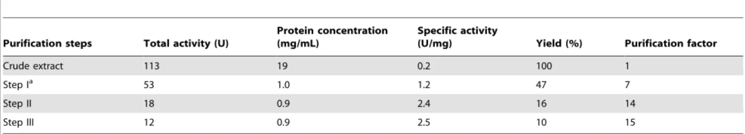
Purification of recombinant 4-HPPD using anion-exchange, hydrophobic interaction and size-exclusion chromatography yielded a pure protein, as judged by SDS-PAGE analyses (Fig. 3A). The purified protein showed a single major band with molecular mass about 45 kDa on SDS-PAGE (Fig. 3B). The yield of purified 4-HPPD was about 10% based on enzyme activity, with about a 15-fold improvement in purity from the cell lysate (Table 2). The five residues of the N-terminus in recombinant 4-HPPD were determined to be TTYSD, consistent with that predicted from the gene sequence of the human liver enzyme [27]. Purified recombinant wild-type and mutant 4-HPPD enzymes were further confirmed to be the same enzyme by Western blotting analysis (Fig. 3C).
Characterisation of recombinant human 4-HPPD
The catalytic activity of purified recombinant 4-HPPD was determined by a HPLC assay, which measures the formation of homogentisate, and the Oxygraph assay which continuously measures oxygen consumption during catalysis. The requirement for a reducing agent in the assay was determined using the HPLC assay. The presence of ascorbate in stoichiometric amounts resulted in an increase in specific activity of the recombinant enzyme byca.2-fold. When the stoichiometry was increased to 4-fold, activity was increased by ca. 2.7-fold compared to in the absence of ascorbate. In contrast, addition of dithiothreitol (DTT) to the assay solution had no significant effect on activity. Addition of tris(2-carboxyethyl)phosphine (TCEP) reduced the activity by ca.30%.
To determine the efficiency of recombinant 4-HPPD to convert 4-HPP substrate to HG product, production of 4-hydroxyphe-nylpyruvate (4-HPA) was determined. No 4-HPA was produced by the wild-type enzyme, indicating that this alternative product was not produced at a significant level. The specific activity of the wild-type enzyme was determined to be 2.660.1 and 2.860.1mmol/ min/mg by Oxygraph and HPLC assays, respectively (Table 3). These results are in agreement with the data reported for native human 4-HPPD [9,27].
Activity of mutant enzymes
To investigate the influence of the C-terminus on the catalytic function of 4-HPPD, several mutants were constructed with different numbers of residues removed: 5 residues (DG388); 8 residues (DE385); 12 residues (DL381); 13 residues (DN380); 14 residues (DG379); and 15 residues (DR378). The R378K, Q375N and R378K/Q375N point mutants were also constructed. All of these proteins were purified to near homogeneity, as judged by
SDS-PAGE analyses (Fig. 3B). Circular dichroism analyses of all mutants suggested no gross changes in the secondary structure as compared to the wild-type enzyme.
Deletion of residues from the C-terminus resulted in a reduction in enzymatic activity (Table 3). The activities of the DG388,
DE385 andDL381 mutants were onlyca.50 to 60% of that of the wild-type enzyme. TheDN380 mutant wasca.20% as active as the wild-type enzyme, and further truncation to R378 abolished all activity. The R378K or E254D mutation reduced activity toca. 5% of the wild-type enzyme, and all activity was abolished in the Q375N and R378K/Q375N mutants, indicating the importance of these residues for catalysis. Production of HPA by these mutants was not detected, showing that the reduction in activity was not due to the formation of an alternative product.
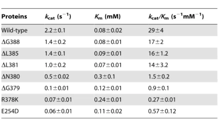
Steady-state kinetic analyses on these mutants were carried out using the Oxygraph assay (Table 4). ThekcatandKmvalues for 4-HPP substrate for wild-type enzyme was determined to be 2.260.1 s21 and 0.0860.02 mM, respectively. No significant change inKmvalue was observed for theDG388,DE385 orDL381 mutants compared to wild-type enzyme, whilstkcatvalues were reduced byca.25 to 50%. However, deletion of further residues (DN380 and DG379 mutants) resulted in increased Km and decreased kcat values. The latter two mutants had a catalytic efficiency of ca. 3% and 1% compared to wild-type enzyme,
Figure 3. SDS-PAGE analyses of wild-type and mutant 4-HPPD enzymes.(A) Fractions are shown for the different purification steps of wild-type 4-HPPD. Lane 1, cell crude lysate; Lane 2, following Q-sepharose anion-exchange chromatography; Lane 3, following SOURCE 15PHE hydrophobic interaction chromatography; Lane 4, concentrated fractions after S-100 Sephacryl gel filtration chromatography. (B) Purified wild-type and mutant 4-HPPD enzymes. (C) Western blotting of (B).
respectively, as judged bykcat/Kmvalues. Substitution in R378 or E254 resulted in about 95% reduction inkcatvalues. In addition, the R378K mutant showed an increasedKmvalue.
Structural modeling
The structure of human 4-HPPD was previously determined without bound substrate (Fig. 4A) [16]. Binding of the 4-HPP substrate into the active site of the enzyme was therefore modeled using a docking routine. The geometry minimization by QM-MM calculations was performed for all enzymes in complex with 4-HPP. Compared to the structure of 4-HPPD, this model with bound 4-HPP was shifted with the Car.m.s.d about 0.83 A˚ . The model of the enzyme in complex with substrate was in agreement with the previously proposed binding mode of 4-HPP [12,25]. The oxygen atoms of the carboxyl anda-keto group of 4-HPP forms a bidentate interaction with iron at distances of 2.0 and 2.03 A˚ , respectively. The substrate aromatic side-chain is sandwiched between the phenyl rings of F336 and F364, and the phenolic 4-hydroxyl group of 4-HPP forms hydrogen bonds with the side-chains of Q265 and Q251 (Fig. 4B).
In the modeled structure of mutant protein, the structure the terminal a-helix was apparently shifted with core r.m.s.d. about 0.46, 0.45 and 0.28 A˚ for the DG379, Q375N and R378K mutants, respectively, as compared to the wild-type model. The side-chains of Q251, E254, Q265, Y295, F336, F364, F368, F371, Q375, L377 and R378 inDG379 mutant model were obviously
moved with r.m.s.d. about 0.32, 1.1, 0.23, 0.54, 0.22, 0.25, 0.37, 0.86, 1.35, 1.39 and 0.78 A˚ , respectively (Fig. 4B). It is noted that the aromatic ring of F371 in this mutant model was particularly rotated by about 60 degrees. Due to the truncation, the bifurcate interactions between the side-chain of Q375 and the oxygen atom of S250 and N380 were disrupted and in turn new hydrogen bonds were formed between the side-chains of Q375 and E254. Similarly, Q375 formed new interactions with Q251 and N380 in the DN380 mutant model (data not shown). In the modeled structure of Q375N mutant, similar movement in the side-chains of these residues were shown. Moreover, the dramatic movement in the side-chains of F347 and F371 (about 0.47 and 2.16 A˚ , respectively) resulted in increased accessibility of the active site cavity. A channel extending from the active site to the C-terminus was shown in the modeled structure (Fig. 4C).
Discussion
Human recombinant 4-HPPD was cloned from HepG2 cells and expressed as soluble protein inE. coli. The purified protein had similar enzymatic activity as the native enzyme. The recombinant enzyme was only activated by ascorbate, and not by other reducing agents. This contrasts with the results for native human liver 4-HPPD, where activation was observed for ascorbate and other reducing cofactors [9,27]. Activation by ascorbate and DTT has been previously reported for rat livera-ketoisocaproate
Table 2.The purification of recombinant human 4-HPPD.
Purification steps Total activity (U)
Protein concentration (mg/mL)
Specific activity
(U/mg) Yield (%) Purification factor
Crude extract 113 19 0.2 100 1
Step Ia 53 1.0 1.2 47 7
Step II 18 0.9 2.4 16 14
Step III 12 0.9 2.5 10 15
aSteps I to III indicate the pooled fractions after the Q-Sepharose column, after the SOURCE 15PHE column and concentrated fractions pooled after the S-100 Sephacryl
column, respectively. The 4-HPPD activity was measured by the formation of HG inmmol/min (U) using the HPLC assay.
doi:10.1371/journal.pone.0069733.t002
Table 3.Activities of the wild-type and mutant enzymes.
Oxygen consumption assay HG product assay
proteins
Specific activity (mmol min21mg21)
Relative activity (%)
Specific activity (mmol min21mg21)
Relative activity (%)
Wild-type 2.660.1 100 2.860.1 100
DG388 1.560.2 56 2.260.1 81
DE385 1.760.1 65 1.660.1 57
DL381 1.160.3 42 1.460.1 50
DN380 0.860.03 31 0.560.02 17
DG379 0.160.02 5 0.0360.01 1
DR378 ND ,0.1 ND ,0.1
R378K 0.0760.01 3 0.0460.01 1
Q375N ND ,0.1 ND ,0.1
R378K/Q375N ND ,0.1 ND ,0.1
E254D 0.0660.01 2 0.260.02 7
oxidase [27,30]. Thisa-ketoisocaproate oxidase was later found to be the same enzyme as 4-HPPD [27,31]. Activation of enzyme activity by ascorbate has also been reported for other oxygenase enzymes, e.g. deacetoxycephalosporin synthase (DAOCS) and prolyl 4-hydroxylase [27,32–34]. In these enzymes, ascorbate is probably used to keep the iron in the ferrous state in the active site. It has also been proposed that ascorbate is able to reduce the ferryl species during uncoupled catalytic cycles performed by prolyl-4-hydroxylase and 2,4-dichlorophenoxyacetic acid/2-oxoglutarate dioxygenase [27,33,34].
The active site of 4-HPPD is enclosed by a C-terminala-helix which is assumed to function as a gate which controls access of substrate [11]. The C-terminus of the reported structure was resolved only as far as Met-384 and the last 9 residues were disordered and not visible in the structure (Fig. 4A) [16]. Arg-378 is located at the end of the finala-helix, and the last 15 residues comprise the disordered C-terminal tail. Previously Leeet. al.[26] identified a mutant rat 4-HPPD gene that encoded a protein with 14 residues deleted from the C-terminus, which was expressed as an inactive enzyme. This mutant showed no brownish pigment formation when expressed in E. coli and had no detectable decarboxylation ability using a-ketoisocaproate as a substrate. This study shows that all activity is lost for human 4-HPPD upon removal of the final 15 residues (DR378) of the C-terminus when 4-HPP was used as substrate. Human and rat 4-HPPD have the same number of amino acids in their sequences and areca.85% identical. The results in this study show the critical role of this disordered tail for enzyme activity in these enzymes. This study also shows that the last 5 residues, VVPGM, of the C-terminus are important for enzyme activity. Deletion of these residues led to loss ofca.50% activity. These mutants had very similarKmvalues to the wild-type enzyme but kcat values were significantly reduced. Even more reduction ofkcatvalues were observed for theDL381 and DN380 and DG379 mutants. The results suggest that the interactions provided by the last five residues, L381 and N380 are critical for the catalysis.
Inspection of the reported structure of human 4-HPPD determined without bound substrate shows that the putative substrate binding site is broad and partly formed by the C-terminus (Fig. 4D). Docking of substrate into the putative binding site appears to induce a conformational change. The simulation model shows that the orientation of the amide group in the side-chain of Q251 is reversed allowing interaction with substrate. In this manner the side-chain of Q251 is located near the aromatic ring of F336 and both residues act as a gate to restrict access to the
substrate binding pocket (Fig. 4A and E). This change seems drive the movement of the final helix (about 2 A˚ ) with formation of a new salt bridge between the side-chains of R338 and E374 and a new hydrogen bond between the side-chains of S366 and Y295. Residues of F347, F371, P339, R378 and Q375 that line the channel from the gate to the C-terminus were shifted about 0.7, 2.1, 1.2, 1.5 and 1.5 A˚ , respectively, to shield the active site from solvent. It is noted that the shift of Q375 led to the switch of its bifurcate interactions with N380 and K249 to N380 and S250 after substrate binding. The tail in the C-terminus is assumed to interact with residues surrounding the active site entrance but it is not visible in this model.
In the structure of human 4-HPPD, residues of N380, L381, T382 and N383 in the tail participate in interactions with residues of E254, Y258, Q375, K247, K248 and K249, which are located around the entrance of active site, to fix the end of the final helix into the appropriate position (Fig. 4A) [16]. The DG379 model showed the truncation of the tail to disrupt these interactions, causing apparent movements of the finala-helix and residues near the active site entrance. In particular a conformational change in the aromatic ring of F371 and large differences in the conforma-tions of E254, R378 and Q375 are predicted (Fig. 4B). The distance between the side-chain of Q251 and F336 was increased about 0.3 A˚ as compared with wild-type model. The broad access might be covered by residues lining the channel. However, due to the change in conformation in the aromatic ring of F371, the positions of F347 and P339 were subtle moved (Fig. 4F). Truncation of the tail might expose the entrance and increases the solvent accessibility to the active site. This change might be the reason for loss of catalytic activity. Results from kinetics of tail truncated enzymes suggest the interactions provided by the last residues together with L381 and N380 are critical for catalysis. The structural models suggest residues of Q375, K248 and D342 located around the active site entrance might interact with these residues in the tail. Furthermore, the crystal structure and models of wild-type or the different truncated mutants showed that Q375 can have various interactions with the tail and residues near the active site entrance, suggesting the dynamic interaction provide by this residue might mediate the position of final helix and C-terminus during the catalytic cycle.
Residues in the terminala-helix of 4-HPPD may also have a role in catalysis. A previous study by Gunsioret al[23] showed that ca.98 and 80% of activity was lost for thesa4-HPPD F337I and F341Y mutants, respectively. The F337I mutant also catalyzed the formation of oxepinone (from an arene oxide-derived intermedi-ate) and hydroxymandelate (HMA) in addition to HG. These two residues correspond to F364 and F368 in the terminal helix ofh 4-HPPD. The results in this study indicate that thep-pinteraction between the 4-HPP substrate and F364 is important for maintaining the benzene ring of 4-HPP in a proper position for oxidation [23,25]. In the present study, the activity lost by substitution of R378 and Q375 indicates a strict requirement of these two residues in the proper position for catalysis. These two residues are not conservative among different species and their equivalent residues in plants are Leu and Glu, respectively (Fig. 2). In theh4-HPPD structure, R378 and Q375 are located close to the C-terminal end of the helix facing the entrance of the active site, and Q375 is situated near the Q251 (about 3.5 A˚ ) [16]. Q251 is a critical catalytic residue which forms hydrogen bonds with the 4-hydroxy group of the 4-HPP substrate. Studies by Raspail et al. [25] showed that the interactions provided by Q272, Q286 and Q358 (corresponding to Q251, Q265 and Q334 in human 4-HPPD) are critical for the formation of the enzyme-4-HPP complex and the first nucleophilic reaction by dioxygen.
Table 4.Apparent kinetic parameters of wild-type and mutant 4-HPPD enzymes measured using the oxygraph assay.
Proteins kcat(s21) Km(mM) kcat/Km(s21mM21)
Wild-type 2.260.1 0.0860.02 2964
DG388 1.460.2 0.0860.01 1762
DL385 1.460.1 0.0960.01 1661.2
DL381 1.060.2 0.0760.01 1463.2
DN380 0.560.02 0.360.1 1.560.2
DG379 0.160.01 0.1260.01 0.960.1
R378K 0.0760.01 0.2460.01 0.2760.01
E254D 0.0660.01 0.1160.02 0.5760.12
Figure 4. Models of 4-HPPD in complex with 4-HPP substrate.Superimposition of the model for the wild-type enzyme and the X-ray crystal structure of human 4-HPPD (PDB code: 3ISQ) [16] (A), and the models ofDG379 (B), and Q375N (C) mutant enzymes (stereo image). The protein is shown as a cartoon and colored grey and cyan for the wild-type model and crystal structure or mutant models, respectively. The metal and 4-HPP present as sphere and stick models and colored green and yellow for wild-type and mutant enzymes, respectively. Hydrogen bonding interactions are shown as dashed black lines in (B). The putative substrate binding cavity is shown as Jacks style and colored green and magenta for wild-type and Q375N mutant models, respectively in (C). (D–G) Presentation of the crystal structure (D), wild-type (E),DG379 (F), and Q375N (G) models as surface styles and colored by interpolated charge. The C-terminus and residues in the final helix are shown as a ribbon and stick model, respectively, and colored in cyan. The cavity for putative substrate binding cavity is colored yellow. The metal and 4-HPP present as sphere and stick models and colored green.
In the wild-type model, Q375 forms bifurcate hydrogen bonds with S250 and N380 to fix the final helix and the tail in the appropriate position, substitution to disrupt these interactions would destabilize the C-terminus. In the Q375N mutant model, the side-chain of Q251 and F336 was separated for about 0.4 A˚ wider than that in the wild-type model and the aromatic ring of F371 was dramatic shifted away from that of F347 (Fig. 4E and G). These changes resulted in opening a solvent accessible channel from the putative substrate binding pocket to the C-terminus. Furthermore, without these interactions to stabilize the tail, it might not function properly to cover the active site entrance. This may account for the complete loss of activity.
R378 is the final residue in the terminal helix. In the structure, this residue interacts with E254 to fix the terminal helix in proper position (Fig. 4A). Mutation of R378 or E254 resulted in most of the activity lost especially the reduction of the kcat value. The R378K mutant model showed that the substitution not only disrupted the interaction between R378 and E254 but also affected the interaction between Q375 and S250 and the hydrogen bonding of Y259 and S366. These results indicating the interactions formed by the two residues are critical for maintaining the terminal helix in a stabile conformation for catalysis. However, inspection the models of truncated and Q375N mutant enzymes, show that although the relative positions of R378 and E254 are shifted, the interactions between the two residues are still retained. The result highlights the roles of R378 and Q375 in stabilization of the C-terminus are different.
The models and kinetic data suggest the bifurcate interactions provided by Q375 are critical to stabilize the finala-helix and the tail in the correct position for catalysis. Hence, truncation of the C-terminus means that the final a-helix cannot appropriately
function as a gate to isolate the active site from solvent and hence impacts on the catalytic reaction performed by mutant 4-HPPD enzymes. Mutant enzymes which are still active produce the expected HG product, suggesting that the orientation of the substrate aromatic ring is still appropriate. Removal of part or all of the C-terminal tail might affect either step in the catalytic cycle including the activation of dioxygen, the nucleophilic attack by dioxygen or the release of the HG product; the latter step has been shown to be rate-limiting for the reaction [35]. However, this hypothesis requires further confirmation. Results from this study suggest that other roles for the C-terminus are also possible. Through the interaction with Q375, the dynamic orientation of the tail might mediate the appropriate position of the finala-helix to ensure that gate is opened during the appropriate steps in the catalytic cycle to allow substrate to bind and the release of product [11,36]. These interactions assume to maintain the integrity of the active site, ensuring correct orientation of substrate and its interaction with residues in the active site which are important for catalysis.
Acknowledgements
We thank Dr M. D. Lloyd (University of Bath, U. K.) for reading and discussing this manuscript before publication and Dr. M. H. Lee for helpful discussions.
Author Contributions
Conceived and designed the experiments: HJL. Performed the experi-ments: JFL YLS NYC CWC CPS. Analyzed the data: HJL JFL YLS TCC. Contributed reagents/materials/analysis tools: HJL TCC. Wrote the paper: HJL.
References
1. Clifton IJ, McDonough MA, Ehrismann D, Kershaw NJ, Granatino N, et al. (2006) Structural studies on 2-oxoglutarate oxygenases and related double-stranded beta-helix fold proteins. J Inorg Biochem 100: 644–669.
2. Hausinger RP (2004) FeII/a-ketoglutarate-dependent hydroxylases and related enzymes. Crit Rev Biochem Mol Biol 39: 21–68.
3. Moran GR (2005) 4-Hydroxyphenylpyruvate dioxygenase. Arch Biochem Biophys 433: 117–128.
4. Tomoeda K, Awata H, Matsuura T, Matsuda I, Ploechl E, et al. (2000) Mutations in the 4-hydroxyphenylpyruvic acid dioxygenase gene are responsible for tyrosinemia type III and hawkinsinuria. Mol Genet Metab 71: 506–510. 5. Goodwin TW, Mercer EI (1983) Introduction to Plant Biochemistry; 2nd ed.,
Sydney: Pergamon Press.
6. Garcia I, Job D, Matringe M (2000) Inhibition of p-hydroxyphenylpyruvate dioxygenase by the diketonitrile of isoxaflutole: a case of half-site reactivity. Biochemistry 39: 7501–7507.
7. Mitchell G, Bartlett DW, Fraser TE, Hawkes TR, Holt DC, et al. (1999) Mesotrione: a new selective herbicide for use in maize. Pest Manage Sci 57: 120– 128.
8. Schulz A, Ort O, Beyer P, Kleinig H (1993) SC-0051, a 2-benzoyl-cyclohexane-1,3-dione bleaching herbicide, is a potent inhibitor of the enzyme p-hydroxyphenylpyruvate dioxygenase. FEBS Lett 318: 162–166.
9. Lindblad B, Lindstedt G, Lindstedt S, Rundgren M (1977) Purification and some properties of human 4-hydroxyphenylpyruvate dioxygenase (I). J Biol Chem 252: 5073–5084.
10. Brownlee JM, Johnson-Winters K, Harrison DH, Moran GR (2004) Structure of the ferrous form of (4-hydroxyphenyl)pyruvate dioxygenase fromStreptomyces avermitilisin complex with the therapeutic herbicide, NTBC. Biochemistry 43: 6370–6377.
11. Fritze IM, Linden L, Freigang J, Auerbach G, Huber R, et al. (2004) The crystal structures of Zea mays and Arabidopsis 4-hydroxyphenylpyruvate dioxygenase. Plant Physiol 134: 1388–1400.
12. Serre L, Sailland A, Sy D, Boudec P, Rolland A, et al. (1999) Crystal structure of Pseudomonas fluorescens 4-hydroxyphenylpyruvate dioxygenase: an enzyme in-volved in the tyrosine degradation pathway. Structure 7: 977–988.
13. Yang C, Pflugrath JW, Camper DL, Foster ML, Pernich DJ, et al. (2004) Structural basis for herbicidal inhibitor selectivity revealed by comparison of crystal structures of plant and mammalian 4-hydroxyphenylpyruvate dioxy-genases. Biochemistry 43: 10414–10423.
14. Han S, Eltis LD, Timmis KN, Muchmore SW, Bolin JT (1995) Crystal structure of the biphenyl-cleaving extradiol dioxygenase from a PCB-degrading pseudomonad. Science 270: 976–980.
15. Kita A, Kita S, Fujisawa I, Inaka K, Ishida T, et al. (1999) An archetypical extradiol-cleaving catecholic dioxygenase: the crystal structure of catechol 2,3-dioxygenase (metapyrocatechase) fromPseudomonas putidamt-2. Structure 7: 25– 34.
16. Pilka ES, Shafqat N, Cocking R, Bray JE, Krojer T, et al. (2009) Crystal structure of human 4-Hydroxyphenylpyruvate dioxygenase. RCSB Protein Data Bank (PDB ID: 3ISQ).
17. Hegg EL, Que LJ (1997) The 2-His-1-carboxylate facial triad: a emerging structural motif in mononuclear non-heme iron(II) enzymes. Eur J Biochem 250: 625–629.
18. Koehntop KD, Emerson JP, Que L Jr (2005) The 2-His-1-carboxylate facial triad: a versatile platform for dioxygen activation by mononuclear non-heme iron(II) enzymes. J Biol Inorg Chem 10: 87–93.
19. Valegard K, Terwisscha van Scheltinga AC, Dubus A, Ranghino G, Oster LM, et al. (2004) The structural basis of cephalosporin formation in a mononuclear ferrous enzyme. Nat Struct Mol Biol 11: 95–101.
20. Valegard K, van Scheltinga AC, Lloyd MD, Hara T, Ramaswamy S, et al. (1998) Structure of a cephalosporin synthase. Nature 394: 805–809. 21. McDonough MA, Kavanagh KL, Butler D, Searls T, Oppermann U, et al.
(2005) Structure of human phytanoyl-CoA 2-hydroxylase identifies molecular mechanisms of Refsum disease. J Biol Chem 280: 41101–41110.
22. You Z, Omura S, Ikeda H, Cane DE, Jogl G (2007) Crystal structure of the non-heme iron dioxygenase PtlH in pentalenolactone biosynthesis. J Biol Chem 282: 36552–36560.
23. Gunsior M, Ravel J, Challis GL, Townsend CA (2004) Engineering p-hydroxyphenylpyruvate dioxygenase to a p-hydroxymandelate synthase and evidence for the proposed benzene oxide intermediate in homogentisate formation. Biochemistry 43: 663–674.
24. Lancaster DE, McNeill LA, McDonough MA, Aplin RT, Hewitson KS, et al. (2004) Disruption of dimerization and substrate phosphorylation inhibit factor inhibiting hypoxia-inducible factor (FIH) activity. Biochem J 383: 429–437. 25. Raspail C, Graindorge M, Moreau Y, Crouzy S, Lefebvre B, et al. (2011)
26. Lee MH, Zhang ZH, MacKinnon CH, Baldwin JE, Crouch NP (1996) The C-terminal of rat 4-hydroxyphenylpyruvate dioxygenase is indispensable for enzyme activity. FEBS Lett 393: 269–272.
27. Ruetschi U, Dellsen A, Sahlin P, Stenman G, Rymo L, et al. (1993) Human 4-hydroxyphenylpyruvate dioxygenase. Primary structure and chromosomal localization of the gene. Eur J Biochem 213: 1081–1089.
28. Garcia I, Rodgers M, Pepin R, Hsieh TF, Matringe M (1999) Characterization and subcellular compartmentation of recombinant 4-hydroxyphenylpyruvate dioxy-genase from Arabidopsis in transgenic tobacco. Plant Physiol 119: 1507–1516. 29. Denoya CD, Skinner DD, Morgenstern MR (1994) AStreptomyces avermitilisgene
encoding a 4-hydroxyphenylpyruvic acid dioxygenase-like protein that directs the production of homogentisic acid and an ochronotic pigment inEscherichia coli. J Bacteriol 176: 5312–5319.
30. Sabourin PJ, Bieber LL (1981) Subcellular distribution and partial character-ization of an a-ketoisocaproate oxidase of rat liver: formation of b -hydroxyisovaleric acid. Arch Biochem Biophys 206: 132–144.
31. Crouch NP, Baldwin JE, Lee MH, MacKinnon CH, Zhang ZH (1996) Initial studies on the substrate specificity of soluble recombinant 4-hydroxyphenylpyr-uvate dioxygenase from rat liver. Bioorg Med Chem Lett 5: 1255–1260.
32. Dubus A, Lloyd MD, Lee HJ, Schofield CJ, Baldwin JE, et al. (2001) Probing the penicillin sidechain selectivity of recombinant deacetoxycephalosporin C synthase. Cell Mol Life Sci 58: 835–843.
33. Majamaa K, Hanauske-Abel HM, Gunzler V, Kivirikko KI (1984) The 2-oxoglutarate binding site of prolyl 4-hydroxylase. Identification of distinct subsites and evidence for 2-oxoglutarate decarboxylation in a ligand reaction at the enzyme-bound ferrous ion. Eur J Biochem 138: 239–245.
34. Myllyla R, Majamaa K, Gunzler V, Hanauske-Abel HM, Kivirikko KI (1984) Ascorbate is consumed stoichiometrically in the uncoupled reactions catalyzed by prolyl 4-hydroxylase and lysyl hydroxylase. J Biol Chem 259: 5403–5405.
35. Johnson-Winters K, Purpero VM, Kavana M, Moran GR (2005) Accumulation of multiple intermediates in the catalytic cycle of (4-hydroxyphenyl)pyruvate dioxygenase fromStreptomyces avermitilis. Biochemistry 44: 7189–7199. 36. Brownlee J, He P, Moran GR, Harrison DH (2008) Two roads diverged: the
structure of hydroxymandelate synthase fromAmycolatopsis orientalisin complex with 4-hydroxymandelate. Biochemistry 47: 2002–2013.
![Figure 2. Alignment of amino acid sequences of the C-terminus of human 4-HPPD with enzymes from other species [37]](https://thumb-eu.123doks.com/thumbv2/123dok_br/18366085.354738/2.918.107.797.900.1001/figure-alignment-amino-sequences-terminus-human-enzymes-species.webp)
![Figure 4. Models of 4-HPPD in complex with 4-HPP substrate. Superimposition of the model for the wild-type enzyme and the X-ray crystal structure of human 4-HPPD (PDB code: 3ISQ) [16] (A), and the models of DG379 (B), and Q375N (C) mutant enzymes (stereo i](https://thumb-eu.123doks.com/thumbv2/123dok_br/18366085.354738/7.918.93.727.93.892/figure-models-complex-substrate-superimposition-crystal-structure-enzymes.webp)
