Other than Evanescent Wave in All-Fiber
Immunofluorescence Biosensor for Quantitative
Detection of
Escherichia coli
O157:H7
Zhonghuan Zhang1., Fei Hua2,3., Ting Liu1
, Yong Zhao2, Jun Li1, Ruifu Yang2, Changxi Yang1*, Lei Zhou2*
1State Key Laboratory of Precision Measurement Technology and Instrument, Department of Precision Instruments, Tsinghua University, Beijing, P.R. of China,
2Laboratory of Analytical Microbiology, State Key Laboratory of Pathogen and Biosecurity, Beijing Institute of Microbiology and Epidemiology, Beijing, P.R. of China,
3Department of Aetiology, Taishan Medical University, Taian, P.R. of China
Abstract
Cylindrical or taper-and-cylinder combination optical fiber probe based on evanescent wave has been widely used for immunofluorescence biosensor to detect various analytes. In this study, in contrast to the contradiction between penetration depth and analyte diameter of optical fiber probe-based evanescent wave, we demonstrate that double-taper optical fiber used in a radiation wave-based all-fiber immunofluorescence biosensor (RWAIB) can detect micron-scale analytes using Escherichia coli O157:H7 as representative target. Finite-difference time-domain method was used to compare the properties of evanescent wave and radiation wave (RW). Ray-tracing model was formulated to optimize the taper geometry of the probe. Based on a commercial multi-mode fiber, a double-taper probe was fabricated and connected with biosensor through a ‘‘ferrule connector’’ optical fiber connector. The RWAIB configuration was accomplished using commercial multi-mode fibers and fiber-based devices according to the ‘‘all-fiber’’ method. The standard sample tests revealed that the sensitivity of the proposed technique forE.coliO157:H7 detection was 103cfu?mL21. Quantitation could
be achieved within the concentration range of 103cfu?mL21to 107cfu
?mL21. No non-specific recognition to ten kinds of
food-borne pathogens was observed. The results demonstrated that based on the double-taper optical fiber RWAIB can be used for the quantitative detection of micron-scale targets, and RW sensing is an alternative for traditional evanescent wave sensing during the fabrication of fiber-optic biosensors.
Citation:Zhang Z, Hua F, Liu T, Zhao Y, Li J, et al. (2014) A Double-Taper Optical Fiber-Based Radiation Wave Other than Evanescent Wave in All-Fiber Immunofluorescence Biosensor for Quantitative Detection ofEscherichia coliO157:H7. PLoS ONE 9(5): e95429. doi:10.1371/journal.pone.0095429
Editor:Miklos S. Kellermayer, Semmelweis University, Hungary
ReceivedSeptember 1, 2013;AcceptedMarch 26, 2014;PublishedMay 7, 2014
Copyright:ß2014 Zhang et al. This is an open-access article distributed under the terms of the Creative Commons Attribution License, which permits unrestricted use, distribution, and reproduction in any medium, provided the original author and source are credited.
Funding:This work was supported by the National Natural Science Foundation of China (Grant No. 81000774) and the Major National Science and Technology Programs of China (Grant Nos. 2011ZX10004 and 2012ZX1004801). The funders had no role in study design, data collection and analysis, decision to publish, or preparation of the manuscript.
Competing Interests:The authors have declared that no competing interests exist. * E-mail: [email protected] (CY); [email protected]. (LZ)
.These authors contributed equally to this work.
Introduction
Since the application of fiber optics in sensing technology in the late 1970s [1–2], optical fiber has been established as an ideal substrate for immunofluorescence sensing because of their reliability, small size, and low cost [3]. Immunofluorescence sensing has great potential for rapid and sensitive analysis of various analytes, ranging from molecules to intact cells, and as an effective technique for biodefense, environmental monitoring, food-security control, etc. [4–5]. Most fiber optic biosensors utilize the evanescent wave (EW) field generated by the total internal reflection (TIR) of light restrained within an optical fiber as sensing region. In this sensing region, specifically bound fluorescence-labeled antibodies can be excited to emit light collected as specific signal, whereas the unbound ones outside the sensing region cannot be excited, thereby adding nothing to the noise. Given this natural separation, fiber-optic biosensors have high signal-to-noise
ratio [6], and they are used for the detection of various small analytes [7] and microorganisms [8–9].
com-bination tapered probes are usually used to generate EW field [7,13] and prepared using a tube-etching technique [14]. To enhance the sensitivity of EW excitation, U-bent optical fiber probes were used [15–16]. However, the fiber probe with definite structure has fixed incident ray angle to stimulate EW and refractive indices of core and cladding and then the characteristics of generated EW cannot be tuned as that of waveguide-based EW [17–20], especially penetration depth (PD). PD provides the inherent advantage of natural separation to EW for fluorescence sensing with high signal-to-noise ratio, but limits its superiority when the diameter of the detected target is larger than the PD [6]. Based on the numerical aperture (NA) of fibers, PD of all-fiber biosensors is usually several hundreds of nanometers [21], which hardly exceeds the size of bacteria (1mm to 5mm diameter, or larger). Therefore, fluorescein-labeled antibodies bound on bacteria surface but beyond the PD of EW field cannot be detected, resulting in an inevitable loss of sensitivity. Although several simulations have demonstrated that the PD value could be several micrometers in a specially designed fiber probe, a large PD value only exists within#5% of the entire probe length, leaving a major portion invalid [22]. This condition might result in poor consistency of quantitation at low concentrations of the detected target. Additionally, EW that penetrates from the probes only accounts for a small part of the entire excitation light power, which leads to a low percentage of utilization of the excitation light [23]. In the present study, finite-difference time-domain (FDTD) method was used to compare the properties of EW and radiation wave (RW). A ray-tracing model was formulated to optimize the taper geometry of the optical fiber probe. Based on a commercial multi-mode fiber, a double-taper optical fiber probe was proposed and fabricated as a low-cost, disposable sensing unit for RW sensing. The sensing unit was connected to the biosensor through a ‘‘ferrule connector (FC)’’ optical fiber connector. Therefore, the consistency and stability of these sensing units for practical tests could be guaranteed by the reliable telecommunication fiber connection technology. Subsequently, a novel RW-based all-fiber immunofluorescence biosensor (RWAIB) was developed to
ana-lyze simultaneously the specific signals within and beyond the reach of EW. The configuration of the RWAIB was developed using commercial multi-mode fibers and fiber-based devices according to the ‘‘all-fiber’’ method. The comprehensive perfor-mance including quantitation ability, sensitivity, and specificity of RWAIB was estimated using Escherichia coli O157:H7 as the representative micron-scale target/analyte.
Materials and Methods
Reagents
3-Aminopropyl-triethoxysilane (APTES), glutaraldehyde, and bovine serum albumin (BSA) were purchased from Sigma-Aldrich (Gillingham, Dorset, UK). Cyanine 5 (Cy5) and HiTrap Desalting prepacked column were obtained from GE Healthcare (Uppsala, Sweden). Unless otherwise specified, all reagents, which were provided by Sinopharm Chemical Reagent Co., Ltd. (Shanghai, China), were of analytical grade and used without further purification. Deionized water was used throughout the experi-ment.
Bacterial Culture and Antibody Preparation
E. coli O157:H7, Salmonella choleraesuis, Salmonella enteritidis, Salmonella paratyphiA,S. paratyphiB,S. paratyphiC,Salmonella typhi, Salmonella typhimurium, Vibrio parahaemolyticus, Vibrio choleraeO1, and
V. choleraeO139 were previously preserved in our laboratory and identified using 16sRNA sequencing. The bacteria were grown until they reached the exponential phase in Luria-Bertani (LB) media at 37uC. The bacteria were harvested by centrifugation at 6000 rpm (Allegra X-22R, Beckman, Germany) for 10 min at 4uC. The bacterial pellets were washed twice and resuspended with sterile normal saline (0.85% salt solution). Bacterial concen-tration was determined using plate count and demonstrated as colony forming units (cfu) per milliliter.
A monoclonal antibody (MAb) specific forE. coliO157:H7 was prepared in our laboratory, and its affinity was determined using
E. coliO157:H7-coated ELISA. MAb was labeled with Cy5 and
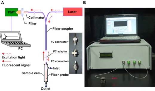
Figure 1. Schematic diagram (A) and prototype (B) of all-fiber biosensor.(A) Twenty percent of the 643 nm excitation light was conducted from the semiconductor laser to the fiber probe through a fiber coupler with FC connectors as the linker. Eighty percent of 668 nm fluorescent signal was collected and transmitted back from the fiber probe to the PMT through a fiber coupler, fiber collimator, and high-pass filter. The signal was processed and displayed on the computer screen. (B) The prototype of all-fiber biosensor consisted of an integrated detecting unit and a controlling unit (computer). RW field produced the visible red light around the fiber probe.
purified with HiTrap Desalting prepacked column to separate and clear free Cy5 and antibody molecules, according to the manufacturer’s instruction.
Double-taper Probe Fabrication, Modification and Activation
The double-taper probe designed to generate RW was made from a length of step-index optical fibers (105mm core/125mm
cladding, Beijing Glass Research Institute, Beijing, China). At the non-sensing end, an FC standard optical fiber connector was fixed to facilitate the alignment of the sensor optical path during the repeated installation-and-uninstallation of the probes. At the sensing end, a double-tapered structure was fabricated using a simple static-and-dynamic etching method with 40% HF as the etchant (experimental details are provided as File S1); this method combined classical static tube etching [24] and dynamic liquid-level-lowering etching [25] and promoted the large-scale prepa-ration of probes with good uniformity. The fabricated double-taper probe was bathed successively in NaOH (1 mol?L21
) and HCl (1 mol?L21
) for 10 min, and then dried for future use. The calibrated optical microscopic images show that the diameter of taper 1 of fabricated double-taper probe was etched from 125mm to ,40mm within the length of ,270mm with the V number matching the diameter [21]; the diameter of taper 2 was reduced to 26mm at distal end within the length of,2.5 cm.
The double-taper probe was silanized by immersing it in 10% APTES (in isopropyl alcohol) for 2 h. As a result, a monolayer silane film was covalently bonded on the silica surface of the probe with the amino functional groups on top. Subsequently, the silanized probe was functionalized using an amine-reactive homo-bifunctional cross-linker glutaraldehyde (12.5%, in deionized water) for 2 h. Residual glutaraldehyde was rinsed with phos-phate-buffered saline (PBS; 135 mmol?L21
NaCl, 15 mmol?L21 sodium phosphate, pH 7.2). The aldehyde group-activated probe was then incubated in MAb solution (0.5 mg?mL21 in PBS) for 4 h at 37uC. Finally, the probe was immersed in BSA solution (1 mg?mL21 in PBS) for 30 min to block non-specific binding cites. The activated sensing probes were stored at 4uC for future use.
RWAIB Structural Design
A semiconductor laser with wavelength of 643 nm and tunable output power of 20 mW (Shanghai Fiblaser Technology Co., Ltd, Shanghai, China) was selected as excitation light source, because the excitation wavelength of Cy5 is at 646 nm (Figure 1). The
pigtail of the laser was linked to one input end of an optical fiber coupler (162, couple ratio: 20/80, Beijing Glass Research Institute, Beijing, China). Twenty percent of the input power could be transmitted to the fiber probe through the coupler. The fiber probe was connected to the coupler using a ‘‘FC’’ optical fiber connector (inset in Figure 1A), which is a common device in optical fiber telecommunication with insertion and return loss that are almost the same in different individuals. The 643 nm light formed an RW field around the fiber probe to excite Cy5 that was bonded on the surface. The fluorescent signal with 668 nm emission wavelength was collected by the fiber probe, and 80% of signal power was sent back to the other input end of the coupler. Fluorescent signal was filtered using a high-pass filter and injected into a photomultiplier tube (PMT-CR131, Beijing Hamamatsu Photon Techniques Inc., Beijing, China) through a fiber collima-tor. The signal was processed by an electronic system and then displayed on a computer screen. During the entire process of detection, the probe was installed in a 4-mm diameter, 50-mm long poly-propylene sample cell.
Evaluation of RWAIB Performance in DetectingE. coli
O157:H7
To determine the sensitivity (detection limit) and quantitation ability of the technique, water samples containing various concentrations (103 cfu?mL21
to 107 cfu?mL21
) of E. coli
O157:H7 were used as standard solutions, with each sample detected in triplicate. The samples were separately injected into the sample cell from its inlet and incubated for 10 min at room temperature to achieve the specific capture of bacteria by the antibody-activated probe. After the transfer of the sample from the outlet of the sample cell, the cleaning buffer (0.1% Tween20 in PBS) was injected into the cell, and the nonspecific binding and residual sample in the cell were removed. Subsequently, Cy5-labelled antibodies (25mg?mL21) were injected into the sample cell and incubated for 10 min at room temperature, then transferred out through the tubule. Immediately after another cycle of the cleaning process, the laser was turned on and maintained for 150 s while signal data was monitored, recorded, and displayed. After biosensor analysis, the surface state of probe with adhering bacteria was observed under a scanning electron microscope (S-3400 N, Hitachi, Japan).
Ten kinds of food-borne pathogens, namely, S. enteritidis, S. paratyphiA,S. paratyphiB,S. paratyphiC,S. typhimurium, S. choleraesuis, S. typhi, V. parahaemolyticus, V. choleraeO1, andV. choleraeO139, were tested using the biosensor at 107 cfu?mL21
to estimate the specificity of the technique.
Results and Discussion
EW and RW Sensing Mechanism
Light transmission within optical fiber is based on the principle of TIR. Light beams propagating inside the fiber core with incident angles (a) greater than the critical incident angle (ac) can be guided along the fiber, where ac is determined using the refractive indices of the fiber core and cladding [ac= sin21(ncl/nco)]. When TIR occurs, EW exists beyond the reflecting interface, and the electric field intensity of EW decays exponentially with the distance from the interface. The PD of EW generally refers to the distance at which the magnitude of electric field at the surface
and (K) are the axial cross-sections (x–zplanes) of the tapered probe as the light under modes (C), (D), and (E) propagates in the probe separately; (L) and (M) are the axial cross-sections (x–zplanes) of the cylindrical and tapered probes as the light under all of the modes propagates in the probes simultaneously. Downward arrows (Q) indicate the outlines of the probes.
doi:10.1371/journal.pone.0095429.g002
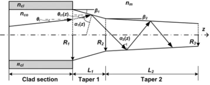
Figure 3. Ray tracing model of a tapered probe.
decays to its 1/evalue [26]. However, if the light beams hit the interface with incident angles less than ac, they will be partially propagated inside the core and partially transmitted into the surrounding medium. In partial transmission, the beams experi-ence reflection and transmission simultaneously and disappear after a short propagating distance. This short-distance propagating wave is called RW [27]. Unlike EW, RW is not restricted near the surface but emitted into surroundings, so that the extended sensing region and added excitation power provide the potential to increase the accuracy and sensitivity of the quantitative detection, especially for analytes with diameter beyond 1mm.
Simulation of EW and RW
Given that incident angles (a) of different beams are constant in a cylindrical probe because of the law of reflection, RW cannot exist for long distances. However, tapered probe is suitable for generating RW because it can gradually decrease incident angles while the beams propagate along it. Thus, all beams can generate RW.
Simplified models of EW and RW generated by cylindrical (Figure 2A) and tapered probes (Figure 2B), respectively, were calculated using FDTD method. The radius of the cylindrical probe was 0.75mm, whereas that of the tapered probe was
reduced from 0.75mm to 0.15mm, and both models had lengths
of 20mm. The refractive indices of the probes (silica) and
surrounding medium (water) were 1.456 and 1.333, respectively. The three guided modes of light in the cylindrical probe in successive order (i.e., the incident angles of these three modes decreased successively) are shown in Figures 2C, 2D, and 2E. The axial cross-sections (x–zplanes) of the cylindrical probe as the light propagated in the probes under modes C, D, and E are shown in Figure 2F, 2H, and 2J, respectively. Downward arrows (Q) indicate the outlines of the probes, whereas the optical power penetrating outside the outlines is the EW field. PD increased as the mode order increased (incident angle decreased), but did not exceed hundreds of nanometers. The axial cross-sections (x–z
planes) of the tapered probe as the light propagated in the probe under modes C, D, and E are shown in Fig. 2G, 2I, and 2K, respectively. The light under fundamental mode C was always restricted within the probe, but its PD increased as light propagated to the distal end. Light under higher mode D changed from guided to radiation mode and then generated RW after a certain distance of propagation. The light under the highest mode E generated RW before the light under mode D because of a smaller incident angle. The axial cross-sections (x–zplanes) of the cylindrical and tapered probes when the light propagates under all
of the modes simultaneously are shown in Figs. 2L and 2M, which demonstrated that the RW could offer a much larger sensing (or excitation) region than the EW. Additionally, the tapered probe, transforming guided mode with various order to radiation mode, enabled the RW distribution throughout the entire length of the probe instead of a short distance (Figures 2M). Therefore, a tapered probe is suitable for generating RW in detecting micron-scale analytes, which could not be thoroughly covered by the EW sensing region.
Ray-tracing in the Tapered Probe
The incident angle (a) is the critical factor that determines the generation of RW of a tapered probe. When a#ac, the guided wave changes into RW; otherwise, only the EW penetrates outside the probe. To characterize the incident anglesa(z) in the tapered fiber probe, a ray-tracing model was established (Figure 3).
The core radius of a step index fiber with core and cladding refractive indices ofncoandncl, respectively, was etched fromR1to
R2to form taper 1, and then successively etched fromR2toR3to form taper 2. The lengths of these two tapers wereL1andL2, and their half cone angles wereb1andb2, respectively. The refractive index of the medium surrounding the tapered probe wasnm.
For continuous tapered probe (i.e., taper 2 is not considered), a guided ray from clad section at an angle ofhiwith respect to thez -axis was launched into taper 1, and reflected at positionz, where the launch angle is changed into hi1(z). According to reference [28],hi1(z) = sin21[R1sinhi/R(z)], whereR(z) =R1–ztanb1. Hence, the incident angle in taper 1,a1(z), can be given as.
a1(z)~900{sin{1 R1sinhi=R1{ztanb1
h i
{b1 ð1Þ
As a1(z) decreased into less than ac after a TIR propagating distance ofL1(a$ac), RW started to appear where the core radius was reduced to R2(a=ac). Given that NA = 0.22, R1= 52.5mm,
nco= 1.456, ncl= 1.444, and nm= 1.333, the same as the actual parameters used in our sensor,L1(a$ac)for differentb1values were calculated. Several results are listed in Table 1, in whichL1(tot)is the complete length of taper 1 at a certainb1if taper 2 does not exist.
Table 1 shows that the continuous tapered probe is not suitable for RW sensing because the invalid length [L1(a$ac)] consists of more than 60% of the total length ranging from 3 mm (3008mm) to 6 cm (60761mm). The taper-and-cylinder combination tapered probe is also unsuitable for RW sensing becausea2(z) is constant in the cylindrical section, and RW disappears at the start of this Table 1.L1(a$ac),R2(a = ac), andL1(tot)at differentb1values.
b1a(6) L1(a$ac)b(mm) R2(a = ac)c(mm) L1(tot)d(mm)
0.05 37591 19.70 60761
0.1 18773 19.73 30080
0.5 3717 20.06 6016
1 1834 20.49 3008
5 318 24.68 600
10 109 33.28 298
aHalf cone angles of taper 1.
bTIR propagating distance alone taper 1 when
ais equal to or greater thanac.
cCore radius of taper 1 whereais equal toac. dComplete length of taper 1.
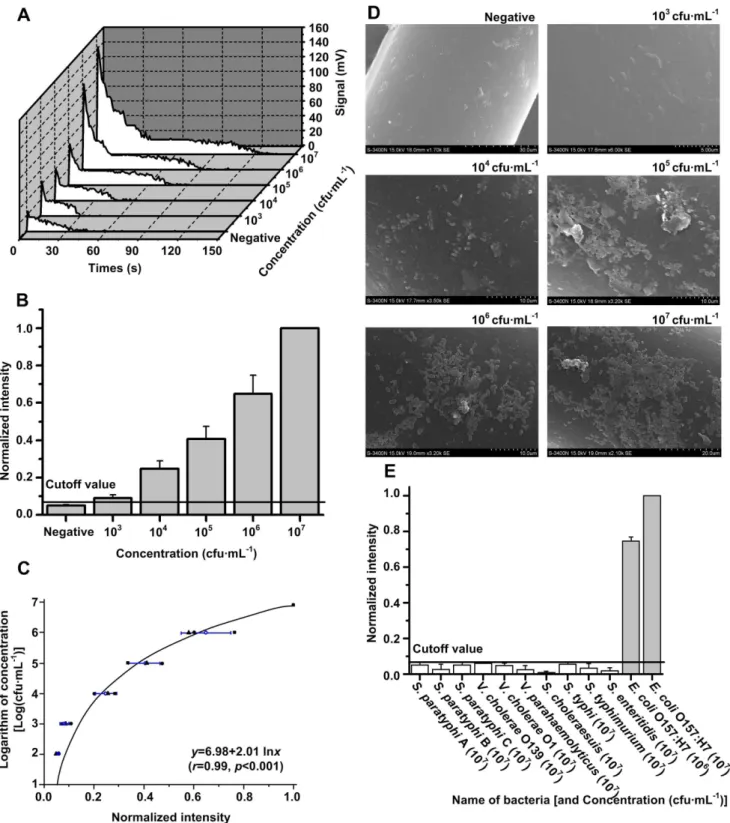
Figure 4. Evaluation of RWAIB for the quantitative detection ofE. coliO157:H7.(A) A group of typical signal-time traces at different concentrations ofE. coliO157:H7 during a complete test cycle (150 s). (B) Sensitivity forE. coliO157:H7 detection, wherexis the concentration of bacteria, andyis the NI. The bar graph revealed a significant difference between positive sample (103cfu?mL21
to 107cfu?mL21
) and negative control with the cutoff value determined as mean+3 SD of the negative control NIs. The sensitivity was 103cfu?mL21. (C) Quantitation ability forE. coli
O157:H7 detection, wherex is the NI, andyis the logarithm of concentration. The correlation and regression analyses revealed an exponent correlation betweenxandy, withr= 0.99 (p,0.05) in the quantitative range of 103cfu
?mL21to 107cfu
?mL21. (D) The scanning electron microscope
images of probes corresponding to negative and positive samples (with concentrations from 103cfu?mL21
to 107cfu?mL21
) proved the direct proportion between the amount of adhering bacteria and the concentration of sample. (E) Specificity forE. coliO157:H7 detection. A significant difference was observed between the NIs of 10 different kinds of food-borne pathogens at 107cfu
?mL21(white bars) and the NIs ofE. coliO157:H7
samples at 106and 107cfu
?mL21(gray bars) with cutoff value as the threshold.
section after a short-distance propagation. Therefore, double-taper probe is necessary for RW sensing. Taper 1 is used to reducea1(z) that is smaller than acin a short length to avoid a long invalid length, whereas taper 2 is used to release RW power gradually and continually reduces thea2(z) of the other beams with lower mode order to generate new RW. The double-taper probe used in this paper can generate RW almost from the beginning of taper 2 to the distal end.
Detection Data Analysis
A group of typical signal-time traces at different concentrations of detected target during a complete test cycle (150 s) are shown in Figure 4A. For each trace, signal intensity reached the maximum value immediately after the laser was turned on (0 s), and then gradually decreased back to the baseline because of fluorescence quenching. The integral intensity within the time of a complete test cycle (150 s) was regarded as an effective result for each test. Given that the batch-to-batch variation in the antibody-activated probe production is inevitable, a normalization of intensity was used to obtain comparable results of different tests. The normalized intensity (NI) is expressed as follows:
NIn~
In{Iblank
Imax{Iblank ð2Þ
where In is the integral intensity for a certain test, Imax is the integral intensity for the maximum concentration of the detected target (107 cfu?mL21 for the detection ofE. coliO157:H7), and
Iblank is the integral intensity for the activated sensing probe in blank solution without any procedure for detection. For a certain batch of activated probe,ImaxandIblankwere definitive.
RWAIB Performance Evaluation for Detection ofE. coli
O157:H7
E. coliO157:H7 was selected as the representative micron-scale target to evaluate the performance of RWAIB, including the sensitivity (detection limit), quantitation ability (correlation anal-ysis), and specificity of the technique. Water samples with 103 cfu?mL21
to 107cfu?mL21
E.coliO157:H7, as well as a negative control (0 cfu?mL21), were detected using RWAIB. NI against the concentration of the bacterium is shown in Figure 4B, with the integral intensity of 107 cfu?mL21
as Imax. NIs of all positive samples (103cfu?mL21to 107cfu?mL21) were significantly higher than the cutoff threshold (mean+3 SD of NIs corresponding to the negative control), which suggests that the sensitivity of RWAIB is 103cfu?mL21. The regression curve for the quantitative detection ofE. coliO157:H7 is shown in Figure 4C. An evident curvilinear correlation (exponent correlation) was found betweeny(logarithm of concentration) andx(NIs), in which the correlation coefficient (r) is equal to 0.99 (p,0.001) from 103cfu?mL21to 107cfu?mL21. Regression analysis can be expressed as follows:
y~6:98z2:01 lnx ð3Þ
The scanning electron microscope images of the probes corresponding to negative and positive samples (with
concentra-tions from 103cfu?mL21
to 107cfu?mL21
) are shown in Figure 4D and the amount of the observed bacteria adhering on the surface of the probe is directly proportional to the increase in concentra-tion. Ten kinds of food-borne pathogens at 107 cfu?mL21
were used to evaluate the specificity of RWAIB, usingE.coliO157:H7 samples (106cfu?mL21
and 107cfu?mL21
) as positive control. The high specificity of RWAIB, in which only twoE. coliO157:H7 samples exhibited strong signals higher than the cutoff threshold, is illustrated in Figure 4E.
Conclusion
Extensive studies have been conducted to develop and optimize static and dynamic chemical etching methods to control the taper length, cone angle, and geometry of the final optical fiber [29–33]. In these studies, the effects of fiber motion, etching rate, meniscus distortion, and etching time among others, have been explored. Geometrically optimized optical fibers, such as double-taper optical fiber, were used to ensure highly efficient light transmission for near-field scanning optical microscopy [29–30] and provide a surface-enhanced Raman scattering sensor with a large active surface and intensive internal reflections at the probe interface [31–33]. Based on the literature, our study first discussed the RW-producing property of double-taper optical fiber to resolve conflicts between PD and analyte diameter during EW-based fiber optic biosensing. We used FDTD calculation to intuitively demonstrate that the sensing region of RW is larger than that of EW.
The bio-active double-taper probes were fabricated from commercial communication multimode optical fibers using the traditional static-and-dynamic etching method, modified, and activated through a covalent method. Eventually, we have presented an all-fiber immunofluorescence biosensor with low-cost, disposable sensing units using double-taper probes for the quantitative detection of E. coliO157:H7. The standard sample tests revealed that the sensitivity of the technique for E. coli
O157:H7 detection was 103cfu?mL21, and quantitation could be achieved within the concentration range of 103cfu?mL21to 107 cfu?mL21. Non-specific recognition to other ten kinds of food-borne pathogens was not observed. Results demonstrate that the RWAIB can be used for the quantitative detection of micron-scale targets, and RW sensing is an alternative for traditional evanescent wave sensing during the fabrication of fiber-optic biosensors.
Supporting Information
File S1 Double-taper probe fabrication. The static-and-dynamic
etching device and its working process for double-taper probe fabrication are shown in details.
(DOC)
Author Contributions
Conceived and designed the experiments: LZ CY RY. Performed the experiments: ZZ FH TL YZ JL. Analyzed the data: ZZ FH. Contributed reagents/materials/analysis tools: TL YZ JL. Wrote the paper: ZZ LZ CY RY.
References
1. Lu¨bbers D, Opitz N (1975) The pCO2/pO2 optrode: A new probe for measuring pCO2 and pO2 of gases and liquids. Z Naturforsch C 30: 532–533. 2. Lu¨bbers DW, Opitz N, Speiser PP, Bisson HJ (1977) Nanoencapsulated fluorescence indicator molecules measuring pH and pO2 down to submicrosco-pical regions on the basis of the optode-principle. Z Naturforsch C 32: 133–134.
3. Wolfbeis OS (2008) Fiber-optic chemical sensors and biosensors. Anal Chem 80: 4269–4283.
5. Skottrup PD, Nicolaisen M, Justesen AF (2008) Towards on-site pathogen detection using antibody-based sensors. Biosens Bioelectron 24: 339–348. 6. Taitt CR, Anderson GP, Ligler FS (2005) Evanescent wave fluorescence
biosensors. Biosens Bioelectron 20: 2470–2487.
7. Long F, He M, Shi HC, Zhu AN (2008) Development of evanescent wave all-fiber immunosensor for environmental water analysis. Biosens Bioelectron 23: 952–958.
8. Ohk S-H, Bhunia AK (2013) Multiplex fiber optic biosensor for detection of
Listeria monocytogenes,Escherichia coliO157: H7 andSalmonella entericafrom ready-to-eat meat samples. Food Microbiol 33: 166–171.
9. Hewitt BM, Singhal N, Elliot RG, Chen AY, Kuo JY, et al. (2012) Novel Fiber Optic Detection Method for in Situ Analysis of Fluorescently Labeled Biosensor Organisms. Environ Sci Technol 46: 5414–5421.
10. Wang CW, Manne U, Reddy VB, Oelschlager DK, Katkoori VR, et al. (2010) Development of combination tapered fiber-optic biosensor dip probe for quantitative estimation of interleukin-6 in serum samples. J Biomed Opt 15: 067005.
11. Wei H, Zhao YK, Bi YJ, Liu HH, Guo ZB, et al. (2007) Direct detection of
Yersinia pestisfrom the infected animal specimens by a fiber optic biosensor. Sensor Actuat B-Chem 123: 204–210.
12. Geng T, Morgan MT, Bhunia AK (2004) Detection of low levels of Listeria monocytogenes cells by using a fiber-optic immunosensor. Appl Environ Microb 70: 6138–6146.
13. Jung CC, Saaski EW, McCrae DA, Lingerfelt BM, Anderson GP (2003) RAPTOR: a fluoroimmunoassay-based fiber optic sensor for detection of biological threats. IEEE Sens J 3: 352–360.
14. Sto¨ckle R, Fokas C, Deckert V, Zenobia R, Sick B, et al. (1999) High-quality near-field optical probes by tube etching. Appl Phys Lett 75: 160–162. 15. Bharadwaj R, Sai V, Thakare K, Dhawangale A, Kundu T, et al. (2011)
Evanescent wave absorbance based fiber optic biosensor for label-free detection ofE. coliat 280 nm wavelength. Biosens Bioelectron 26: 3367–3370. 16. Cao WQ, Duan YX (2006) Optical fiber evanescent wave sensor for oxygen
deficiency detection. Sensor Actuat B-Chem 119: 363–369.
17. Horvath R, Lindvold LR, Larsen NB (2002) Reverse-symmetry waveguides: theory and fabrication. Appl Phys B 74: 383–393.
18. Horvath R, Pedersen HC, Skivesen N, Selmeczi D, Larsen NB (2005) Monitoring of living cell attachment and spreading using reverse symmetry waveguide sensing. Appl Phys Lett 86: 071101.
19. Horvath R, Cottier K, Pedersen HC, Ramsden JJ (2008) Multidepth screening of living cells using optical waveguides. Biosens Bioelectron 24: 799–804. 20. Horvath R, Pedersen HC, Skivesen N, Selmeczi D, Larsen NB (2003) Optical
waveguide sensor for on-line monitoring of bacteria. Opt Lett 28: 1233–1235. 21. Golden JP, Anderson GP, Rabbany S, Ligler F (1994) An evanescent wave
biosensor. II. Fluorescent signal acquisition from tapered fiber optic probes. IEEE T Biomed Eng 41: 585–591.
22. Ahmad M, Hench LL (2005) Effect of taper geometries and launch angle on evanescent wave penetration depth in optical fibers. Biosens Bioelectron 20: 1312–1319.
23. Gloge D (1971) Weakly Guiding Fibers. Appl Optics 10: 2252–2258. 24. Turner DR (1983) Etch procedure for optical fibers. US patent 4469554 A. 25. Muramatsu H, Homma K, Chiba N, Yamamoto N, Egawa A (1999) Dynamic
etching method for fabricating a variety of tip shapes in the optical fibre probe of a scanning near-field optical microscope. J Microsc 194: 383–387.
26. Mirabella FM (1985) Internal Reflection Spectroscopy. ApplSpectrosc Rev 21: 45–178.
27. Mynbaev DK, Scheiner LL (2001) Fiber-optic communications technology: Prentice Hall NJ, USA.
28. Ankiewicz A, Pask C, Snyder A (1982) Slowly varying optical fibers. J Opt Soc Am 72: 198–203.
29. Lazarev A, Fang N, Luo Q, Zhang X (2003) Formation of fine near-field scanning optical microscopy tips. Part I. By static and dynamic chemical etching. Rev Sci Instrum 74: 3679–3683.
30. Haber LH, Schaller RD, Johnson JC, Saykally RJ (2004) Shape control of near-field probes using dynamic meniscus etching. J Microscopy 214: 27–35. 31. Lucotti A, Pesapane A, Zerbi G (2007) Use of a geometry optimized fiber-optic
surface-enhanced raman scattering sensor in trace detection. Appl Spectrosc 61: 260–268.
32. Pesapane A, Lucotti A, Zerbi G (2010) Fiber-optic SERS sensor with optimized geometry: testing and optimization. J Raman Spectrosc 41: 256–267. 33. Foti A, D’Andrea C, Bonaccors F, Lanza M, Calogero G, et al. (2013) A

![Table 1 shows that the continuous tapered probe is not suitable for RW sensing because the invalid length [L 1(a $ ac) ] consists of more than 60% of the total length ranging from 3 mm (3008 m m) to 6 cm (60761 m m)](https://thumb-eu.123doks.com/thumbv2/123dok_br/18303854.347986/5.918.97.826.118.280/table-continuous-tapered-suitable-sensing-invalid-consists-ranging.webp)