Article
Printed in Brazil - ©2013 Sociedade Brasileira de Química0103 - 5053 $6.00+0.00
A
*e-mail: [email protected]
Electropolymerized Supramolecular Tetraruthenated Porphyrins Applied as a
Voltammetric Sensor
Monize M. da Silva,a Gabriel H. Ribeiro,a Alzir A. Batista,b Anizio M. de Faria,a
André L. Bogadoa and Luis R. Dinelli*,a
aFaculdade de Ciências Integradas do Pontal, Universidade Federal de Uberlândia, Rua Vinte, 1600, 38304-402 Ituiutaba-MG, Brazil
bDepartamento de Química, Universidade Federal de São Carlos, Rodovia Washington Luís (SP 310), km 235, 13565-905 São Carlos-SP, Brazil
A porfirina 5,10,15,20-Tetra(4-piridil)manganês(III), [Mn-TPyP(H2O)2]PF6, e a
porfirina supramolecular eletropolimerizável (ESP), {Mn-TPyP(H2O)2[RuCl3(dppb)]4}PF6
(dppb = 1,4-bis(difenilfosfina)butano), foram sintetizadas e caracterizadas. Um filme fino da PSE foi obtido na superfície do eletrodo de carbono vítreo utilizando o método de voltametria cíclica. Foi observado um aumento da corrente de pico com o aumento do número de ciclos voltamétricos, mostrando um comportamento típico de espécies sendo adsorvidas na superfície do eletrodo. O eletrodo modificado com a ESP foi utilizado para a quantificação de acetoaminofeno por voltametria cíclica. O eletrodo modificado apresentou resposta linear de corrente de pico anódico em função da concentração de acetoaminofeno na faixa de concentração entre 0,05 e 0,70 mmol L–1. O eletrodo
modificado foi utilizado para a determinação de acetoaminofeno em uma amostra comercial e os resultados foram satisfatórios, pois apresentaram concordância quando comparado pelo método de HPLC.
Porphyrin 5,10,15,20-Tetra(4-pyridyl)manganese(III), [Mn-TPyP(H2O)2]PF6, and
electropolymerized supramolecular porphyrins (ESP), {Mn-TPyP(H2O)2[RuCl3(dppb)]4}PF6
(dppb = 1,4-bis(diphenilphosphine)butane), were synthesized and characterized. A thin solid film of ESP was obtained on a glass carbon electrode surface by a cyclic voltammetry method. The peak current increased with the number of voltammetric cycles, which shows a typical behavior of the species being adsorbed on the surface of the electrode. Cyclic voltammetry was also employed for acetaminophen quantification using an ESP modified electrode. The modified electrode shows a linear relationship between the anodic peak current and the concentration of acetaminophen (in the rage 0.05 to 0.7 mmol L–1). The performance of the modified electrode was verified by the
determination of acetaminophen in a commercial pharmaceutical product and the results were in good agreement with those obtained by a control HPLC method.
Keywords: tetraruthenated porphyrins, supramolecules, acetaminophen determination,
voltammetric sensor
Introduction
The importance of porphyrins is not limited to their participation in the transport of oxygen to heme proteins and photosynthetic activities. In fact, they also have valuable contributions in various fields, such as liquid crystalline materials,1 due to their remarkable
electro-optical properties, oxygen measurements in vivo,2,3
photodynamic therapy,4,5 malaria treatment,6 molecular
wires,7,8 energy conversion,9,10 nonlinear optical,11 optical
limiter,12 Langmuir and Langmuir-Blodgett (LB) films,13,14
as well as switching fluorescence.15
Porphyrins are also present in many applications of chemical analyses,16 such as electrochemical and optical
sensors,17,18 modified electrodes,19,20 spectrophotometric
reagents,21,22 stationary phase in the HPLC column,23,24
and as modifiers in open tubular electrochromatography.25
electrochemical deposition allow for film formation on small surface electrodes as well as on electrodes with complex geometry.
The use of modified electrodes has been considered the best strategy for the determination of several analytes, particularly active principles in pharmaceutical formulations. Accordingly, methods for acetaminophen determination in pharmaceutical preparations have been developed for many years due to the fact that an overdose of acetaminophen can cause fulminating hepatic necrosis and other toxic effects.26 Techniques typically employed for
the determination of acetaminophen in clinical laboratories are titrimetry, chromatography, spectrophotometry and immunoassays. However, determination using electrochemical techniques has been demonstrated to be promising.27-34 One of the first studies reported the use of
glassy carbon electrodes modified with 4-vinylpyridine.35
Shortly after, the electropolymerized porphyrin was already being used as the modified electrode for the determination of acetaminophen.36
Porphyrins can also be modified by peripheral groups, which increase the size of the macromolecules. The 5,10,15,20-Tetra(4-pyridyl)-21H,23H-porphyrin (TPyP) has been used effectively in the formation of supramolecules due to their reaction with peripheral metal complexes. Such supramolecules have been used successfully as modifiers for preparation of electrochemical sensors.37-42
The study of TPyP containing ruthenium peripheral complexes was boosted up by the discovery of exceptional electrocatalytic activity of 5,10,15,20-Tetra(piridyl) porphyrin cobalt (II) in the tetra electron reduction of O2 to
H2O, this complex has four groups of pentamim ruthenium
in peripheral positions, in the tetra electron reduction of O2 to H2O.43
In a study conducted by our research group, we synthesized the tetraruthenated porphyrins {H2TPyP[RuCl3(dppb)]4}
a n d { M - T P y P [ R u C l3( d p p b ) ]4} w h e r e d p p b =
1,4-bis(diphenylphosphino)butane and M = nickel or cobalt, from the reaction of 5,10,15,20-Tetra(4-pyridyl)-21H,23H-porphyrin with the mer-[RuCl3(dppb)(H2O)]
complex in a ratio of 1:4.44 Additionally, the X-ray structure
of tetraruthenated porphyrin [Ni-TPyP[RuCl3(dppb)]4} was
reported. The great advantage of these series of complexes as modifiers was the possibility of film deposition through successive voltammetric cycling of different electrodes such as platinum, ITO and glassy carbon. The proposed mechanism for the formation of films, which necessarily depends on the peripheral group “RuCl3(dppb),” involves
the formation of binuclear mixed-valence complexes (RuII/RuIII) with bridging chlorides between the molecules.
The modified glassy carbon electrode with porphyrin
{Co-TPyP[RuCl3(dppb)]4} was successfully used as an
electrochemical sensor for the detection of many analytes such as catechol.
Understanding the mechanism of the film formation for these classes of molecules {M-TPyP[RuCl3(dppb)]4}
(M = transition metal) allowed for the development of new classes of materials containing supramolecules. One great advantage in the use of electropolymerized electrode is the control of the number of layers deposited by controlling the number of voltammetric cycles. In towards of this view, the present work describes the synthesis of the new supramolecule {Mn-TPyP(H2O)2[RuCl3(dppb)]4}PF6,
which was immobilized on a glassy carbon electrode by electropolymerization and used as a sensor for acetaminophen.
Materials and methods
Materials
The chemicals employed were of reagent-grade quality (Aldrich or Flucka) and used as received. Reagent-grade solvents (Merck) were distilled prior to use. Doubly distilled deionized water was used for all aqueous solutions. Purified argon atmosphere was used in all procedures described herein for the removal of dissolved oxygen.
Measurements
UV-Vis spectra were recorded in CH2Cl2 on a
Perkin Elmer (Lambda 25) spectrophotometer. Electron paramagnetic resonance (EPR) spectra were measured at –160 oC with a Varian E-109 Instrument operating
at the X-band frequency, within a rectangular cavity (E-248) fitted with a temperature controller. Cyclic voltammetry was carried out at room temperature in freshly distilled dichloromethane containing 0.1 mol L–1
Bu4N+PF6– (TBAH) or 0.1 mol L–1 sodium acetate (NaAc)
solution, using a µautolab III potentiostat/galvanostat. The voltammetric measurements were performed in a cell with three electrodes, using a modified glass carbon or bare glass carbon (geometric diameter = 0.2 cm) as the working electrode (previously polished with alumina), a saturated Ag/AgCl as the reference electrode, a platinum electrode as the auxiliary electrode. The glass carbon and the platinum electrode were polished with alumina before use. Under these conditions, ferrocene is oxidized at 0.43 V (Fc+/Fc). Elemental analyses were performed at
were carried out with a CDM230 (Meter Lab). Solutions were prepared in CH2Cl2 or methanol at a concentration of
3.1 mol L–1. Magnetic susceptibility measurements were
used on a scale of Magnetic Susceptibility JM (Johnson Matthey). The value of magnetic susceptibility was found using the following equations: Xm = Xg × MW; µ = K (Xm × T)1/2; µ = [n(n+2)]1/2 (Xm: Molar susceptibility;
Xg: susceptibility in grams (value obtained directly in the balance); MW: molecular weight; µ: magnetic moment; K constant: 2.84; T: temperature in Kelvin and n: number of unpaired electrons).45
Atomic-force microscopy (AFM) measurements were performed at the Department of Material Science of the Federal University of São Carlos, São Carlos (Brazil), using a Nanoscope V Veeco/Bruker with a scan assist. The chromatographic evaluations were performed using a Varian ProStar HPLC comprising a ProStar 210 binary pump, a ProStar 325 UV/Vis Detector (at 254 nm), and a Rheodyne model 7125i injection valve with a 5µl loop. Experiments were carried out at 25 °C. Data were processed using Galaxie software for data acquisition. The mobile phases were prepared volumetrically from individually measured amounts of each solvent. All solvents were filtered and degassed before use. All measurements were performed in a reversed C18 column
phase (150×4.6 mm ID; particle size, 5 µm), with a mixture of methanol: H2O (70:30, v/v) as the mobile phase
for the detection of acetaminophen.
Syntheses
T h e m e r - [ R u C l3( d p p b ) ( H2O ) ] [ d p p b =
1,4-bis(diphenylphosphino)butane] complexes were prepared according procedures reported in the literature.46,47
H2TPyP was synthesized by a modification of a
procedure described in the literature:48 freshly distilled
pyrrole (3.35 g, 0.05 mol) and 4-pyridinecarboxaldehyde (5.35 g, 0.05 mol) were added to 350 mL of refluxing reagent grade acetic acid. After refluxing for 2 h, the solution was cooled down to room temperature and filtered. The purple crystals were washed sequentially with methanol, hot water and dried in vacuum to remove the absorbed acid. The H2TPyP was then purified in an alumina
column using chloroform as a solvent and 5% methanol in chloroform as an eluent to yield 2.00 g (26%).
The [Mn-TPyP(H2O)2]PF6 was synthesized by a
modification of a procedure described in the literature:48
0.150 g (0.247 mmol) of 5,10,15,20-tetra(pyridyl)porphyrin was dissolved in 100 mL glacial acetic acid and had 0.149 g of manganese acetate (0.617 mmol) and then the same molar amount of KPF6 (0.113g), slowly added to it. The
system was refluxed for 6 hours, which was followed by an UV/Vis spectroscopy. After that, the solvent was evaporated and the resulting product was dried under vacuum for 24 hours. To purify the porphyrin, the obtained solid was dissolved in distilled water at 62 °C, filtered off, and reprecipitated with a sodium acetate (2 mol L–1) solution,
washed with cold water and then dried under vacuum. Finally, the product was eluated through a chromatography column, using alumina as a stationary phase and the mixed solvent chloroform (95%)/methanol (5%) as eluent. Yield: 181 mg (85.5%); C40H28F6MnN8O2P.CHCl3 found (theoretical) / %:
C 50.26 (50.66); H 3.69 (3.01); N 11.14 (11.53). UV/Vis (CH2Cl2) λmax / nm 475 (ε = 8.93 × 10³ mol−1 L cm−1,
Soret Band); 579 (ε = 9.19 × 10² mol–1 L cm–1); 613
(ε = 7.04 × 10² mol–1 L cm–1); IR (1% KBr solution)
νmax/cm–1 1610 (νN=C); 1435 (νC=C); 1011 (γC–H); 843 (PF6);
696 (γC–H); 557 (γC–H); 247 (νMn–N–); cyclic voltammetry: redox pair Mn(II)/Mn(III), E
1/2 = 107.5 mV, Eap/cp = 0.97
(ap = anodic peak; cp = cathodic peak).
The supramolecular tetraruthenated porphyrins {Mn-TPyP(H2O)2[RuCl3(dppb)]4}PF6 was synthesized by
a modification of a procedure described literature:44 15 mg
(21.1 µmol) of [Mn-TPyP(H2O)2]PF6 and 59 mg (91 µmol)
of mer-[RuCl3(dppb)H2O] reacted in 10 mL of a mixture
of chloroform (95%) and methanol (5%). The mixture was stirred for 4 h, then had its volume reduced under vacuum until approximately 2 mL and had diethyl ether added to it in order to result in a reddish-brown powder. The excess of mer-[RuCl3(dppb)(H2O)] was removed by dissolving
the reaction product in CH2Cl2 followed by its filtration.
The filtrate was reduced to 1 mL, and ether was added to achieve the desired compound. Yield: 58 mg (82%); C152H140Cl12F6MnN8O2P9Ru4 found (theoretical) / %:
C 54.17 (53.88); H 4.10 (4.16); N 3.36 (3.31); UV-Vis (CH2Cl2) λmax / nm 470 (ε = 1.61 × 10⁵ mol−1 L cm−1), 575
(ε = 2.12 × 10⁴ mol−1 L cm−1), 615 (ε = 9.27 × 10³ mol−1 L cm−1).
The band at 522 nm (ε = 1.98 × 10⁴ mol–1 L cm–1) is
characteristic of the peripheral complex; IR (1% KBr solution) νmax/cm–1 1611 (ν
C=N), 1433 (νP–C), 1097 (νP–C),
1010 (νP–C), 844 (PF6), 744 (γC–H), 697 (γC–H), 514 (νRu–P),
340 (νRu–Cl), 266 (νMn–N); cyclic voltammetry: redox pair Ru(II)/Ru(III), E
1/2 = 615.5 mV, Eap/cp=1.01.
E le c tr o de m o di f ie d by e l ec t r op o ly m er iz a t io n of {Mn-TPyP(H2O)2[RuCl3(dppb)]4}PF6
Electropolymerization of {Mn-TPyP(H2O)2
[RuCl3(dppb)]4}PF6 on the glassy carbon electrode surface
was carried out in CH2Cl2 solutions containing 10–4 mol L–1
of monomer and 0.1 mol L–1 TBAH by cycling (100 mV s–1)
–0.4 V and +1.0 V (vs Ag/AgCl). Therefore, a film was obtained on the glassy carbon electrode surface after 4 voltammetric cycles. Finally, the modified electrode was washed with dichloromethane in order to remove the non-electropolymerized porphyrin on the electrode surface. This electrode is named ESPE (electropolymerized supramolecular porphyrin electrode).
Detection of acetaminophen
Acetaminophen was determined by cyclic voltammetry using the electropolymerized supramolecular porphyrin electrode (ESPE). Cyclic voltammograms were recorded in the range of 0.4 to 1.0 V at a scan rate of 100 mV s–1 in
0.1 mol L–1 acetate buffer. An analytic curve ranging from 0.05
to 1.0 mmol L–1 acetaminophen was prepared. Samples were
analyzed by the standard addition method. Acetate buffer solution (pH 4.75) 0.1 mol L–1 was prepared from 0.1 mol L–1
of acetic acid (HAc) and 0.1 mol L–1 sodium acetate. The pH
(2-8) of the solutions were adjusted to the required value by addition of aliquots of 1.0 mol L–1 HAc or 1.0 mol L–1 sodium
hydroxide. Commercial acetaminophen (syrup, 100 mg mL–1
of acetaminophen) was obtained from a drugstore. The standard sample of acetaminophen was purchased from Aldrich. All electrochemical experiments were in triplicate.
Results and discussion
C h a r a c t e r i z a t i o n o f m a n g a n e s e p o r p h y r i n – [Mn-TPyP(H2O)2]PF6
Several studies in the literature show the syntheses and characterizations of porphyrins containing the manganese ion (III) as the central metal.49-51 Herein, only unpublished
results about [Mn-TPyP(H2O)2]PF6 will be discussed.
The magnetic susceptibility measurements revealed a value of 1.08 × 10⁻⁵ (This value was obtained using
the formula shown in the experimental section) for [Mn-TPyP(H2O)2]PF6, which is in agreement with the four
unpaired electrons.
This measurement shows that the manganese ion (III) has a configuration of type t2g3eg1, which is characteristic
of the tetragonal geometry with a strong Jahn-Teller effect. The tetragonal geometry has been proposed because the elemental analysis suggests two additional water molecules in the experimental composition when compared to the theoretical formulation. The infrared spectrum of the complex showed bands related to the P–F bond at 844 cm–1, due to the PF
6 stretching as the counter ion, which
provides the ionic behavior of this specimen. Conductivity measurements (42.4 µS cm–1) made in methanol confirmed
that the ionic complex has a 1:1 ratio. Therefore, this result supports a tetragonal geometry for [Mn-TPyP(H2O)2]PF6.
Characterization of tetraruthenated porphyrin {Mn-TPyP(H2O)2[RuCl3(dppb)]4}PF6
The UV/Vis spectroscopy data were useful f o r t h e c h a r a c t e r i z a t i o n o f p o r p h y r i n s . T h e f r e e b a s e [ T P y P ] a n d t h e m e t a l l o p o r p h y r i n [Mn-TPyP(H2O)2]PF6 showed typical absorption spectra.48
The {Mn-TPyP(H2O)2[RuCl3(dppb)]4}PF6 complex
showed a small difference in the electronic spectrum, when compared to the porphyrin [Mn-TPyP(H2O)2]PF6
spectrum, except for the band at 522 nm, that showed a small increase in the absorbance, which is due to the contribution of the “RuCl3(dppb)(py)” moiety to the
porphyrin complex.46 This implies that the peripheral
complex does not interfere in the local symmetry (D4h) of
the porphyrin and, therefore, the electronic spectrum of the tetraruthenated porphyrin is the sum of the porphyrin with the ruthenium complex electronic spectrum.44 The structure
of {MnTPyP(H2O)2[RuCl3(dppb)]4}PF6 is shown in Figure 1.
T h e i n f r a r e d s p e c t r u m o f t h e {Mn-TPyP(H2O)2[RuCl3(dppb)]4}PF6 showed characteristic
bands of the porphyrin ring, bands related to the ruthenium complexes and the P–F band at 844 cm–1, due to the PF
6 as
the contra ion, which provides the ionic behavior of this specimen. Conductivity measurements made in methanol (61.9 µS cm–1) confirmed that the ratio of the ionic complex
is 1:1.
The magnetic susceptibility measurement revealed the value of 0.95 × 10⁻⁵, which is characteristic of eight
unpaired electrons. Therefore, the manganese ion (III) and
the ruthenium (III) complexes have a configuration of type t2g3, eg1 and t2g5, respectively.
M o d i f i e d e l e c t r o d e by e l e c t r o p o l y m e r i z a t i o n o f supramolecular tetraruthenated porphyrin.
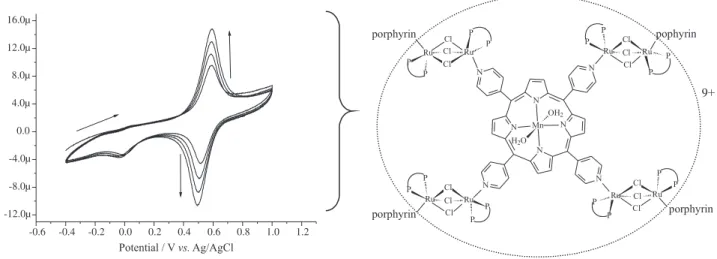
The film was formed from repetitive voltammetric sweeps between the –0.4 V and +1.0 V range, with a scan rate of 0.1 V s–1, using 1.0 × 10⁻⁴ mol L–1
{Mn-TPyP(H2O)2[RuCl3(dppb)]4}PF6, which resulted in
the behavior shown in Figure 2A.
The peak current increased with the number of cycles, which shows a typical behavior of the species being adsorbed on the surface of the electrode.
The mechanism of the electropolymerization has already been reported in the literature and involves the reduction of “RuCl3(dppb)” moiety (RuIII→RuII) at 0 V,
with the formation of a mixed binuclear valence complex (Figure 2B) (RuII/RuIII) at E
1/2 = 0.55 V.44 The film thickness
can be controlled by the number of voltammetric cycles and films formed with a very high number of cycles are very thick and have a passive electrode. Table 1 shows the behavior of the ESPE in the acetaminophen detection with different layers (between 1-7 voltammetric cycles). It is possible to observe the maximum Iap and minimum
Eap when the modified electrode was obtained with four
cycles. After that the voltammetric cycles does not improve those electrochemical parameters. For that reason, four voltammetric cycles were used to make the film on the voltammetric sensor to be used in the next measurements.
The film was also characterized by Atomic Force Microscopy (AFM) measurement (Figure 3) and its thickness can be estimated as thick as 4.5 nm, but unfortunately its thickness was not determined. However
useful information may still be gleaned from it, especially the roughness of the film (1.16 nm in a 100 µm2 area).
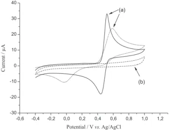
Electrochemical behavior of acetaminophen
Figure 4a shows the electrochemical behavior of acetaminophen using the modified and unmodified electrode. It was possible to observe in the modified electrode
Figure 2. (A) Repetitive voltammograms (4 cycles) of {Mn-TPyP(H2O)2[RuCl3(dppb)]4}PF6 and (B) electropolymerized supramolecular tetraruthenated
porphyrins, 0.1mmol L–1 {Mn-TPyP[Ru(dppb)]
4(µCl3)2}2n(4n2+1)+, 0.1 mol L–1 TBAH, CH2Cl2, glassy carbon as working electrode, scan rate = 100 mV s–1.
Table 1. Iap and Eap of the acetaminophen at a constant concentration
(1.2 × 10⁻⁴ mol L–1) using ESPE with different layers. The sodium acetate
solution was used as the support electrolyte and at a scan rate = 100 mV s–1
Nº of voltammetric cycles Iap / µA Eap / mV
1 12.1 420
2 12.4 400
3 11.7 408
4 16.6 394
5 14.6 403
6 15.5 403
7 12.4 426
Figure 3. AFM image of a {Mn-TPyP[Ru(dppb)]4(µCl3)2}2n(4n2+1)+ film
response a well defined oxidation peak of acetaminophen, with higher current and also a shift of the cathodic potential from 589 to 518 mV. Also the electrochemistry process seems to be more reversible (∆Ep = 80 mV) compared to the bare carbon glassy electrode (CG). As can be seen in Figure 4a the acetaminophen oxidation process associated is quasi reversible on the modified electrode, differently of the process showed by the bare glassy carbon electrode, showing that kinetically the oxidation processes on the electrode surfaces are different. Figure 4b shows the electrochemical behavior at ESPE before addition of acetaminophen in the electrolyte solution. It is possible to observe that the modified electrode does not show any electrochemical process in the electrolyte acetate buffer solution between –0.4 to 1.0 V.
The variation of peak current with scan rate, from 20 to 500 mV s–1, was investigated using 7.05 × 10⁻⁴ mol L–1
acetaminophen in 0.1 mol L–1 acetate buffer solution
pH 4.75 (Figure S1 in the Supplementary Information section). The results showed that anodic peak currents change linearly with the square root of the scan rate (ν1/2) for acetaminophen which indicates a
diffusion-controlled process for electrooxidation of acetaminophen on the surface of the EPSE according to the following equation:52
Iap (µA) = 1.37258 + 2.7899v1/2 (mV s–1)1/2, (R2 = 0.999)
pH dependence study
The electrochemical behavior of acetaminophen was studied in sodium acetate as a function of pH as shown in
Figure 5. It was observed (Figure 5A) that the oxidation potential (Eap) of acetaminophen decreased as the pH
increased. This behavior indicates that the acetaminophen is hydrolyzed in alkaline medium which brought more reducing compounds such as p-hydroxyaniline.52 The
dependence of the anodic peak potential (Eap) with pH can
be described by the follow equation:
Eap (mV) = –56.18 pH + 883.55, (R = 0.999)
The slope of −56.18 mV pH–1 was obtained in these
experiments, which is very close to the theoretical Nernstian value of –59 mV for electrochemical processes involving the same number of protons and electrons. 52At
pH higher than 9.0 the oxidation become kinetically less favorable. This may be explained by the partial formation of the phenoxide, which is negatively charged and for that reason is preferentially attracted to the positively polarized electrode surface.28,53 Figure 5B shows the effect of pH on
the peak current of acetaminophen, which the value of Iap
increases with the increase pH, reaching a maximum at pH 4.75, and then decreases at alkaline pH. Therefore, pH 4.75 was selected for further studies.
Figure 4. (a) Cyclic voltammograms of 0.25 mmol L–1 acetaminophen in
0.1 mol L–1 acetate buffer solution (pH 4.75) at the ESPE (solid line); GC
electrode (dot line). (b) Cyclic voltammogram at the ESPE in 0.1 mol L–1
acetate buffer solution (pH 4.75).
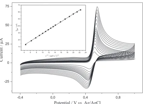
Determination of acetaminophen
Figure 6 shows cyclic voltammograms obtained from the increasing additions of acetaminophen in 0.1 mol L–1
acetate buffer solution (pH 4.75) at scan a rate of 0.10 V s–1
using the ESPE. A linear relationship was found between the anodic peak current and the acetaminophen concentration. Peak currents as a function of concentration in the range of 50 to 700 µmol L–1 is shown in inset of Figure 6 for which
was obtained a regression equation of Iap (µA) = 0.47 +
0.0416 C (µmol L–1) (R2 = 0999). The estimated detection
limit was 5.32 µmol L–1 (three times the blank standard
deviation/slope). The relative standard deviation (RSDs) of 0.17% and 0.86 % for 10 measurements of 50 µmol L–1
and 700 µmol L–1 acetaminophen, respectively, suggested
that the ESPE has a high level of reproducibility.
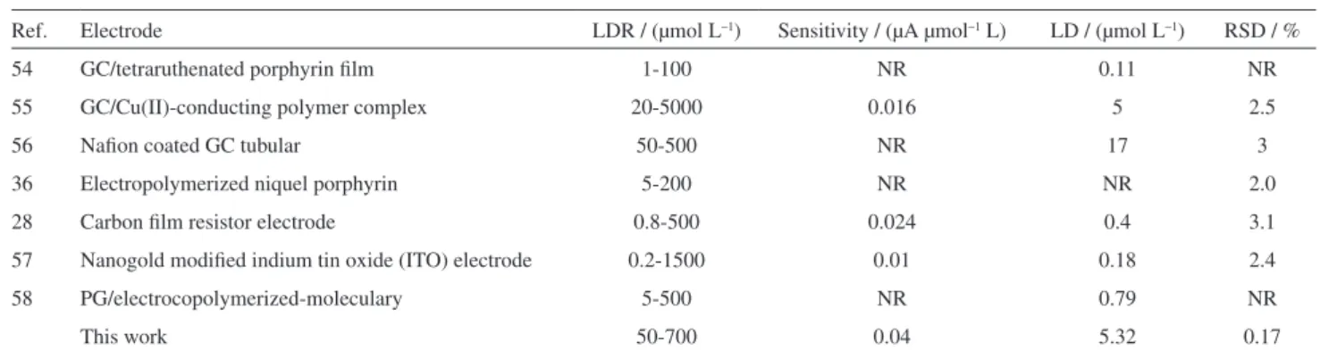
The linear dynamic range (LDR), sensitivity, detection limit (LD) and relative standard deviation (RSD) of the proposed method were compared with other systems
for determination of acetaminophen (Table 2). The results of ESPE show that the proposed method can be efficiently used for the identification and determination of acetaminophen by cyclic voltammetric. The RSD value for the present method suggests that the ESPE has a high level of reproducibility.
Application: acetaminophen detection in real samples
The modified electrode was used for the detection of acetaminophen in commercial drugs by cyclic voltammetry. A standard calibration curve was obtained with a commercial drug containing acetaminophen, and with the information contained on the bottle, a solution was prepared supposedly with 42.3 mmol L–1. From the
regression equation, obtained by the standard addition method, the concentration of acetaminophen was found to be 40.45 ± 1.86 mmol L–1. The detection range obtained
by t-test with a confidence interval of 95% varies from 35.83 to 45.08 mmol L–1. Therefore, the result found for
the sample shows that the modified electrode is efficient for the quantitative determination of acetaminophen.
The voltammetric results obtained with the new sensor were compared with those from the HPLC method. Five different concentrations of standard acetaminophen solutions (132.3; 264.6; 396.9; 529.2 and 661.6 µmol L–1)
were analyzed by reversed-phase HPLC in order to plot the analytical curve, Aacetaminophen peak (a.u.) = 0.18 Cacetaminophen
(µmol L–1) + 13.27, R2 = 0.98. The determination of
acetaminophen in commercial drugs was performed by the injection, in triplicate, of an aqueous solution (1:100, v/v) previously prepared from commercial drug. The concentration of acetaminophen, determined by HPLC, was of (40.57 ± 1.31) mmol L–1. At a 95% confidence level, there
was no significant difference in accuracy (evaluated by the Student t-value, confidence interval 37.32-43.82 mmol L–1)
and also no significant difference in precision (evaluated by
Table 2. Comparison of voltammetry methods for determination of acetaminophen
Ref. Electrode LDR / (µmol L–1) Sensitivity / (µA µmol–1 L) LD / (µmol L–1) RSD / %
54 GC/tetraruthenated porphyrin film 1-100 NR 0.11 NR
55 GC/Cu(II)-conducting polymer complex 20-5000 0.016 5 2.5
56 Nafion coated GC tubular 50-500 NR 17 3
36 Electropolymerized niquel porphyrin 5-200 NR NR 2.0
28 Carbon film resistor electrode 0.8-500 0.024 0.4 3.1
57 Nanogold modified indium tin oxide (ITO) electrode 0.2-1500 0.01 0.18 2.4
58 PG/electrocopolymerized-moleculary 5-500 NR 0.79 NR
This work 50-700 0.04 5.32 0.17
LDR: Linear dynamic range; LD: Detection limit; RSD: Relative standard deviation; NR: Not Reported.
Figure 6. Cyclic voltammograms of acetaminophen as a function of concentration in the range of 50-700 µmol L–1 in 0.1 mol L–1 acetate
the variance ratio F-value) between the cyclic voltammetry method and the HPLC method.
Conclusions
T h i s w o r k r e p o r t s o n t h e s y n t h e s i s a n d characterization of a new supramolecule containing manganese (III) and peripheral ruthenium (III) complexes {Mn-TPyP(H2O)2[RuCl3(dppb)]4}PF6. This polymetalated
porphyrin was electropolymerized in a glassy carbon electrode by the cyclic voltammetry and subsequently used as a voltammetric sensor for detection and quantification of acetaminophen. The sensor presented high sensitivity and stability. The film was characterized by Atomic Force Microscopy, which revealed a thin film in the indium tin oxide surface (1.16 nm in 100 µm2 of area). When
the modified electrode was used for the detection of acetaminophen in real samples, satisfactory results were obtained compared to other methods such as HPLC.
Supplementary Information
Supplementary information (Figure S1) is available free of charge at http://jbcs.sbq.org.br as a PDF file
Acknowledgements
The authors gratefully acknowledge the LERMAC (Laboratório de Energias Renováveis, Materiais e Catálise) and LCI (Laboratório de compostos Inorgâcicos FACIP/ UFU), the FAPEMIG, FINEP (ctinfra 03/2007) and Rede Mineira de Química for financial support and Professor Jeosadaque José de Sene for English review.
References
1. Li, J.; Tang, T.; Li, F.; Li, M.; Dyes Pigments2008, 77, 395. 2. Swartz, H. M.; Clarkson, R. B.; Phys. Med. Biol.1998, 43,
1957.
3. Zheng, W.; Shan, N.; Xu, L.; Wang, X.; Dyes Pigments2008,
77, 153.
4. MacDonald, I. J.; Dougherty, T. J.; J. Porphyr. Phthalocya.
2001, 5, 105.
5. Dudkowiak, A.; Teslak, E.; Habdas, J.; J. Mol. Struct.2006,
792-793, 93.
6. Robert, A.; Benoit-Vical, F.; Dechy-Cabaret, O.; Meunier, B.;
Pure Appl. Chem.2001, 73, 1173.
7. Crossley, M. J.; Burn, P. L.; J. Chem. Soc. Chem. Commun. 1991, 21, 1569.
8. Ishida, T.; Morisaki, Y.; Chujo, Y.; Tetrahedron Lett.2006, 47, 5265.
9. Wrobel, D.; Lukasiewicz, J.; Goc, J.; Waszkowiak, A.; Ion, R.;
J. Mol. Struct. 2000, 555, 407.
10. Takechi, K.; Shiga, T.; Motohiro, T.; Akiyama, T.; Yamada, S.; Nakayama, H.; Kohama, K.; Solar Cells2006, 90, 1322. 11. Barbosa Neto, N. M.; De Boni, L.; Mendonça, C. R.;
Misoguti, L.; Queiroz, S. L.; Dinelli, L. R.; Batista, A. A.; Zílio, S. C.; J. Phys. Chem. B2005, 109, 17340.
12. Barbosa Neto, N. M.; Oliveira, S. L.; Misoguti, L.; Mendonça, C. R.; Gonçalves, P. J.; Borissevitch, I. E.; Dinelli, L. R.; Romualdo, L. L.; Batista, A. A.; Zílio, S. C.; J. Appl. Phys. 2006, 99, 123103.
13. Pavinatto, F. J.; Gameiro Jr., A. F.; Hidalgo, A. A.; Dinelli, L. R.; Romualdo, L. L.; Batista, A. A.; Barbosa Neto, N. M.; Ferreira, M.; Oliveira Jr., O. N.; Appl. Surf. Sci.2008, 254, 5946. 14. Ferreira, M.; Dinelli, L. R.; Wohnrath, K.; Batista, A. A.;
Oliveira Jr., O. N.; Thin Solid Films2004, 446, 301. 15. Yang, L.; Lifeng, C.; He, T.; J. Org. Chem.2006, 71, 8279. 16. Biesaga, M.; Pyrzynska, K.; Trojanowicz, M.; Talanta2000,
51, 209.
17. Itagaki, Y.; Deki, K.; Nagashima, S. I.; Sadaoka, Y.; Sens. Actuators, B2006, 117, 302.
18. Di Marco, G.; Lanza, M.; Sens. Actuators, B2000, 63, 42. 19. Dobson, D. J.; Saini, S.; Anal. Chem.1997, 69, 3532. 20. Guerra, S. V.; Xavier, C. R.; Nagagaki, S.; Kubota, L. T.;
Electroanalysis1998, 10, 462.
21. Kilian, K.; Pyrzynska, K.; Talanta 2003, 60, 669. 22. Tabata, M.; Morito, H.; Talanta1997, 44, 151.
23. Kitamura, Y.; Kawata, K.; Tanaka, K.; Furuyasshiki, Y.; Mifune, M.; Tsukamoto, I.; Saito, M.; Haginaka, J.; Saito, Y.;
Talanta2006, 69, 1260.
24. Hu, Q. F.; Yang, G. Y.; Yin, J. Y.; Yao, Y.; Talanta2002, 57, 751. 25. Deyl, Z.; Mitsik, I.; Eckhardt, A.; Kasicka, V.; Kral, V.; Curr.
Anal. Chem.2005, 1, 103.
26. Bosch, M. E.; Sánchez, A. J. R.; Rojas, F. S.; Ojeda, C. B.;
J. Pharmaceut. Biomed.2006, 42, 291.
27. Babaei, A.; Afrasiabi, M.; Mirzakhani, S.; Taheri, A. R.; J. Braz. Chem. Soc.2011, 22, 344.
28. Felix, F. S.; Brett, C. M. A.; Angnes, L.; J. Pharmaceut. Biomed. 2007, 43, 1622.
29. Habibi, B.; Jahanbakhshi, M. ; Pournaghi-Azar, M. H.; Anal. Biochem.2011, 411, 167.
30. Kachoosangi, R. T.; Wildgoose, G. G.; Compton, R. G.; Anal. Chim. Acta2008, 618, 54.
31. Song, J.; Yang, J.; Zeng, J.; Tan, J.; Zhang, L.; Sens. Actuators, B2011, 155, 220.
32. Özcan, A. ; Şahin, Y.; Anal. Chim. Acta2011, 685, 9. 33. Lourenção, B. C.; Medeiros, R. A.; Rocha-Filho, R. C.; Mazo,
L. H. ; Fatibello-Filho, O.; Talanta2009, 78, 748.
34. Ghorbani-Bidkorbeha, F.; Shahrokhiana, S.; Mohammadi, A.; Dinarvand, R.; Electrochim. Acta2010, 55, 2752.
36. Huang, S. S.; Tang, H.; Li, B. F.; Mikrochim. Acta. 1998, 128, 37.
37. Angnes, L.; Azevedo, C. M. N.; Araki, K.; Toma, H. E.; Anal.
Chim. Acta1996, 329, 91.
38. Knoll, J.; Swavey, S.; Inorg. Chim. Acta2009, 2989.
39. Calfumán, K.; Aguirre, M. J.; Villagra, D.; Yañez, D.; Arévalo, C.; Matsuhiro, B.; Mendoza, L.; Isaacs, M.; J. Solid
State Electrochem2010, 14, 1065.
40. Trofimova, N. S.; Safronov, A. Y.; Ikeda, O.; Electrochim. Acta
2005, 50, 4637.
41. Durantini, J.; Otero, L.; Funes, M.; Durantini, E. N.; Fungo, F.; Gervaldo, M.; Electrochim. Acta 2011, 56, 4126.
42. Gómez-Caballero, A.; Ugarte, A.; Sánchez-Ortega, A.; Unceta, N.; Goicolea, M. A.; Barrio, R. J.; J. Electroanal. Chem. 2010, 638, 246.
43. Steiger, B.; Shi, C.; Anson, F. C.; Inorg. Chem.1993, 32, 2107. 44. Dinelli, L. R.; Poelhsitz, G. V.; Castellano, E. E.; Ellena, J.;
Galembeck, S. E. ; Batista, A. A.; Inorg. Chem.2009, 48, 4692. 45. Carlin, R. L.; Magnetochemistry; Springer-Verlag: Berlin, 1986. 46. Dinelli, L. R.; Batista, A. A.; Wohnrath, K.; Araujo, M. P.;
Queiroz, S. L.; Bonfadini, M. R.; Oliva, G.; Nascimento, O. P.; Cyr, P. W.; Macfarlane, K. S., James, B. R.; Inorg. Chem.1999,
38, 5341.
47. Barbosa, M. I. F.; dos Santos, E. R.; Graminha, A. E.; Bogado, A. L.; Teixeira, L. R.; Beraldo, H.; Trevisan, M. T. S.; Ellena, J.; Castellano, E. E.; Rodrigues, B. L.; de Araujo, M. P.; Batista, A. A.; Polyhedron2011, 30, 41.
48. Fleischer, E. B.; Inorg. Chem. 1962, 3, 493.
49. Fagadar-Cosma, E.; Mirica, M. C.; Balcu, I.; Bucovicean, C.; Cretu, C.; Armeanu, I.; Fagadar-Cosma, G.; Molecules2009,
14, 1370.
50. Horvátha, O.; Valicsek, Z.; Harrach, G.; Lendvay, G.; Fodor, M. A.; Coordin. Chem. Rev.2012, 256, 1531.
51. Romualdo, L. L.; Bogado, A. L.; Valle, E. M. A.; Moreira, I. S.; Ellena, J.; Castellano, E. E.; de Araujo, M. P.; Batista, A. A.;
Polyhedron2008, 27. 53.
52. Xu, Z.; Yue, Q.; Zhuang, Z.; Xiao, D.; Microchim. Acta2009,
164, 387.
53. Wangfuengkanagul, N.; Chailapakul, O.; J. Pharmaceut. Biomed.2002, 28, 841.
54. Quintino, M. S. M.; Araki, K.; Toma, H. E.; Angnes, L.;
Electroanalysis 2002, 14, 1629.
55. Boopathi, M.; Won, M. S.; Shim, Y. B.; Anal. Chim. Acta 2004,
512, 191.
56. Silva, M. L. S.; Garcia, M. B. Q.; Lima, J. L. F. C.; Barrado, E.;
Anal. Chim. Acta 2006, 573, 383.
57. Goyal, R. N.; Gupta, V. K.; Oyama, M.; Bachheti, N.;
Electrochem. Commun. 2005, 7, 803.
58. Ozcan, L.; Sahin, Y.; Sens. Actuators, B 2007, 127, 362.
Submitted: June 7, 2013
Published online: September 11, 2013
Supplementary Information
0103 - 5053 $6.00+0.00S
I
*e-mail: [email protected]
Electropolymerized Supramolecular Tetraruthenated Porphyrins Applied as a
Voltammetric Sensor
Monize M. da Silva,a Gabriel H. Ribeiro,a Alzir A. Batista,b Anizio M. de Faria,a
André L. Bogadoa and Luis R. Dinelli*,a
aFaculdade de Ciências Integradas do Pontal, Universidade Federal de Uberlândia, Rua Vinte, 1600, 38304-402 Ituiutaba-MG, Brazil
bDepartamento de Química, Universidade Federal de São Carlos, Rodovia Washington Luís (SP 310), km 235, 13565-905 São Carlos-SP, Brazil
Figure S1. CVs of 7.05 × 10⁻⁴ mol L–1 acetaminophen in 0.1 mol L–1 acetate buffer solution (pH 4.75) at the ESPE with different scan rates. The inset
![Figure 1. Structure of {Mn-TPyP(H 2 O) 2 [RuCl 3 (dppb)] 4 }PF 6 (P–P = dppb).](https://thumb-eu.123doks.com/thumbv2/123dok_br/18996988.462514/4.892.484.822.556.845/figure-structure-mn-tpyp-rucl-dppb-pf-dppb.webp)