Revista Brasileira de Fisioterapia
Electromyographic activity of vastus medialis
obliquus and vastus lateralis muscles
during functional activities in subjects with
patellofemoral pain syndrome
Atividade eletromiográfica do vasto medial oblíquo e vasto lateral durante
atividades funcionais em sujeitos com síndrome da dor patelofemural
Santos EP, Bessa SNF, Lins CAA, Marinho AMF, Silva KMP, Brasileiro JS
Abstract
Objective: The purpose of this study was to evaluate the amplitude and onset of electrical activation of the vastus medialis obliquus (VMO), vastus lateralis longus (VLL) and vastus lateralis obliquus (VLO) during functional activities in individuals with patellofemoral pain syndrome (PFPS). Methods: Twenty women participated in the study: ten in a control group and ten in the group with PFPS. The electromyographic signal of the quadriceps muscle was detected using simple active differential surface electrodes and a four-channel electromyography system, during open kinetic chain activities (using an isokinetic dynamometer) and closed kinetic chain activities (step and squat maneuvers). For the statistical analysis, Student’s t test and ANOVA with Tukey’s post-hoc method were used, with a significance level of p≤0.05. Results: The results suggested that the electrical activation of the VMO was less intense than in the VLO (p=0.04) and that there was greater delay in VMO onset (p=0.0023) in the group with PFPS, with regard to all of the functional activities evaluated. There was a significant difference between the VMO and VLO in relation to the activities of isokinetic extension at 30º/s (p=0.042) and step down with knee flexion at 75º (p=0.038) in the group with SDPF, and in the activities of rising from a bench (p=0.041), single-leg hop (p=0.046) and heel raising (p=0.004) in the control group. Conclusions:Under the experimental conditions used, this study suggests that there is an imbalance in the electric activity and abnormal recruitment patterns among the VMO, VLL and VLO muscles in individuals with PFPS, with greater delay and lower amplitude of activation of the VMO in this group.
Key words: electromyography; quadriceps muscle; functional activities; patellofemoral pain syndrome.
Resumo
Objetivo: O propósito deste estudo foi avaliar a amplitude e o tempo de ativação elétrica do vasto medial oblíquo (VMO), vasto lateral longo (VLL) e vasto lateral oblíquo (VLO) durante atividades funcionais em portadoras da síndrome da dor patelofemural (SDPF).
Métodos: Participaram do estudo 20 mulheres, sendo dez do grupo controle e dez do grupo com SDPF. O sinal eletromiográfico do quadríceps foi detectado por eletrodos ativos diferenciais simples de superfície e um eletromiógrafo de quatro canais, durante atividades em cadeia cinética aberta (em um dinamômetro isocinético) e fechada (através de um step e durante o agachamento). Na análise estatística foram utilizados o teste t de student e uma análise de variância (ANOVA), com método pos-hoc de Tukey, com
nível de significância de p≤0,05. Resultados: Os resultados sugerem uma menor intensidade na atividade elétrica do VMO em relação ao VLO (p=0,04) e maior retardo no tempo de ativação do VMO (p=0,0023) no grupo com SDPF considerando todas as atividades avaliadas. Houve diferença significativa do VMO em relação ao VLO nas atividades de extensão isocinética à 30º/s (p=0,042) e descida do step com 75º de flexão de joelho (p=0,038) no grupo com SDPF, e nas atividades de levantar-se de um banco (p=0,041), salto unipodal (p=0,046) e elevação dos calcanhares (p=0,004) no grupo controle. Conclusões:Nas condições experimentais realizadas, o estudo sugere um desequilíbrio na atividade elétrica e um padrão de recrutamento anormal entre os músculos VMO, VLL e VLO em sujeitos com SDPF, com maior retardo e menor amplitude de ativação do VMO neste grupo de sujeitos.
Palavras-chave: eletromiografia; músculo quadríceps; atividades funcionais; síndrome da dor patelofemural.
Received: 27/07/07– Revised: 05/12/07 – Accepted: 12/06/08
Department of Physical Therapy, Health Sciences Center, Universidade Federal do Rio Grande do Norte (UFRN) – Natal (RN), Brazil
Correspondence to: Jamilson Simões Brasileiro, Avenida Jaguarari, 5.250, Bloco A, apto. 801, Candelária, CEP 59065-500, Natal (RN), Brasil, e-mail: [email protected]
Introduction
The knee is considered to be one of the joints that is most injured in sport activities1. Because it is incapable of
dissipating excessive forces, it becomes susceptible to inju-ries and the development of infirmities resulting from the absorption of these forces2. Among the large variety of
pa-thological conditions affecting this joint, the patellofemoral pain syndrome (PFPS) is the most common3. It accounts for
25% of the diagnoses in orthopedic clinics4,5 and 30 to 33%
of the cases within sports medicine and at rehabilitation centers2,6. These disorders involve the extensor mechanism
of the knee and are characterized by diffuse anterior or retropatellar pain. Such pain is commonly reported after performing activities like going up or down stairs, kneeling, squatting and remaining seated for a long time, and during sport activities2,7,8. It affects one in every four individuals9 and
most commonly affects adolescents and young adults aged between 15 and 35 years9,10, especially female athletes3,5,11.
Even though the etiological factors are not clear, some researchers have correlated the appearance of patellofemoral dysfunctions to anatomical and biomechanical abnormalities of the lower limb or soft tissue restrictions (static imbalance), along with knee muscle disorders (dynamic imbalance)3,12.
Among these abnormalities, dynamic imbalance has been stu-died by several authors, who have reported the appearance of abnormal patterns of patellar alignment with changes in the activity of the medial and lateral stabilizers of the patellofemo-ral joint and the vastus medialis obliquus (VMO), vastus late-ralis longus (VLL)13,14 and vastus lateralis obliquus (VLO)2,4,5,15
muscles. Other studies have shown the inluence of hip rota-tion on the recruitment patterns of the VMO and VL (vastus lateralis) muscles16,17.
he electromyographic activity of the VMO and VL among individuals with PFPS has been thoroughly investigated through open kinetic chain (OKC) and closed kinetic chain (CKC) exer-cises. However, few studies have observed the intensity and the time required to achieve muscular activation in functional ac-tivities. Some authors have suggested that there are no spatial (activation amplitude)18-22or temporal alterations (beginning time
of activation)7,9,19-21in the electromyographic femoral quadriceps
muscle (m.) patterns during functional tasks. On the other hand, studies comparing subjects with and without patellofemoral dys-functions have found spatial VMO and/or VL abnormalities in step activities or going up and down steps2,12,23, walking on a treadmill24
or tapping of the patellar tendon25, along with asynchrony in the
muscular activation patterns in step tasks2, step tasks associated
with therapeutic patellar tapping22,26, standing on tiptoes and on
the heels27 and walking on treadmills in the horizontal and
incli-ned surface activities24,28.
Although studies have investigated the myoelectric activity of the femoral quadriceps muscle of subjects with and without PFPS in functional tasks, there is no consensus in the literature regarding changes in the activation patterns of the three por-tions of this muscle. Moreover, there is a scarcity of scientiic studies evaluating the role of the VLO in patellar stabilization.
In this light, the purpose of this study was to investigate the recruitment patterns of the VMO, VLL and VLO in relation to patellar control among individuals with PFPS, analyzing the intensity and start time (onset) of electric activity when func-tional activities are performed.
Materials and methods
Characterization of the study
his study was a randomized blind controlled clinical trial, conducted between December 2006 and April 2007 in the elec-tromyography laboratory of a local hospital.
Subjects
Twenty sedentary women, or who did not practice regular physical activity (over three times a week), took part in the study. he volunteers were divided into two groups, composed of ten clinically asymptomatic individuals (22.4±1.65 years; 51.3±6.77kg; 1.60±0.06m) and ten individuals with a clinical diagnosis of PFPS (24.7±4.35 years; 61.6±12.27kg; 1.66±0.05m). he statistical tests did not reveal any anthropometric diferences between these two groups. Only women were evaluated in this study, in view of the biomechanical diferences between the genders and the greater incidence of this pathological condition among women4. Among
subjects who demonstrated bilateral knee pain, the knee in which the greatest pain was reported was evaluated.
he healthy volunteers were selected from among students attending the physical therapy course at a public university, by means of a physical evaluation consisting of clinical tests. he volunteers with PFPS were recruited from a university hospi-tal and three local rehabilitation centers, and they performed a clinical diagnosis carried out by their orthopedist. For both groups, there was an identifying evaluation and an analysis of whether they were in a condition to take part in the study, in accordance with the inclusion and exclusion criteria.
he subjects in the PFPS group were included if they sho-wed pain in the anterior or retropatellar region of the patellofe-moral joint, reported in at least two of the following functional activities: squatting for a long period of time, going up and/or down stairs, remaining seated for a long time, kneeling, run-ning, isometrically contracting the quadriceps and practicing
sports7. Furthermore, they had to report pain of at least 2 on
the Visual Analog Scale (VAS) in the patellofemoral joint in the week before the evaluation2.
hese subjects did not demonstrate a history of surgery in the knee joint, subluxation or patellar dislodgment, clinical evidence of meniscal injury, acute inlammatory processes, trauma or pain in other evaluated joints of the lower limb, liga-ment instability, pathological condition of the patellar tendon, chondral injuries, osteoarthritis7, use of medications or
pre-vious physical therapy consultations within the last semester. Subjects who did not report any histories of knee pain, di-seases, surgery, trauma or injury of the osteomyoarticular sys-tem of the lower limb evaluated were included in the control group2. he volunteers also had to have indicated a VAS pain
score of zero in relation to the patellofemoral articulation on the evaluation day and during the preceding week2.
All subjects were previously informed of the objectives of the study and were asked to sign an informed consent state-ment, in accordance with the guidelines of resolution 196/96 of the National Health Council. he study had previously been approved by the Research Ethics Committee of the Onofre Lo-pes University Hospital (HUOL, protocol 017/06).
Instruments
For the study, a four-channel conditioner module was used (EMG System do Brasil Ltda), interfaced with a microcom-puter that received the electromyographic signals and iled them, along with the AqDados version 5.0 software for digital signal analysis. To capture the electrical activity of the muscles, simple diferential active surface electrodes were used (EMG System do Brasil Ltda), composed of two rectangular parallel bars of Ag\AgCl. One claw type reference electrode was used to eliminate external interference. he signal gain was 1.000x (20 on the active electrode and 50 on the A/D converter). he signal acquisition rate was 1.000Hz, with a iltering of between 20 and 500Hz. he signal was normalized in accordance with the maximum voluntary isometric contraction (MVIC), with the knee at 60º lexion.
To determine the muscle activity onset, initially the subject was instructed to remain in a state of absolute muscular re-laxation, and the RMS value was taken as the reference. After asking the subject to initiate the exercise, the beginning of muscle activity was found when the reference values exceeded three standard deviations (sd) of the mean baseline value that had been found while the subject was resting. When the values exceeded this limit, the time immediately before this point (in ms) was considered to be the beginning of muscular activity. he duration of data collection was set at seven seconds, from which four seconds were reserved to record the activity levels
while resting. he magnitude of the muscle activity was recor-ded as the area under the linear envelope (the integral) during muscular activity. he linear envelope has been shown to be reliable for determining the muscular activation magnitude during functional activities. he EMG activity magnitude du-ring the activities was calculated as percentages of MVIC.
he knee joint movement amplitude was measured using a universal goniometer, positioned with the ixed arm aligned with the greater trochanter of the femur and the axis in the region of the knee joint interline. An isokinetic dynamometer (Medina£, Spain) was used for the OKC exercises. To simu-late going up and down stairs, a wooden step was used and its height could be regulated at angles of 45 and 75º of knee lexion2. To simulate the activity of getting up from a chair,
a bench of a height that favored a 90º angle of hip and knee lexion was used.
Procedures
Initially, the subjects warmed up on a stationary bicycle for ive minutes with the saddle positioned at the height of the greater trochanter of the femur. Soon afterwards, they perfor-med sustained passive stretching of the hamstrings, leg triceps, thigh adductors and femoral quadriceps: two series of 30 se-conds with an interval of 30 sese-conds.
After these initial procedures, the volunteers were posi-tioned on the isokinetic dynamometer (trunk and hip at 90º lexion) and were stabilized by pelvic and axillary belts, secured using Velcro. he subjects’ skin was duly prepared at the elec-trode locations, involving shaving the area and cleaning using 70% alcohol, with the aim of reducing the tissue impedance. he electrodes were covered with an electrically conducting gel; were positioned over the VMO, VLL and VLO of the lower limb under evaluation; and were attached using adhesive tape. he electrodes for the VMO and VLL were positioned in accor-dance with the Seniam29 standards. For the VLO electrode, an
imaginary line was traced out from the antero-superior iliac spine to the center of the patella. It was attached at the center of the venter of the VLO, approximately 2.2cm from the femur lateral epicondyle, at a 50º inclination4. he reference electrode
was positioned over the medial malleolus of the non-evaluated, contralateral limb. Two velcro bands were placed over the elec-trode region to avoid dislodging them during the activities. he evaluated activities were:
1. Maximum isometric extension on the isokinetic dynamo-meter, with 60º of knee lexion;
2. Isokinetic extension with the equipment adjusted to 30º/s, going from 60 to 0º;
3. Squatting on the limb under evaluation, from standing up to 45º of knee lexion;
4. Going up onto the step with 45º of knee lexion, starting with the limb under evaluation;
5. Going down from the step with 45º of knee lexion, starting with the non-evaluated, contralateral limb;
6. Mounting the step with 75º of knee lexion, starting with the limb under evaluation;
7. Descending from the step with 75º of knee lexion, starting with the non-evaluated, contralateral limb;
8. Standing up from a bench without support, up to a stan-ding position;
9. Unipedal jump, starting from a doorstop (24cm in height) and landing on the ground;
10. Heel elevation from the ground;
11. Maintaining the body vertical while standing on the heels.
The activities were administered in a random sequence that was drawn for each subject. The subjects were familia-rized with all of the tasks and instructed to perform them in a natural manner. Each task was performed once, with a one minute interval between them. The OKC activities followed a standardized verbal command, in which the subjects were instructed to perform a maximum voluntary muscular con-traction. For the other activities, they were asked to start and end the movements upon the investigator’s command. The data gathering for all the activities took place over a five second period.
Statistical analyses
he normalization of the data was investigated through des-criptive statistical procedures using the Shapiro-Wilks test. his analysis was done by means of parametric tests. he diferences between the vastus muscles were assessed using ANOVA with normalized RMS values, followed by Tukey’s test, when indicated. To investigate the onset among the quadriceps muscles, between each of the exercises, the Student’s t-test for independent samples was used. he data were analyzed using the Statistical Package for Social Sciences (SPSS) software (version 14.0). In all of the analyses, a signiicance level of p≤0.05 was used.
Results
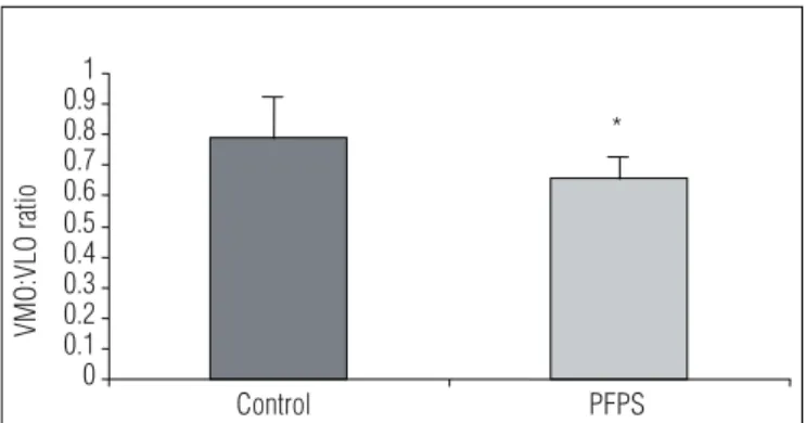
Comparing the groups, the results showed signiicant dife-rences in the ratios of VMO/VLO electric activity, such that the control group demonstrated a mean of 0.79 and the group with PFPS showed a mean of 0.66 (p=0.04). hus, the individuals with PFPS showed decreased VMO intensity in relation to VLO (Figure 1). he data did not show any signiicant diferences in the VMO/VLL ratios between the groups (p≥0.05).
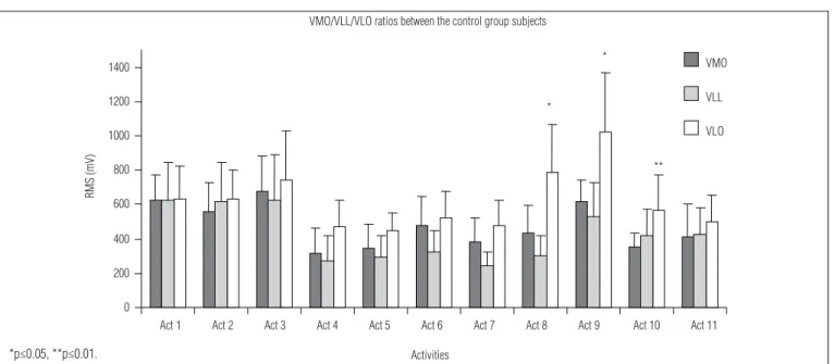
Comparing the muscles in the control group for each acti-vity, the data showed diferences in the VMO/VLO ratios during the activities 8 (p=0.041), 9 (p=0.046) and 10 (p=0.004) (Figure 2). On the other hand, among the individuals with PFPS, the data showed diferences in the activities 2 and 7 (p=0.042 and p=0.038, respectively), (Figure 3). All of these activities demons-trated signiicant diferences with greater activation of VLO.
Considering the subjects in all the functional tasks, the results showed a signiicant diferences in the relative onset of VMO-VLO between the groups (p=0.0023). his reveals a grea-ter delay in the time taken to activate the VMO in relation to the VLO, among individuals with PFPS (Figure 4).
However, no signiicant diferences in the VMO-VLL onset (p≥0.05) were observed in any of the activities, between the two groups. Although no signiicant diferences were found when considering each activity in isolation, the subjects with PFPS showed a delay in VMO onset in relation to VLO, and anticipa-tion of the VLL in most evaluated activities.
Discussion
Electromyographic activity ratios
he results showed a signiicantly lower VMO/VLO ratios in the PFPS group and VMO/VLL ratios that were similar to that of the control group. his suggested diferentiated activation be-tween the VLL and VLO muscles, in subjects with patellofemoral dysfunctions. hese results concurred with the study by Pulzatto2,
who studied the inluences of the activities of going up and down a step with 45 and 75º knee lexions, on the electrical activity of the VMO, VLL and VLO muscles. hey reported lower values for the VMO/VLO ratios and no diferences between the VMO/VLL ratios among individuals with PFPS.
0 0.1 0.2 0.3 0.4 0.5 0.6 0.7 0.8 0.9 1
Control PFPS
*
VMO:VLO ratio
Figure 1. Means and standard deviations of the vastus medialis obliquus (VMO)/ vastus lateralis obliquus (VLO) ratios during the functional activities evaluated in the control and patellofemoral pain syndrome (PFPS) groups.
*decreased intensity in the VMO in relation to the VLO; significantly greater in the PFPS group than in the control group (p≤0.05).
Figure 4. Means and standard deviations of the vastus medialis obliquus (VMO)-vastus lateralis obliquus (VLO) relative onset (millisecs) during the functional activities in the control and patellofemoral pain syndrome (PFPS) groups.
In addition to the absence of activity prioritizing the VMO mus-cle shown in the literature or in this study8, these results suggested
that the electrical activity in the VLO muscle was boosted2,id est,
this muscle participated more actively in patella lateralization among individuals with PFPS. Although some authors disagree with these results3,8,24, this inding demonstrated that signiicant
imbalances of the medial and lateral stabilizers of the patella were associated with individuals with PFPS.
In agreement with the data of the present study, Bevilaqua-Grossi, Monteiro-Pedro and Bérzin4 showed that not only
are VLL and VLO muscles physiologically diferent15, but
also they demonstrated diferences in recruitment patterns in the static knee extension exercises at 15º. Likewise, in an Figure 2. Amplitude of electromyographic activity in the vastus medialis obliquus (VMO), vastus lateralis longus (VLL) and vastus lateralis obliquus (VLO) muscles (mV) during the functional activities performed by the control group.
0 200 400 600 800 1000 1200 1400
Act 2 Act 3 Act 4 Act 5 Act 6 Act 7 Act 8 Act 9 Act 10 Act 11 VMO
VLL
VLO VMO/VLL/VLO ratios between the control group subjects
*
** *
Activities
RMS (mV)
Act 1
Figure 3. Amplitude of electromyographic activity in the vastus medialis obliquus (VMO), vastus lateralis longus (VLL) and vastus lateralis obliquus (VLO) muscles (mV) during the functional activities performed in the patellofemoral pain syndrome (PFPS) group.
0 200 400 600 800 1000 1200 1400
Act 1 Act 2 Act 3 Act 4 Act 5 Act 6 Act 7 Act 8 Act 9 Act 10 Act 11 *
*
RMS (mV)
Activities
VMO/VLL/VLO ratio between the PFPS group subjects
VMO
VLL
VLO
0 0.02 0.04 0.06 0.08 0.1 0.12 0.14 0.16 0.18
Control PFPS
Relative onset (ms)
**
**delay in the activation of the VMO in relation to the VLO in the PFPS group; signifi-cantly greater than in the control group (p≤0.01).
*p≤0.05, **p≤0.01.
*p≤0.05.
1. Gonçalves RS, Pinheiro PJ. Co-activação dos músculos flexores e extensores da articulação do joelho em condições isocinéticas. Rev Port Cien Desp. 2005;2(5):215-23.
2. Pulzatto F. Atividade elétrica dos músculos estabilizadores da patela em indivíduos portadores da síndrome da dor femoropatelar durante exercícios realizados no step. (tese de mestrado). São Carlos (SP): UFSCar; 2005.
3. Gramani-Say K. Atividade elétrica dos estabilizadores dinâmicos da patela no exercício de agachamento associado a diferentes posições do quadril em indivíduos normais e portadores de síndrome de dor femoropatelar. (tese de mestrado). São Carlos (SP): UFSCar; 2005.
electromyographic study on activities of mounting onto a step backwards, Pulzatto et al.12 found greater recruitment of the
VLO than of the VLL as the knee lexed. his corroborated the present indings that during 30% isokinetic extension and eccentric activity on the step at 75º among the subjects with PFPS. According to Owings and Grabiner10, the performance of
eccentric actions may cause changes to the normal myoelec-tric patterns of the quadriceps and contribute towards lateral dislodgement of the patella.
In a similar manner, healthy individuals demonstrated di-ferent activation intensities between the VMO and VLO mus-cles, as observed in the activities of getting up from the bench, unipedal jumping and rising on the heels. To our knowledge, no previous study has evaluated these activities in regards to diferences in activation amplitudes between these muscles. his study suggested that performing these functional tasks may favor patella lateralization and predispose these subjects towards developing patellofemoral disorders.
herefore, the results from this study reinforced the notion that the VLO had an antagonistic function in relation to the VMO, given that in most activities for both groups, the acti-vation observed in the VMO and VLL muscles was no greater than in the VLO. On the other hand, greater activation was observed in the VLO than in the other muscles of subjects with PFPS, thus possibly making this muscle the main agent respon-sible for the patellar dynamic imbalance.
Electrical activation onsets
he results from the present study showed that there was a delay in VMO activation in relation to the VLO in both groups, considering all the activities. he control group demonstrated a mean delay in the VMO of around 4ms in relation to the VLO. By implementing experimental models, Neptune, Wright and Bogert30 showed that a delay of 5ms in VMO muscular activation,
in relation to the VL, resulted in signiicantly greater lateral loa-ding of the patellofemoral joint. his corroborates with what was
found in the present study, in which a delay of 10ms in the VMO, in relation to the VLO, was recorded among individuals with PFPS. According to Souza and Gross23, neuromuscular synchronism is
an important factor in normal movement, thus implying that strength is not the only criterion for determining precise move-ments. herefore, asynchrony in the activation time of the qua-driceps may contribute towards lateral patella contact with the trochlear groove, which suggests that there may be an imbalance in the neuromuscular control in subjects with PFPS.
It must emphasize that, to our knowledge, this has been the only study that observed earlier activation of the VLO in relation to the other portions of the quadriceps, followed by the VMO and then the VLL in the PFPS group. hese results do not corroborate the indings of Santos et al.24, who studied
treadmill walking on a horizontal surface and at an inclination of 5º between subjects with and without PFPS. hey showed that the electrical activity of the VLL preceded the activation of the VMO and VLO in individuals with PFPS. Similarly, Pul-zatto2showed VLL activation preceding VMO and, inally VLO
activation in step activities at 45 and 75º among individuals with PFPS. In addition to these methodological diferences, the divergences of these indings from the present study may have been due to the sample size and the fact that the studies evaluated speciic functional tasks.
Although there is no consensus between the recent studies on PFPS, the present study showed that there were diferences in this group of subjects regarding the amplitude and onset of femoral quadriceps muscle activity while performing functio-nal activities. hus, it is suggested that there is an imbalance of electrical activity and abnormal recruitment patterns between the VMO, VLL and VLO muscles, with greater delays and lower activation amplitudes of the VMO in the group with PFPS. Ho-wever, more studies need to be conducted to investigate possi-ble changes of the three supericial components of the femoral quadriceps muscle in activities that simulate functional tasks, and to investigate the real function of the parts of this muscle in the dynamic behavior of the patella.
4. Bevilaqua-Grossi D, Monteiro-Pedro V, Bérzin F. Análise funcional dos estabilizadores patelares. Acta Ortop Bras. 2004;12(2):99-104.
5. Bevilaqua-Grossi D, Felício LR, Simões R, Coqueiro KRR, Monteiro-Pedro V. Avaliação eletromiográfica dos músculos estabilizadores da patela durante exercício isométrico de agachamento em indivíduos com síndrome da dor femoropatelar. Rev Bras Med Esporte. 2005;11(3):159-63.
6. Bevilaqua-Grossi D, Monteiro-Pedro V, Vasconcelos RA, Arakaki JC, Bérzin F. The effect of hip abduction on the EMG activity of vastus medialis obliquus, vastus lateralis longus and vastus lateralis obliquus in healthy subjects. J Neuroengineering Rehabil. 2006;3(13):3-13.
309
7. Cowan SM, Bennell KL, Hodges PW, Crossley KM, Mcconnel J. Delayed onset of electromyographic activity of vastus lateralis compared with vastus medialis obliquous in subjects with patellofemoral pain syndrome. Arch Phys Med Rehabil. 2001;82(2):183-9.
8. Ribeiro DC, Loss JF, Cañeiro JPT, Lima CS, Martinez FG. Análise eletromiográfica do quadríceps durante a extensão do joelho em diferentes velocidades. Acta Ortop Bras. 2005;13(4):189-93.
9. Witvrouw E, Cambier D, Danneels L, Bellemans J, Werner S, Almqvist F et al. The effect of exercise regimens on reflex response time of the vasti muscles in patients with anterior knee pain: a prospective randomized intervention study. Scan J Med Sci Sports. 2003;13(4):251-8.
10. Owings TM, Grabiner MD. Motor control of the vastus medialis oblique and vastus lateralis muscles is disrupted during eccentric contractions in subjects with patellofemoral pain. Am J Sports Med. 2002;30(4):483-7.
11. Nunes CV, Monteiro-Pedro V. Efeito do exercício isométrico de extensão do joelho associado à adução isométrica do quadril na atividade elétrica dos músculos vasto medial oblíquo e vasto lateral oblíquo em indivíduos com disfunção femoropatelar. Rev Bras Fisioter. 2003;7(2):145-50.
12. Pulzatto F, Gramani-Say K, Siqueira ACB, Santos GM, Bevilaqua-Grossi D, Oliveira AS et al. Influência da altura do step no exercício de subida posterior: estudo eletromiográfico em indivíduos sadios e portadores da síndrome da dor femoropatelar. Acta Ortop Bras. 2005;13(4):168-70.
13. Sacco Ide C, Konno GK, Rojas GB, Arnone AC, Pássaro Ade C, Marques AP et al. Functional and EMG responses to a physical therapy treatment in patellofemoral syndrome patients. J Electromyogr Kinesiol. 2006;16(2):167-74.
14. Tang SF, Chen CK, Hsu R, Chou SW, Hong WH, Lew HL. Vastus medialis obliquus and vastus lateralis activity in open and closed kinetic chain exercises in patients with patellofemoral pain syndrome: an electromyographic study. Arch Phys Med Rehabil. 2001;82(10):1441-5.
15. Bevilaqua-Grossi D, Monteiro-Pedro V, Souza GC, Silva Z, Bérzin F. Contribution to the anatomical study of the oblique portion of the vastus lateralis muscle. Braz J Morphol Sci. 2004;21(1):47-52.
16. Gramani-Say K, Pulzatto F, Santos GM, Vassimon-Barroso V, Oliveira AS, Bevilaqua-Grossi D et al. Efeito da rotação do quadril na síndrome da dor femoropatelar. Rev Bras Fisioter. 2006;10(1):75-81.
17. Serrão FV, Cabral CMN, Bérzin F, Monteiro-Pedro V. Effect of tibia rotation on the electromyographical activity of the vastus medialis oblique and vastus lateralis longus muscles during isometric leg-press. Phy Ther In Sports. 2005;6:15-23.
18. Sheehy P, Burdett RG, Irrgang JJ, VanSwearingen J. An electromyographic study of vastus medialis oblique and vastus lateralis activity while ascending and descending steps. J Orthop Sports Phys Ther. 1998;27(6):423-9.
19. Powers CM, Landel R, Perry J. Timing and intensity of vastus muscle activity during functional activities in subjects with and without patellofemoral pain. Phys Ther. 1996;76(9):946-55.
20. McClinton S, Donatell G, Weir J, Heiderscheit B. Influence of step height on quadriceps onset timing and activation during stair ascent in individuals with patellofemoral pain syndrome. J Orthop Sports Phys Ther. 2007;37(5):239-44.
21. Cowan SM, Hodges PW, Bennell KL. Anticipatory activity of vastus lateralis and vastus medialis obliquus occurs simultaneously in voluntary heel and toe raises. Phy Ther In Sport. 2001;2:71-9.
22. Cowan SM, Hodges PW, Crossley KM, Bennell KL. Patellar taping does not change the amplitude of electromyographic activity of the vasti in a stair stepping task. Br J Sports Med. 2006;40(1):30-4.
23. Souza DR, Gross MT. Comparison of vastus medialis obliquus: vastus lateralis muscle integrated electromyographic ratios between healthy subjects and patients with pain. Phys Ther. 1991;71(4):310-6.
24. Santos GM, Gramani-Say K, Pulzato F, Oliveira AS, Bevilaqua-Grossi D, Monteiro-Pedro V. Relação eletromiográfica integrada dos músculos vasto medial oblíquo e vasto lateral longo na marcha em sujeitos com e sem síndrome de dor femoropatelar. Rev Bras Med Esporte. 2007;13(1):17-21.
25. Christou EA. Patellar taping increases vastus medialis oblique activity in the presence of patellofemoral pain. J Electromyogr Kinesiol. 2004;14(4): 495-504.
26. Cowan SM, Hodges PW, Bennell KL, Crossley KM. Altered vastii recruitment when people with patellofemoral pain syndrome complete a postural task. Arch Phys Med Rehabil. 2002;83(7):989-95.
27. Cowan SM, Bennell KL, Hodges PW, Crossley KM, McConnell J. Simultaneous feedforward recruitment of the vasti in untrained postural tasks can be restored by physical therapy. J Orthop Res. 2003;21(3):553-8.
28. Lange GW, Hintermeister RA, Schlegel T, Dillman CJ, Steadman JR. Electromyographic and kinematic analysis of graded treadmill walking and the implications for knee rehabilitation. J Orthop Sports Phys Ther. 1996;23(5):294-301.
29. Surface electromyography non-invasive assessment of muscles (Seniam), (página da Internet). European Recommendations for Surface Electromyography. Disponível em: http://www.seniam.org/.
30. Neptune RR, Wright IC, van der Bogert AJ. The influence of orthotic devices and vastus medialis strength and timing patellofemoral on loads during running. Clin Biomech. 2000;15(8):611-8.