A
RTIGOC
IENTÍFICO Revista Brasileira de FisioterapiaAnalysis of the reflex response time of the
patellar stabilizer muscles in individuals with
patellofemoral pain syndrome
Análise do tempo de resposta reflexa dos músculos estabilizadores patelares em
indivíduos com síndrome da dor patelofemural
Bevilaqua-Grossi D1, Felicio LR2, Leocádio LP3
Abstract
Objective: To investigate the reflex response time (RRT) of the vastus medialis obliquus (VMO), vastus lateralis obliquus (VLO) and vastus lateralis longus (VLL) muscles in clinically healthy individuals and subjects with patellofemoral pain syndrome (PPS). Methods: Twelve clinically health women and twelve women with PPS were evaluated. Electromyography (EMG) records were obtained using active electrodes connected to an electromyograph that was activated by an external sensor attached to the medial portion of the patella ligament, by means of percussion. The RRT was analyzed by measuring the time, in seconds, between zero and peak electrical response of the VMO, VLO and VLL muscles, for both groups. The statistical analysis consisted of analysis of variance (ANOVA, p< 0.05) and the Tukey post-hoc test (p< 0.05) to compare the response between muscles, and Student’s t test (p< 0.05) to compare the response between groups. Results: Both groups presented lower RRT for the VMO muscle than for the VLO and VLL muscles. However, no significant difference was seen between the VLO and VLL muscles. There was no significant difference in RRT between the groups. Conclusions: According to these results, it can be suggested that the RRTs in the different portions of the quadriceps muscle do not distinguish between subjects with PPS and clinically healthy individuals. The RRT for the VMO muscle was lower than the RRT for the VLO and VLL muscles, for both groups.
Key words: patellofemoral pain syndrome; response time; electromyography.
Resumo
Objetivo: Avaliar o tempo de resposta reflexa (TRR) dos músculos vasto medial oblíquo (VMO), vasto lateral oblíquo (VLO) e vasto
lateral longo (VLL) em indivíduos clinicamente saudáveis e portadores de síndrome da dor patelofemural (SDPF). Métodos: Foram avaliadas 12 mulheres clinicamente saudáveis e 12 mulheres com SDPF. Os registros eletromiográficos foram obtidos por eletrodos ativos simples conectados a um eletromiógrafo, acionados por um sensor externo fixado sobre a porção média do ligamento da patela a partir de sua percussão. A análise do TRR foi realizada por meio da medida do tempo zero ao pico da resposta elétrica dos músculos VMO, VLO e VLL, em segundos, para ambos os grupos. A análise estatística empregada foi o teste de análise de variância (ANOVA, p< 0,05) e teste Tukey post hoc (p< 0,05) para comparação entre os músculos, e o teste t de Student (p< 0,05) para a comparação entre os grupos. Resultados: Ambos os grupos apresentaram um TRR menor para o músculo VMO, quando comparado aos músculos VLO e VLL; entretanto, não se observou diferença significativa entre os músculos VLO e VLL. Na comparação do TRR entre os grupos, não se observou diferença significativa. Conclusões: De acordo com esses resultados, pode-se sugerir que o TRR das porções do músculo quadríceps não diferencia indivíduos com SDFP dos indivíduos clinicamente saudáveis, sendo que o VMO apresenta um TRR menor em relação ao VLO e VLL para ambos os grupos.
Palavras-chave: síndrome da dor patelofemural; tempo de resposta; eletromiografia.
Recebido: 24/01/2007 – Revisado: 13/07/2007 – Aceito: 27/09/2007
1 Department of Biomechanics, Medicine and Rehabilitation of the Locomotor Apparatus, Faculdade de Medicina de Ribeirão Preto, Universdiade de São Paulo, Ribeirão Preto (SP), Brazil 2 Postgraduate in Orthopedics, Traumatology and Rehabilitation of the Locomotor Apparatus Program, FMRP-USP
3 Hospital Orthomed Center, Uberlândia (MG), Brazil
Correspondence to: Débora Bevilaqua-Grossi, Faculdade de Medicina de Riberão Preto, USP, CEP 14049-900, Riberão Preto (SP), Brazil, e-mail: [email protected]
27
Introduction
he patellofemoral pain syndrome (PPS) frequently afects female athletes, the female sedentary population, and young women are most affected1-3. This syndrome is present in
approximately 25% of orthopedic diagnoses4 and is deined as
a pain at the front of the knee and/or rear of the patella4-6. It is
aggravated during physical activities and when going up and down stairs, walking on slopes, squatting and remaining seated for long periods of time7.
Although the etiological factors of PPS are not well-deined, some authors have pointed towards biomechanical abnor-malities in the lower limbs as the main cause1-3. Among the
biomechanical factors most frequently correlated with the development of PPS, dynamic disequilibrium stands out1,2.
Disequilibrium between the medial dynamic stabilizers, vastus medialis obliquus (VMO), vastus lateralis obliquus (VLO) and vastus lateralis longus (VLL) muscles can also cause patellar misalignment, leading to PPS1,8-10. Bevilaqua-Grossi et
al.11 reported that the VLO has an important patellar
stabili-zing function, opposing the VMO. herefore, studies that also evaluate the electrical activity or relex response time of this muscle are necessary.
he equilibrium in neuromuscular activity between VMO, VLO and VLL may be considered to be an important factor during patellar kinematics12. Some studies have analyzed the
beginning of voluntary electrical activity of the median and la-teral stabilizing muscles of the patella in clinically healthy and individuals with PPS, and have demonstrated synchronism of these muscles under diferent functional conditions13.
Howe-ver, other authors have not observed this synchronism when comparing the beginning of the electrical activity of the (VMO) and vastus lateralis (VL) muscles between normal individuals and those with PPS2,7. he beginning of this activity has been
used to evaluate the neuromuscular response time of the pa-tellar stabilizing muscles and also how the efect of carrying out treatment protocols on individuals with PPS inluences this parameter12,14,15.
Voight and Wieder14 evaluated the beginning of the VMO
and VL muscle relex activity in men and women (both clini-cally healthy individuals and individuals with PPS) and found that, for the clinically healthy individuals, the VMO muscle was activated before the VL muscle. On the other hand, for the individuals with PPS, the inverse occurred. Likewise, Witvrow et al.12 suggested that PPS is associated with neuromuscular
control disturbances of the patellar stabilizers. However, these authors did not evaluate the VLO muscle and included both genders in their sample.
he relex response time was also evaluated by Moore et al.16 before and after inducing fatigue protocols. hey reported
that men and women demonstrated diferent relex response times for the quadriceps muscle. herefore, when evaluating the relex response time, the volunteers’ gender must be taken into consideration.
Karst and Willett17 and Powers et al.13 reported that
these time differences in the activation of the VMO and VL muscles are not of great significance and therefore do not influence the patellar kinematics leading to PPS. However, these authors did not evaluate the obliquus portion of the VL muscle. No studies evaluating the neuromuscular con-trol over the VMO, VLO and VLL muscles by means of the reflex response time among clinically healthy and women with PPS were found in the literature consulted. There-fore, the objective of this study was to evaluate the reflex response time of the VMO, VLO and VLL muscles among clinically healthy and individuals with PPS.
Materials and methods
Subjects
Twenty-four sedentary female volunteers who were not doing any physical activities were selected. hey underwent functional evaluations and were divided into two groups: a control group of clinically healthy women (n= 12), with a mean age 22.7 years (±2.25) and mean height of 165 cm; and a group with PPS (n= 12), with a mean age 22.0 (±2.04) years and mean height of 158 cm. he inclusion criteria for the PPS group were: previous reports of pain at the front of the knee during functional activities2; no pain reported for at least the
last two months; and presence of three or more clinical signs and symptoms observed during the functional evaluation18.
he exclusion criteria for the PPS group were: reports of a history of surgery, trauma and osteomyoarticular system injuries to the hip, ankle and foot; use of medications or pre-vious physical therapy treatment less than six months before the time of the present study; and neurological diseases. he study was conducted in accordance with National Health Council Resolution 196/96 and was approved by the Research Ethics Committee of the Centro Universitário do Triângulo (UNITRI) on April 11, 2002. All volunteers signed a free and informed consent statement.
Equipment
28
times. hese was connected to the Myosystem(São Paulo, SP), electromyograph with a 12-bit A/D converter and 100 time ampliication, thus giving a total gain of 2000 times. he common-mode rejection ratio (CMRR) was 93 dB and the acquisition frequency was 2 kHz. A reference electrode of 3 cm2 was connected to the equipment and attached to
the lateral malleolus of the lower limb that was analyzed. A lexible sensor was also attached to the median portion of the patellar ligament, which allowed immediate detection after percussion using the relex hammer.
Procedures
Before placing the electrodes, the area was shaved and anti-septic treatment with 70% alcohol was applied. he electrodes were positioned on the VMO, VLO and VLL muscles with the patient in dorsal decubitus.
On the vastus medialis obliquus (VMO) muscle, the elec-trode was positioned four cm above the superomedial edge of the patella, with an inclination of 55° in relation to the center of the patella and the anterosuperior iliac spine. In relation to the vastus lateralis longus (VLL), the electrode was positioned 15 cm from the superolateral edge of the patella, with an incli-nation of 13.6º. To position the electrode on the vastus lateralis obliquus (VLO), the lateral epicondyle of the femur needed to be located and the beginning and middle of the muscle belly needed to be followed, with an inclination of 50.4º11. he
refe-rence electrode was positioned over the anterior tibial tubero-sity of the limb to be tested (Figure 1).
he volunteers were put in a seated position with the hips at 90° of lexion and the knees supported on the bed with the feet hanging, in accordance with the recommendations from the SENIAM project19. In the seated position, a function test
on the quadriceps muscle was performed to verify the positio-ning of the electrodes. hree percussions were performed on the patellar ligament, at intervals of 30 seconds between per-cussive actions. As the relex hammer touched the sensor, the discharge system instantly picked up the electrical activity of the VMO, VLL and VLO muscles through the electrodes.
he relex response time analysis was done by measuring from time zero to the peak electrical response of the VMO, VLO and VLL muscles, in seconds, through the Myosystem-Br1 software version 2.9 b (Uberlândia, Minas Gerais) (Figure 2). Because the analysis was performed in relation to the relex response time, no bandpass ilter was applied, because this, especially the high pass ilter, could alter the initial relex response time.
Statistical analyses
For the statistical analysis, the means of the three percus-sive actions performed on each volunteer were calculated. To compare the relex response time between the VMO, VLL and VLO muscles in the PPS and control groups, ANOVA and the Tukey post-hoc test were used. To compare the PPS and control groups, Student’s t test for unpaired measurements was used. Both of these tests used a signiicance level of less than 5%.
Results
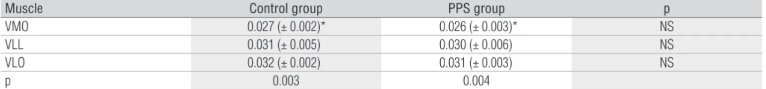
he results revealed that for both the control group and the PPS group there were lower relex response times for the VMO muscle than for the VLL and VLO muscles. However, no sig-niicant diferences were observed between the VLO and VLL muscles. Comparing the control and PPS groups, there were no signiicant diferences in the VMO, VLL and VLO muscles (Table 1).
ASIS
VMO VLL
VLO
Figure 1. Positioning of the electrodes on the vastus medialis obliquus (VMO), vastus lateralis longus (VLL) and vastus lateralis obliquus (VLO) muscles, in accordance with the inclination of each portion in relation to
the anterosuperior iliac spine (ASIS) and the center of the patella (C)11.
Figure 2. Electromyographic activity during percussion of the patellar tendon. Onset of response and latency period (A), rise time (B) and reflex response peak (C).
C
B A 8.0 6.0 4.0 2.0 0.0 -2.0 -4.0
0.01 0.02 0.03 0.04 0.05 0.06 0.07 Microvolts
29
Discussion
he data revealed that, for both the control and the PPS groups, the medial stabilizer (VMO) reached its relex peak ac-tivation earlier than did the lateral stabilizers (VLO and VLL). According to Cowan et al.7, the VMO presents a biomechanical
advantage over the VL due to the oblique orientation of its muscle ibers. However, the neuromuscular response of the VMO may relate to the attempt by this stabilizer to resist the lateral patellar forces, given that the patella tends to lateralize.
Despite the methodological diferences, these data concur with what was found by Crossley et al.20. hose authors found
that the VMO muscle had a shorter activation time than did the VL muscle, among individuals with PPS, although they were in-vestigating the activation time when going up and down a step.
It could be seen when comparing between groups that the relex response time patterns of the patella stabilizer muscles were similar. However, it must be emphasized that the multi-factorial nature of PPS, the lack of validated diagnostic research instruments for improving the nature of the sample, and the number of volunteers may have afected the characterization of the individuals with PPS.
his study is in agreement with the data presented by Voight and Wieder14 and Witvrow et al.12, who reported diferences in
relex response time between the VMO and VL muscles that was less than six milliseconds for the control group. However, these authors demonstrated that, among the individuals with PPS, the VL muscle showed a lower relex response time than did the VMO. hus, these authors suggested that this inversion could have been caused by neuromuscular disequilibrium in the VMO and VL muscles, thereby altering the patellar
Table 1. Means and standard deviations (in seconds) for the reflex response times of the vastus medialis obliquus (VMO), vastus lateralis obliquus (VLO) and vastus lateralis longus (VLL) muscles, for the control and PPS groups.
* Significant difference between VMO muscle and the VLL and VLO muscles: statistical analysis using ANOVA and post-hoc Tukey tests.
Muscle Control group PPS group p
VMO 0.027 (± 0.002)* 0.026 (± 0.003)* NS
VLL 0.031 (± 0.005) 0.030 (± 0.006) NS
VLO 0.032 (± 0.002) 0.031 (± 0.003) NS
p 0.003 0.004
kinematics. However, these authors did not evaluate the obli-quus portion of the VL muscle. he data from the present study do not conirm this airmation, because the present study did not demonstrate lower relex response times for the VLO and VLL muscles, in comparison with the VMO muscle for the PPS and control groups, thus suggesting that relex response time was not a parameter that was able to distinguish between indi-viduals with and without PPS.
Although Karst and Willett17 did not ind any diferences in
the activation times for the VMO and VLL muscles, either under relex conditions or during voluntary contraction, these authors also suggested that alterations in the relex response times of these muscles are not capable of predisposition towards PPS. he results from the present study are in agreement with this, thereby reinforcing the idea that disequilibrium between the medial and lateral portions was not an etiological factor for this syndrome.
However, it must be taken into consideration that the sample evaluated in the present study was asymptomatic, since the pain in individuals with PPS is usually insidious and intermittent at irst21. hus, studies evaluating neuromuscular disequilibrium of
the patellar stabilizers among symptomatic individuals need to be conducted. herefore, under these experimental conditions, alterations in the relex response time cannot be considered to be indicative of PPS, because no diferences between the groups were observed. herefore, relex response time must not be used in phy-siotherapeutic evaluations to characterize individuals with PPS.
herefore, it can be concluded that the relex response time cannot be used as a diferential factor between people with PPS and clinically healthy individuals. For both groups, the relex response times in the VMO muscle were lower than what was observed in the VLO and VLL muscles.
References
1. Tang SFT, Chen CK, Hsu R, Chou SW, Hong WH, Lew HL. Vastus medialis obliquus and vastus lateralis activity in open and closed kinetic chain exercise in patients with patellofemoral pain syndrome: an electromyographic study. Arch Phys Med Rehabil. 2001;82:1441-5.
2. Cowan SM, Bennell KL, Crossley KM, Hodges PW, McConnell J. Physical therapy alters recruitment of the vasti in patellofemoral pain syndrome. Arch Phy Med Rehabil. 2002;34:1879-85.
3. Baker V, Bennell K, Stillman B, Cowan S, Crossley K. Abnormal knee joint position sense in individuals with patellofemoral pain syndrome. J Orthop Res. 2002;20:208-14.
4. Powers CM, Maffucci R, Hampton S. Rearfoot posture in subjects with paellofemoral pain. J Orthop Sports Phys Ther. 1995;22:155-60.
30
6. Bierdert RM, Warnke K. Correlation between the Q angle and the patella position: a clinical and axial tomography evaluation. Arch Orthop Trauma Surg. 2001;121:346-9.
7. Cowan SM, Bennell KL, Hodges PW, Crossley KA, McConnell J. Delayed onset of electromyographic activity of vastus medialis obliquus relative to vastus lateralis in subjects with patellofemoral pai syndrome. Arch Phys Med Rehabil. 2001;82:183-9.
8. McGinty G, Irrgang JJ, Pezzullo D. Biomechanical considerations for rehabilitation of the knee. Clin Biomech. 2000;15:160-6.
9. Callaghan MJ, McCarthy CJ, Oldham JA. Electromyographic fatigue characteristics of the quadriceps in patellofemoral pain syndrome. Man Ther. 2001;6:27-33.
10. Cowan SM, Bennell KL, Hodges PW, Crossley KM, McConnell J. Simultaneous feedforward recruitment of the vasti in untrained postural tasks can be restored by physical therapy. J Orthop Res. 2003;21:553-8.
11. Bevilaqua-Grossi D, Monteiro-Pedro V, Bérzin F. Análise funcional dos estabilizadores da patela. Acta Ortop Bras. 2004;12:99-104.
12. Witvrouw E, Sneyers C, Lysens R, Victor J, Bellemans J. Reflex response times of vastus medialis oblique and vastus lateralis in normal subjects and in subjects with patellofemoral pain syndrome. J Orthop Sports Phys Ther. 1996;24:160-5.
13. Powers CM, Landel R, Perry J. Timing and intensity of vastus muscle activity during functional activities in subjects with and without patellofemoral pain. Phys Ther. 1996;76:946-55.
14. Voight ML, Wieder DL. Comparative reflex response times of vastus medialis obliquus and vastus lateralis in normal subjects and subjects with extensor mechanism dysfunction. Am J Sports Med. 1991;19:131-7.
15. Witvrouw E, Cambier D, Danneels L, Bellemans J, Werner S, Almqvist F, et al. The effect of exercise regimens on reflex response time of the vasti muscles in patients with anterior knee pain: a prospective randomized intervention study. Scand J Med Sci Sports. 2003;13(4):251-8.
16. Moore BD, Drouin J, Gansneder BM, Schultz SJ. The differential effects of fatigue on reflex response timing and amplitude in males and females. J Electromyogr Kinesiol. 2002;12:351-60.
17. Karst GM, Willett GM. Onset timing of Electromyographic activity in the vastus medialis oblique and vastus lateralis muscle in subjects with and without patellofemoral pain syndrome. Phys Ther. 1995;75:813-23.
18. Coqueiro KRR, Bevilaqua-Grossi D, Bérzin F, Soares AB, Candolo C, Monteiro-Pedro V. Analysis on the activation of the VMO and VL muscles during semisquat exercises with and without hip adduction in individuals with patellofemoral pain syndrome. J Electromyogr Kinesiol. 2005;15(6)596-603.
19. Hermes HJ, Freriks B, Stegman D, Block J, Rau G, Disslhorst-Klug, et al. European recommendations for surface electromyography – Results of the SENIAN Project. Netherlands: Roessingh Research and Development; 1999.
20. Crossley KM, Cowan SM, McConnell J, Bennell KL. Physical therapy improves knee flexion during stair ambulation in patellofemoral pain. Med Sci Sports Exerc. 2005;37(2):176-83.