http://dx.doi.org/10.1590/bjpt-rbf.2014.0161 Braz J Phys Ther. 2016 May-June; 20(3):231-239 231
Delayed effect of Kinesio Taping on neuromuscular
performance, balance, and lower limb function in healthy
individuals: a randomized controlled trial
Caio A. A. Lins1, Daniel T. Borges1, Liane B. Macedo1, Karinna S. A. Costa1, Jamilson S. Brasileiro1
ABSTRACT | Background: Kinesio Taping (KT) is an elastic bandage that aims to improve neuromuscular performance,
although there is no consensus as to its beneits. Objective: To analyze the immediate and delayed effects of KT on the
neuromuscular performance of the femoral quadriceps, on balance, and lower limb function in healthy subjects. Method: This is a randomized controlled trial. Thirty-six women with a mean age of 22.2±3.6 years and BMI of 22.5±2.3 Kg/m2 were
divided into three groups: control, with ten minutes of rest (control, n=12), application of Kinesio Taping without tension
(placebo, n=12) and with tension (KT, n=12) on the quadriceps. The primary outcome was isokinetic performance, while secondary outcomes were the single-hop test, one-footed static balance, and electromyographic activity. The evaluations were carried out in ive stages: 1) before application of KT, 2) immediately after the application of KT, 3) after 24h, 4) after 48h, and 5) after 72h. Mixed ANOVA was used to determine differences between groups. Results: There was no
change in one-footed static balance, electromyographic activity of the VL in the lower limb function, nor in isokinetic performance between groups. Conclusion: KT promotes neither immediate nor delayed changes in neuromuscular
performance of the femoral quadriceps in healthy women. Keywords: torque; electromyography; bandages.
Clinical Trials Identiier: NCT02431910.
BULLET POINTS
• The study evaluated immediate and delayed effects of the application of Kinesio Taping. • KT did not change immediate or delayed neuromuscular performance.
• KT application effects do not depend on the time/duration of application.
• The results do not support the hypothesis that the application of KT results in performance improvement.
HOW TO CITE THIS ARTICLE
Lins CAA, Borges DT, Macedo LB, Costa KSA, Brasileiro JS. Delayed effect of Kinesio Taping on neuromuscular performance, balance, and lower limb function in healthy individuals: a randomized controlled trial. Braz J Phys Ther. 2016 May-June; 20(3):231-239 . http://dx.doi.org/10.1590/bjpt-rbf.2014.0161
1 Laboratório de Análise da Performance Neuromuscular (LAPERN), Departamento de Fisioterapia, Universidade Federal do Rio Grande do Norte (UFRN), Natal, RN, Brazil
Received: May 12, 2015 Revised: Sept. 04, 2015 Accepted: Nov. 20, 2015
Introduction
Kinesio Taping (KT) is an elastic bandage developed
by Kenzo Kase. According to its creator, it has speciic
features, ranging from its design to its elongation1, that improve functional performance. In practice, this
technique has been widely used by healthy people in order to prevent injuries and increase neuromuscular
performance, seeking better performance during
physical activities, whether at the professional or amateur level2.
KT consists of a thin elastic tape which can be
stretched up to 50% of its original length, resulting
in lower restriction compared to conventional tapes1,
thereby proposing to increase joint stability and improve muscular performance3. However, the mechanisms by which the application of KT reaches such goals
are not well understood. One such mechanism
would be by increasing muscle activity during the implementation of KT through neurofacilitation, where the tactile stimulation provided by the tape activates cutaneous receptors, thus promoting alpha motor neuron stimulation4. Furthermore, due to its characteristics, the bandage could provide increased interstitial space, promoting better blood and lymph
232 Braz J Phys Ther. 2016 May-June; 20(3):231-239
In this context, the effect of applying KT has been the subject of research to evaluate its inluence on both
balance and function of the lower limbs, as well as on
muscle activation (EMG) and strength (dynamometry) in patients and in healthy people, but with conlicting
results8-15. Recently, a meta-analysis on the effect of KT on increasing muscle strength showed that its implementation does not promote improvement in healthy adults16. Another meta-analysis on the
inluence of KT on the treatment and prevention of
sports injuries showed that this technique has little
beneicial effect on muscle strength, muscle activation,
or active range of motion17. However, the studies
included in both meta-analyses are classiied as being
of moderate methodological quality and only a few
of them found signiicant effects. In addition, the authors make it clear that more research needs to be
conducted, particularly blind randomized controlled
studies that include a placebo group.
Two other systematic reviews investigated the clinical effects of KT and reported that there are
few high-quality studies and therefore insuficient
evidence to support the use of this technique in clinical practice18,19. A study by Słupik et al.20 noted that there was no increase in the electromyographic activity of
the vastus medialis (VM) during isometric contraction of the knee extensors immediately after applying KT to this muscle. Nevertheless, the same study noted an increase in electromyographic activity of the VM at 24 and 72 hours after applying KT and 24 hours after removal of the bandage. These indings raise a
hypothesis of the possible delayed effects of applying KT, suggesting that an adjustment period would be needed in the application technique in order to meet
the expected goals of healthy people. However, the
same study did not use a placebo or control group, in addition to only observing the effect of KT on
one variable.
Thus, there is no consensus in the literature about the real effects of KT, although this technique is
being widely used by healthy people seeking better performance during physical activities. In addition,
few studies have evaluated its chronic effects on neuromuscular performance, both on patients and
on healthy people. Given the above, this study aimed
to analyze the immediate and delayed effects of KT
application on isokinetic knee extensor performance, electromyographic activity of the vastus lateralis (VL), one-footed static balance, and lower limb function for healthy subjects.
Method
Subjects
This is a randomized controlled trial consisting of
36 healthy women with a mean age of 22.2±3.6 years
and body mass index (BMI) of 22.5±2.3 Kg/m2.
They were non-probabilistically recruited and randomly distributed using the website www.randomization. com. Only female subjects were included due to the
large biomechanical differences that occur between
genders. The inclusion criteria were: age between 18 and 28 years; being recreationally active21; hip, knee, and ankle joint integrity; no history of musculoskeletal injury in the last 6 months; no previous surgical
history of their lower limbs; uncorrected neurological,
vestibular, visual, and/or auditory deicits; allergy to the adhesive material. Subjects who incorrectly executed the assessment procedures or missed any evaluations were excluded from the study.
The participants received information about the research objectives and signed a free and informed
consent form, according to Resolution 466/12 of the National Health Council and the Declaration of Helsinki. The study was approved by the Ethics Committee of Universidade Federal do Rio Grande do Norte (UFRN), Natal, RN, Brazil (protocol number 752.302). This study was registered at www.clinicaltrials.gov under registration number NCT02431910.
Procedures
A pilot study was conducted in order to adjust all
the research procedures and to train the researchers
involved. Two evaluators participated in the study:
evaluator 1 was responsible for carrying out the evaluation of all of the subjects, while the second evaluator
was responsible for implementing the intervention. However, due to the presence of a group that did not
apply the bandage, the subjects and evaluator 1 were
not blinded to the intervention performed.
Initially, all of the subjects illed out an evaluation
form with anthropometric data (age, weight, height,
and BMI), personal information, and questions about physical activity frequency. Next, they performed a warm up on a stationary bicycle for ive minutes (ErgoFit Cycle 167, Ergo-Fit, Pirmasens, Germany), with a 15W load at a constant speed of 20 km/h, and
with their seat adjusted to the height of the greater
trochanter of the femur.
After the warm up, the isokinetic performance
evaluation was performed, considered as the primary
233
Braz J Phys Ther. 2016 May-June; 20(3):231-239
balance, lower limb function, and VL electromyographic
activity were also assessed and considered as secondary
outcomes. The evaluations were always conducted using the non-dominant limb, which was set from the subject’s account by asking which leg they use to kick a ball.
The evaluations were performed at ive distinct
time points: before the intervention protocol (pre),
immediately after (post), and 24h, 48h, and 72h after the intervention protocol. The last evaluation (72h) was performed 24h after the removal of KT.
Isokinetic performance evaluation
To carry out this evaluation, the subject was placed in the sitting position in the chair of a computerized
isokinetic dynamometer (Biodex Multi-Joint System 4™, Biodex Medical Systems Inc., Shirley, NY, USA). The dominant thigh was ixed by a strap, as were the pelvis and thorax region. On the non-dominant limb, the dynamometer rotation axis was aligned with the
lateral epicondyle of the femur and the lever arm
was adjusted to the distal region of the leg and ixed at 5 cm above the medial malleolus of the ankle.
The gravity correction factor was carried out by the dynamometer itself, adjusted by the weight of the
relaxed leg at 30° of knee lexion.
The isokinetic performance evaluation was performed by ive concentric knee extension contractions at 60°/s. This evaluation started from 90° lexion up to full extension of the knee and recorded the peak torque normalized by body weight (PT/BW), expressed as percentage and average power. The return to lexed position was done passively.
During the evaluation, verbal encouragement and visual feedback were provided by the computer.
To become familiarized with the equipment, the
subjects performed three submaximal contractions at 60º/s, followed by a 60-second interval until the start of testing.
One-footed static balance evaluation
For this evaluation, subjects were assessed on a computerized baropodometry platform (Eclipse 3000, Guy-Capron SAS, Montchanin, France) with a 40×40 cm surface and acquisition frequency of 20Hz. They were positioned standing on the platform to support the non-dominant limb and with their knee lexed at 20° (considering 0° to be full knee extension), veriied by a universal goniometer. The subject was then instructed to keep their head in a neutral position looking at a ixed point, with their
spine erect and upper limbs supported on their hips.
The dominant lower limb remained with the hip at
0° and the knee at 90° lexion. Data acquisition time was ten seconds. The assessment was repeated three
times, with the average of the two repetitions that
showed the least luctuation being considered for analysis. The rest time was one minute between each
test, and the analyzed variables were the displacement velocity of the pressure center in the anteroposterior
and mediolateral directions.
Lower limb function evaluation
The single-hop test was performed, considered
testing measures of functional performance22. They were instructed to start the hop without the support of the
contralateral limb to avoid impulse movements.
The subjects were encouraged to perform a single hop as far as possible without any type of footwear, and
the hallux-hallux distance was measured using a tape measure. To allow for a comparison of values between
the subjects, the data were normalized as a function of
the height of each subject (hop distance/height × 100).
The test was repeated twice, and the further of
the two measurements was recorded. For the hop
to be considered valid, the subject should remain balanced for two seconds after completing the hop
and the contralateral limb could not touch the ground. One minute of rest was allowed between tests.
Electromyographic activity records
For electromyographic activity analysis of the VL muscle, the skin was shaved and cleansed with 70% alcohol before electrode placement. An 8-channel signal conditioning module with 16-bit resolution (TeleMyo Transmitter, Noraxon Inc., Scottsdale, AZ, USA) was used for signal acquisition and common-mode rejection ratio (CMRR) >100 Db. Signals were captured on a sampling frequency set at 1500 Hz, iltered at a frequency between 10 and 500 Hz and ampliied 1000 times. Signals were captured using passive adhesive surface electrodes (Noraxon Inc.) 4 cm long and 2.2 cm wide, separated by an inter-electrode distance of 2 cm. The electrode was placed on the VL muscle belly, according to recommendations of the SENIAM (Surface Electromyography for the Non-Invasive Assessment of Muscles) project23. The software myoResearch 3.2 (Noraxon Inc.) was used for analysis of the digital signals.
The electromyographic activity recording was
234 Braz J Phys Ther. 2016 May-June; 20(3):231-239
during the concentric evaluation was considered as the electromyographic signal of higher torque from the
ive recorded on the isokinetic dynamometer, being carried out with a 1-s window during contraction for the analysis. Normalization was performed by the RMS peak value during maximal voluntary isometric
contraction, as the subjects were instructed to perform
two knee extension contractions at an angle of 60° lex for 5 seconds, with a 60-second rest interval between them. The contraction that generated the most torque was used for normalization.
Interventions
After the baseline assessment, the subjects were randomly assigned to one of three groups.
The second evaluator applied the protocol according
to randomization: control group (n=12) - remained 10 minutes at rest (time required for applying the
bandages in the other groups); placebo group (n=12)
- application of KT (kinesio tex Gold) to the femoral quadriceps (FQ) muscle without tension; and KT group (n=12) - application of KT on the FQ muscle with tension.
Subjects from the KT group were submitted to KT application on the FQ of the non-dominant limb as suggested by Kase et al.1 to increase muscle performance. Thus, the bandage was applied to the rectus femoris (RF), VL, and VM longitudinally, from proximal to distal. For the RF muscle, the proximal anchor was applied 5 cm below the anterior
superior iliac spine and the distal anchor was placed
on the upper edge of the patella. For the VL muscle, the proximal and distal anchors were placed on the
greater trochanter of the femur and on the lateral edge
of the patella, respectively. As for the VM muscle, the proximal anchor was placed on the middle third
of the medial thigh region and the distal anchor on
the medial edge of the patella. For the three muscles in question, the anchors were applied with 0%
tension and the therapeutic area (area between the anchors) was followed on the belly of muscles with
50% tension, in order to promote greater muscle
activation1. This application was carried out with the subjects standing on one foot, with the hip of
the non-dominant limb at 0° and the knee lexed, as suggested by Lins et al.14, keeping the muscle in a
stretched position. For the placebo group, the same protocol was followed, except that the application of the bandage was maintained at 0% tension on the anchor and also in the treatment zone.
Statistical analysis
Based on initial values obtained from a pilot study conducted with 15 subjects, a sample of 36 subjects with 12 in each group was adequate to detect a clinically signiicant difference of 12.0%
between groups, assuming a standard deviation of
41.0 for the PT/BW outcome during the concentric contraction. A statistical power of 80%, an alpha of 5%, and a loss rate of 10% were considered for the sample calculation. The sample size calculation was performed for the ANOVA repeated measures statistical test with interactions between groups. The software Gpower3.1 was used for the calculation.
Statistical analysis was performed using the Statistical Package for the Social Sciences software (SPSS) version 20.0. The normal distribution of data and homogeneity of variance were veriied by the Kolmogorov-Smirnov (KS) and Levene tests, respectively. Estimates of
average effect (differences between groups) for all
variables were calculated using the ANOVA mixed model. This analysis model incorporated the intervention groups (control, placebo, and kinesio taping), time (pre, post, 24h, 48h, and 72h), and the group × time interaction. When a signiicant F value was found, the Bonferroni post-hoc test was applied in order to identify the differences. A signiicance level of 5% was adopted for all statistical analyses (P<0.05), which were conducted by an independent researcher.
Results
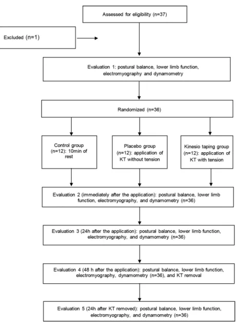
One subject was excluded from the study because
she felt pain at the time of initial evaluation (Figure 1). Table 1 shows the homogeneity for the analyzed
variables between the groups at baseline.
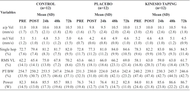
Table 2 shows the mean values and standard
deviation of the analyzed variables at the ive time points of evaluation (pre, post, 24h, 48h, and 72h) for the three groups.
Table 3 presents the analysis between groups for
the comparisons post and 24h after the intervention,
while Table 4 also shows the analysis between groups,
235 Braz J Phys Ther. 2016 May-June; 20(3):231-239
Table 1. Mean values and standard deviation (SD) of age, height, body mass index (BMI), anteroposterior velocity (A/P VEL), mediolateral
velocity (M/L VEL), single hop, RMS of VL muscle (RMS VL), peak torque normalized by body weight (PT/BW), and average power of all groups evaluated at baseline.
VARIABLES CONTROL PLACEBO KINESIO TAPING
n=12 n=12 n=12
Age (years) 21.4 (3.6) 22.3 (3.8) 23.3 (3.1)
Height (m) 1.63 (0.06) 1.64 (0.03) 1.65 (0.07)
BMI (Kg/m2) 22.3 (2.4) 22.7 (2.3) 22.5 (2.3)
A/P VEL (mm/s) 11.1 (1.6) 10.2 (1.6) 11.5 (3.0)
M/L VEL (mm/s) 5.1 (1.2) 4.6 (0.7) 5.2 (1.0)
Single Hop (%) 72.7 (7.6) 72.8 (11.7) 76.2 (9.6)
RMS VL (%) 62.2 (14.1) 63.6 (23.5) 58.2 (16.8)
PT/BW (%) 254.7 (33.9) 231.3 (32.3) 240.2 (47.4)
Power (W) 82.3 (14.5) 76.4 (19.4) 84.1 (24.4)
236 Braz J Phys Ther. 2016 May-June; 20(3):231-239
Discussion
This study aimed to evaluate the immediate and delayed effects of KT application on the neuromuscular
knee extension performance in one-footed static balance and lower limb function of healthy subjects.
The results indicated that the application of KT does not promote immediate or delayed changes to
displacement velocity of the pressure center in the anteroposterior or mediolateral directions, the distance of the single hop, the electromyographic amplitude
of VL, the normalized peak torque or the average power of knee extensors.
Corroborating the results of this study, Nunes et al.24
evaluated the effects of applying KT to the sural triceps
on vertical jump, drop jump, and single-leg stance in Table 2. Mean values and standard deviation (SD) of the variables: anteroposterior velocity (a/p vel), mediolateral velocity (m/l vel),
single hop, RMS of VL muscle (RMS VL), peak torque normalized by body weight (PT/BW) and average power, in ive stages of evaluation (pre, post, 24h, 48h, and 72h), of all groups.
Variables
CONTROL (n=12)
PLACEBO (n=12)
KINESIO TAPING (n=12)
Mean (SD) Mean (SD) Mean (SD)
PRE POST 24h 48h 72h PRE POST 24h 48h 72h PRE POST 24h 48h 72h
a/p Vel
(mm/s) (1.7)11.0 (1.7)10.8 (2.1)10.6 (1.8)10.8 (2.8)10.5 (1.6)10.1 (1.7)9.8 (2.4)9.5 (2.0)10.5 (2.4)10.0 (3.0)11.5 (2.8)10.0 (2.6)10.1 (2.8)10.5 (1.8)9.6 m/l Vel
(mm/s) (1.2)5.1 (1.0)5.1 (1.1)4.8 (1.2)5.3 (1.5)5.0 (0.7)4.6 (0.6)4.2 (0.8)4.4 (0.8)4.9 (1.0)4.6 (1.0)5.2 (1.0)4.6 (1.0)4.8 (1.2)5.1 (0.9)4.5 Single hop
(%) (7.6)72.7 (7.4)79.4 (8.3)81.2 (7.5)81.7 (9.5)82.0 (11.7)72.8 (11.2)77.3 (11.2)81.0 (9.9)84.0 (10.5)84.6 (9.6)78.3 (9.9)82.2 (10.7)83.0 (9.3)86.3 (11.4)84.5 RMS VL
(%) (14.1)62.2 (14.1)65.4 (13.0)75.0 (7.2)67.8 (8.6)70.2 (23.5)63.6 (18.1)66.1 (18.6)66.0 (23.1)66.2 (21.4)69.0 (16.8)58.1 (20.3)63.0 (17.6)59.0 (18.4)63.0 (18.7)61.7 PT/BW
(%) (33.9)254.7 (30.7)250.2 (35.7)253.5 (46.6)247.4 (37.1)256.0 (32.3)231.3 (31.0)230.0 (41.0)224.0 (42.1)245.6 (23.2)242.4 (47.4)240.2 (47.4)239.1 (42.7)230.3 (44.3)245.7 (42.7)246.6 Power
(W) (14.5)82.3 (13.0)84.6 (17.3)85.5 (19.6)85.7 (19.0)88.1 (19.4)76.3 (12.7)74.1 (14.7)76.4 (14.7)81.2 (11.0)82.8 (24.4)84.0 (21.8)81.8 (23.8)85.6 (22.2)86.6 (21.4)86.7
Data expressed as mean and standard deviation (SD).
Table 3. Differences between groups immediately and 24 hours after intervention in all groups (control, placebo, and Kinesio Taping)
for all analyzed variables: anteroposterior velocity (a/p vel), mediolateral velocity (m/l vel), single hop, RMS of VL muscle (RMS VL), peak torque normalized by body weight (PT/BW), and average power.
Variables
Mean differences between groups Conidence interval (95% CI) Immediately after intervention
(95% CI), p 24 hours after intervention(95% CI), p
Control vs Placebo
p
Control vs Kinesio
p
Kinesio vs Placebo
p
Control vs Placebo
p
Control vs Kinesio
p
Kinesio vs Placebo
p
a/p Vel
(mm/s) (–1.3-3.5)1.1 0.78 (–1.4-3.0)0.8 0.90 (–2.0-2.6)0.2 0.95 (–1.5-3.7)1.1 0.89 (–1.9-2.8)0.4 0.96 (–1.8-3.2)0.6 0.94 m/l Vel
(mm/s) (–0.1-1.9)0.9 0.08 (–0.3-1.4)0.5 0.40 (–0.6-1.3)0.3 0.94 (–0.7-1.4)0.3 0.92 (–0.9-0.9)0.1 0.98 (–0.6-1.4)0.3 0.90 Single hop
(%) (–8.2-12.4)2.1 0.99 (–12.2-6.7)–2.8 0.96 (–5.1-14.8)4.9 0.68 (–10.7-11.0)0.2 0.99 (–11.8-8.2)-1.8 0.90 (–8.6-12.5)1.9 0.92 RMS VL
(%) (–19.9-18.6)–0.6 0.96 (–15.3-20.2)2.4 0.92 (–21.7-15.6)–3.1 0.98 (–8.8-26.8)8.9 0.64 (–0.3-32.4)16.0 0.06 (–24.2-10.2)–7.0 0.93 PT/BW
(%) (–20.5-63.2)21.3 0.62 (–27.3-49.5)11.0 0.99 (–30.2-50.7)10.2 0.90 (–13.5-72.6)29.5 0.28 (16.4-62.8)23.2 0.44 (–35.3-48.0)6.3 0.99 Power
(W) (–7.8-28.7)10.4 0.48 (–14.0-19.5)2.7 0.95 (–10.0-25.4)7.7 0.84 (–12.0-30.2)9.1 0.85 (–19.4-19.3)–0.4 0.95 (–11.2-29.5)9.1 0.80
237 Braz J Phys Ther. 2016 May-June; 20(3):231-239
athletes and did not observe changes in these variables. Lins et al.14 found no change in distance for the single
and triple hop in healthy subjects after applying
KT to the FQ. In addition, Huang et al.25 analyzed
vertical jump height 30 minutes after applying KT
to the sural triceps in healthy subjects and found no
signiicant change in that variable. However, unlike the present study, Nakajima and Baldridge26 observed
that the application of KT to the ankle did not change
the vertical jump height, but increased the dynamic
postural control in healthy subjects. They say it is
possible that the tension supplied by KT may have
increased the neural feedback during ankle motion,
improving balance, but the tactile stimulus was not strong enough to increase muscle power while
performing the jump.
Thus, we suggest that the application of KT in
healthy people does not inluence one-footed static balance or lower limb function. A possible explanation
for these results could be the application of KT only to the quadriceps muscle, since other muscles and
joints, such as the hip and ankle, are also involved in these activities. Therefore, the application to just
one muscle group cannot provide enough incentive
to change these variables in healthy women. It is worth emphasizing that, unlike other studies, this
study evaluated the delayed effect of KT on these
variables and found no signiicant changes even after 48 hours of application and 24 hours after its removal,
thus demonstrating that “an adaptation period” is not necessary for the application technique to achieve the
expected goals, as suggested in previous studies20,27.
A study by Słupik et al.20 noted that there was
no increase in the electromyographic activity of the
VM immediately after the application of KT to this muscle. However, they observed an increase in VM electromyographic activity 24h and 72h after KT application and 24h after removal of the bandage. Mohammadi et al.27 observed an increase in grip
strength immediately after KT application to the
elbow lexors and extensors and 90 minutes after application of the technique. The results of these
studies raise the hypothesis of possible delayed effects of KT application on neuromuscular performance, which differs from the results of this study where we
observed no signiicant changes in any of the variables in any of the assessed time points.
Studies evaluating the delayed effects of the
technique are rare and have different methodologies,
especially relating to the duration of KT application. Generally, the immediate effect of KT on neuromuscular performance is evaluated, as noted by Lins et al.14 and
Oliveira et al.15. Those studies noted the immediate
effect of KT application on the FQ in healthy subjects
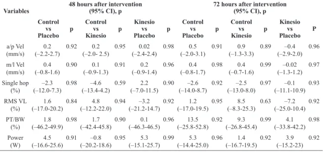
and in subjects undergoing reconstruction of the anterior cruciate ligament, respectively, verifying that
applying the technique did not signiicantly change the Table 4. Differences between groups at 48 hours and 72 hours after intervention in all groups (control, placebo and Kinesio Taping) for
all analyzed variables: anteroposterior velocity (a/p vel), mediolateral velocity (m/l vel), single hop, RMS of VL muscle (RMS VL), peak torque normalized by body weight (PT/BW), and average power.
Variables
Mean differences between groups Conidence interval (95% CI) 48 hours after intervention
(95% CI), p 72 hours after intervention(95% CI), p
Control vs Placebo
p
Control vs Kinesio
p
Kinesio vs Placebo
p
Control vs Placebo
p
Control vs Kinesio
p
Kinesio vs Placebo
P
a/p Vel
(mm/s) (–2.2-2.7)0.2 0.92 (–2.0- 2.5)0.2 0.95 (–2.4-2.4)0.02 0.98 (–2.0-3.1)0.5 0.91 (–1.3-3.3)0.9 0.89 (–2.9-2.0)–0.4 0.96 m/l Vel
(mm/s) (–0.8-1.6)0.4 0.90 (–0.9-1.3)0.1 0.91 (–0.9-1.4)0.2 0.96 (–0.8-1.7)0.4 0.98 (–0.7-1.6)0.4 0.99 (–1.3-1.2)–0.02 0.97 Single hop
(%) (–12.0-7.3)–2.3 0.98 (–13.4-4.2)–4.6 0.59 (–7.0-11.5)2.2 0.90 (–14.0-8.7)–2.6 0.92 (–13.0-8.0)–2.5 0.97 (–11.1-10.9)–0.1 0.93 RMS VL
(%) (–17.0-20.2)1.6 0.84 (–12.2-22.0)4.8 0.94 (–21.2-14.7)–3.2 0.92 (–17.0-19.5)1.2 0.95 (–8.3-25.3)8.5 0.63 (–25.0-10.4)–7.2 0.92 PT/BW
(%) (–46.2-49.9)1.8 0.98 (–42.4-45.8)1.7 0.90 (–46.3-46.5)0.1 0.96 (–25.8-52.8)13.5 0.92 (–26.8-45.4)9.3 0.99 (–33.8-42.2)4.1 0.98 Power
(W) (–16.6-25.6)4.5 0.91 (–20.2-18.6)–0.8 0.95 (–15.1-25.7)5.3 0.99 (–14.4-25.0)5.3 0.96 (–16.7-19.5)1.4 0.92 (–15.2-23)3.9 0.92
238 Braz J Phys Ther. 2016 May-June; 20(3):231-239
electromyographic activity of the VL or the isokinetic knee extensor performance.
In this study, the application of KT did not promote
any changes in the analyzed parameters, suggesting that the tactile stimulation promoted by KT did not
suficiently alter neuromuscular performance in healthy people. In addition, our study evaluated the delayed
effects of KT on these variables, showing that there
were also no signiicant changes compared to previous
values, therefore there is no need for an adjustment period for the application technique to promote greater
activation of the proposed mechanisms of action, i.e.
neurofacilitation4 and increase in local blood low1. Thus, we suggest that there is no evidence to support the application of this technique for this population
or in order to improve athletic performance. It is worth emphasizing that the results of this
study should be limited to healthy and active women
who practice recreational physical activity. Thus,
it is suggested that further studies are conducted to evaluate the chronic effects of KT on function, balance, and neuromuscular performance of patients
in the rehabilitation process.
Conclusion
The results of this study suggest that the application of KT to the quadriceps muscle is not able to promote immediate or delayed changes to neuromuscular performance, balance, or lower limb function in
healthy, active women.
References
1. Kase K, WallisJ, Kase T. Clinical therapeutic applications
of the kinesio taping method. 2nd ed. Tokyo: Kinesio Taping
Association; 2003.
2. Kneeshaw D. Shoulder taping in the clinical setting.J
Bodyw Mov Ther. 2002;6(1):2-8. http://dx.doi.org/10.1054/
jbmt.2001.0233.
3. Thelen MD, DauberJA, StonemanPD. The clinical efficacy of
kinesio tape for shoulder pain: a randomized, double-blinded, clinical trial.J Orthop Sports Phys Ther. 2008;38(7):389-95.
http://dx.doi.org/10.2519/jospt.2008.2791. PMid:18591761. 4. Konishi Y. Tactile stimulation with Kinesiology tape
alleviates muscle weakness attributable to attenuation of Ia afferents.J Sci Med Sport. 2013;16(1):45-8. http://dx.doi.
org/10.1016/j.jsams.2012.04.007. PMid:22682093. 5. Cools AM, WitvrouwEE, DanneelsLA, Cambier DC.
Does taping influence electromyographic muscle activity
in the scapular rotators in healthy shoulders? Man Ther.
2002;7(3):154-62. http://dx.doi.org/10.1054/math.2002.0464.
PMid:12372312.
6. Halseth T, McChesneyJW, DebelisoM, VaughnR, Lien
J. The effects of Kinesio Taping on proprioception at the
ankle.J Sports Sci Med. 2004;3(1):1-7. PMid:24497814.
7. Macgregor K, GerlachS, MellorR, HodgesPW. Cutaneous stimulation from patella tape causes a differential increase
in vasti muscle activity in people with patellofemoral pain. J Orthop Res. 2005;23(2):351-8. http://dx.doi.org/10.1016/j.
orthres.2004.07.006. PMid:15734248.
8. MurrayH, HuskL.Effects of Kinesio taping on proprioception
in the ankle.J Orthop Sports Phys Ther. 2001;31:1-7.
9. OsterhuesDJ. The use of Kinesio Taping in the
management of traumatic patella dislocation: a case study. Physiother Theory Pract. 2004;20(4):267-70. http://dx.doi.
org/10.1080/09593980490888370.
10. Fu TC, WongAM, PeiYC, WuKP, Chou SW, LinYC.
Effect of Kinesio Taping on muscle strength in athletes: a pilot study.J Sci Med Sport. 2008;11(2):198-201. http://
dx.doi.org/10.1016/j.jsams.2007.02.011. PMid:17588814.
11. FirthBL, DingleyP, DaviesER, Lewis JS, Alexander
CM. The effect of kinesiotape on function, pain, and motoneuronal excitability in healthy people and people with achillestendinopathy.Clin J Sport Med. 2010;20(6): 416-21. http://dx.doi.org/10.1097/JSM.0b013e3181f479b0.
PMid:21079436.
12. VithoulkaI, BenekaA, MalliouP, AggelousisN, Karatsolis K, DiamantopoulosK.The effects of Kinesio-Taping on
quadriceps strength during isokinetic exercise in healthy non athlete women.Isokinet Exerc Sci. 2010;18:1-6. 13. - AytarA, OzunluN, SurenkokO, BaltaciG, OztopP, Karatas
M. Initial effects of Kinesio Taping in patients with
patellofemoral pain syndrome: a randomized, double-blind study.Isokinet Exerc Sci. 2011;19(2):135-42.
14. LinsCA, Locks FNo, AmorimAB, MacedoLB, BrasileiroJS. Kinesio Taping does not alter neuromuscular performance of femoral quadriceps or lower limb function in healthy
subjects: Randomized, blind, controlled, clinical trial. Man Ther. 2013;18(1):41-5. http://dx.doi.org/10.1016/j.
math.2012.06.009. PMid:22796389.
15. OliveiraAKA, BorgesDT, LinsCAA, Cavalcanti RL,
MacedoLB, BrasileiroJS. Immediate effects of Kinesio Taping on neuromuscular performance of quadriceps and balance in individuals submitted to anterior cruciate
ligament reconstruction: a randomized clinical trial.J Sci Med Sport. 2016;19(1):2-6. http://dx.doi.org/10.1016/j.
jsams.2014.12.002. PMid:25601016.
16. Csapo R, AlegreLM. Effects of Kinesio Taping on skeletal
muscle strength: a meta-analysis of current evidence.J Sci Med Sport. 2015;18(4):450-6. http://dx.doi.org/10.1016/j.
jsams.2014.06.014. PMid:25027771.
17. WilliamsS, Whatman C, HumePA, Sheerin K. Kinesio Taping in treatment and prevention of sports injuries: a
meta-analysis of the evidence for its effectiveness.Sports Med. 2012;42(2):153-64.
http://dx.doi.org/10.2165/11594960-000000000-00000. PMid:22124445.
18. MorrisD, JonesD, RyanH, RyanCG. The clinical effects of Kinesio Tex taping: a systematic review.Physiother
Theory Pract. 2013;29(4):259-70. PMid:23088702.
19. MostafavifarM, WertzJ, BorchersJ. A systematic review
239 Braz J Phys Ther. 2016 May-June; 20(3):231-239 injury.Phys Sportsmed. 2012;40(4):33-40. http://dx.doi.
org/10.3810/psm.2012.11.1986. PMid:23306413.
20. SłupikA, DwornikM, BiałoszewskiD, ZychE. Effect of Kinesio Taping on bioelectrical activity of vastus medialis
muscle: preliminary report.Ortop Traumatol Rehabil. 2007;9(6):644-51. PMid:18227756.
21. PinciveroDM, GandaioCM, ItoY. Gender-specific knee
extensor torque, flexor torque, and muscle fatigue responses during maximal effort contractions.Eur J Appl Physiol. 2003;89(2):134-41.
http://dx.doi.org/10.1007/s00421-002-0739-5. PMid:12665976.
22. Keays SL, Bullock-SaxtonJ, Keays AC. Strength and function
before and after anterior cruciate ligament reconstruction. Clin Orthop Relat Res. 2000;373:174-83. http://dx.doi.
org/10.1097/00003086-200004000-00021. PMid:10810475.
23. HermensHJ, FreriksB, Disselhorst-Klug C, RauG.
Development of recommendations for SEMG sensors and sensor placement procedures.J Electromyogr Kinesiol. 2000;10(5):361-74.
http://dx.doi.org/10.1016/S1050-6411(00)00027-4. PMid:11018445.
24. NunesGS, NoronhaM, Cunha HS, Ruschel C, BorgesNG
Jr. Effect of kinesio taping on jumping and balance in athletes: a crossover randomized controlled trial.J Strength
Cond Res. 2013;27(11):3183-9. http://dx.doi.org/10.1519/
JSC.0b013e31828a2c17. PMid:23439339.
25. HuangCY, HsiehTH, LuSC, SuFC. Effect of the kinesio tape to muscle activity and vertical jump performance in
healthy inactive people.Biomed Eng Online. 2011;10(1):70.
http://dx.doi.org/10.1186/1475-925X-10-70. PMid:21831321. 26. NakajimaMA, Baldridge C. The effect of Kinesio Tape
on vertical jump and dynamic postural control.Int J Sports Phys Ther. 2013;8(4):393-406. PMid:24175126.
27. MohammadiHK, Kalantari KK, NaeimiSS, PouretezadM,
ShokriE, Tafazoli M, et al. Immediate and delayed effects of
forearm Kinesio Taping on grip strength.Iran Red Crescent Med J. 2014;16(8):e19797. PMid:25389492.
Correspondence
Jamilson Simões Brasileiro
Universidade Federal do Rio Grande do Norte Departamento de Fisioterapia
Avenida Senador Salgado Filho, 3000, Campus Universitário, Lagoa Nova