Rev Bras M ed Esporte _ Vol. 11, Nº 3 – M ai/Jun, 2005
155e
1. PhD in Physiotherapy. Professor of the Department of Biomechanics,M edicine and Rehabilitation of the Locomotor Apparatus and Coordina-tor of the Physiotherapy Course – Ribeirão Preto School of M edicine – São Paulo University – USP.
2. Physiotherapist. M S underw ay in Orthopedics, Traumatology and Re-habilitation of the Locomotive Organs – FM RP-USP.
3. Physiotherapy graduation course underw ay – UFSCar. 4. Physiotherapist. M S in Physiotherapy – UFSCar.
5. Physiotherapist. Assistant Professor of the Department of Physiothera-py – UFSCar.
Received in 30/11/04. 2nd version received in 14/3/05. Approved in 16/3/05. Correspondence to: Débora Bevilaqua-Grossi, Faculdade de M edicina de Ribeirão Preto-USP, Campus Universitário – 14049-900 – Ribeirão Preto, SP, Brasil. Tel./fax: (16) 602-4413/633-0336. E-mail: [email protected]
Electromyographic activity evaluation of the
patella muscles during squat isometric exercise
in individuals w ith patellofemoral pain syndrome
Débora Bevilaqua-Grossi1, Lílian Ramiro Felicio2, Rebeca Simões3,
Kelly Rafael Ribeiro Coqueiro4 and Vanessa M onteiro-Pedro5
O
RIGINALA
RTICLEKey w ords: Patellofemoral pain syndrome (PPS). Exercise. Electromyography.
Phys-iotherapy. ENGLISH VERSION
ABSTRACT
The objective of this study w as to compare the electromyograph-ic (EM G) activity of vastus medialis obliquus (VM O), vastus latera-lis longus (VLL) and vastus lateralatera-lis oblíquus (VLO) during w all slide squat isometric exercises at 45° (WS 45°) and at 60 ° (WS 60°) of knee flexion. Fifteen healthy control w omen and fifteen w omen w ith patellofemoral pain syndrome (PPS) participated in this study. The EM G activity w as registered during WS 45° and W S 60° per-formed at maximal isometric voluntary contraction (M IVC) using surface differential electrodes connected to an EM G system. The EM G signals w ere analyzed using the root mean square (RM S) values and w ere normalized by M IVC obtained at 75° of knee flex-ion. To compare data betw een groups and exercises, the ANOVA-tw o-w ay and Duncan post hoc tests w ere applied (p < 0.05). The results demonstrated higher EM G activity for all muscles studied at WS 60° w hen compared to WS 45° in both control a nd PPS groups. There w ere not significant differences betw een muscles during WS 60° in the control group, although a high er activity of VLL in relation to VM O and VLO w as observed during WS 45° in control group. For the PPS group, no statistical difference w as ob-served betw een muscles during both exercises. Thus, strengthen-ing programs usstrengthen-ing WS 60° could be more effective f or healthy w omen; how ever, both exercises could be indicated for rehabilita-tion programs aimed at w omen w ith PPS. In addirehabilita-tion, the absence of significant differences betw een muscles in PPS group verified in this study suggests that muscle unbalance could not be a pre-disposing factor for PPS in w omen.
INTRODUCTION
One of the most frequent knee musculoskeletal disorders is the
patellofemoral pain syndrome (PPS)(1-3), including approximately
25% of the orthopedic diagnosis(2), being defined as an anterior
and/or retropatellar knee pain as result of structural and biome-chanical alterations of the joint(4-6). This dysfunction frequently
af-fects athletes and female sedentary population, w here young adult individuals are the most afflicted(5,7-9).
Although the etiological factors for PPS are still unknow n, some authors point the biomechanical alterations of the low er limb as
the main cause(5,7,8,10). Among the most frequent biomechanical
fac-tors related to the PPS development, the static and dynamic un-balance stands out(5).
Among the alterations on the static unbalance, some authors assure that abnormalities such as excessive subtalar pronation, increase on the Q angle, external tibia torsion, retraction of the lateral retinaculum and improper patellar behavior can cause
ante-rior knee pain(11,12). How ever, other authors relate PPS w ith the
unbalance on the dynamic stabilizer muscles, especially among the medialis and lateralis components(5,11-14) and, more recently, the
oblique portion of the vastus lateralis, the vastus lateralis obliquus (VLO)(15).
The conservative treatment is alw ays the first choice for individ-uals w ith PPS(3,16). These rehabilitation programs are based on closed
kinetic chain (CKC) and open kinetic chain (OKC) exercises(5,8,11,14,17).
How ever, many authors report that CKC exercises in the first 60o
of knee flexion are more tolerated by individuals w ith PPS(11,17,18).
Among the CKC exercises, the squat exercise is considered as safe and effective due to the stabilizing effect of the quadriceps
and ischiotibial muscle co-contraction(16,17). The squat exercise is
frequently used in conditioning programs of many sports that re-quire high pow er and strength levels(19).
Rehabilitation protocols for individuals w ith PPS are aimed at the selective strengthening of the VM O muscle in order to rees-tablish the normal function of the patellofemoral joint(3,16,20,21).
Anderson et al.(22) evaluated the EM G activity of VM O and VL
muscles in healthy individuals during squat exercises at 0o to 30o,
0o to 60o and 0o to 90o, and verified that the VM O:VL relation
tend-ed to increase w ith the increase on the knee flexion, therefore suggesting that this increase on the knee flexion leads to an in-crease on the VM O activity in relation to VL.
Earl et al.(17) also analyzed the EM G activity of VM O and VL
(vas-tus lateralis) muscles in female and male normal athletes during squat exercises at 0o-30o knee flexion w ith and w ithout hip
adduc-tion. The authors found no significant differences betw een VM O and VL muscles during exercises. How ever, those authors evalu-ated athlete individuals w ithout PPS only. Besides, the physical activity, not specified by the authors, could lead to alterations on the EM G activity of the muscles evaluated.
Similarly, Tang et al.(5) found no significant difference betw een
the EM G activity of VM O and VL muscles during squat eccentric
and concentric phases betw een 15o-75o knee flexion in normal
in-dividuals w ith symptoms of PPS.
156e
Rev Bras M ed Esporte _ Vol. 11, Nº 3 – M ai/Jun, 2005Fig. 2 – Wall slide squat exercise at 45o of knee flexion (A) and w all slide
squat exercise at 60o of knee flexion (B).
A
B
Thus, the objective of this w ork w as to evaluate and to compare the EM G activity of VM O, VLO and VLL muscles during w all slide squat exercise at 45o and 60o knee flexion in clinically healthy
indi-viduals and in indiindi-viduals w ith PPS.
M ETHODS
Volunteers
Thirty female untrained volunteers w ere analyzed, being divided into tw o groups: control group (n = 15) w ith average age of 20.93 ± 3.15 years, average w eight of 58.38 ± 5.88 kg and average stat-ure of 165 ± 4.3 cm and group w ith PPS (n = 15) average age of 21.8 ± 3.12 years, average w eight of 50.53 ± 5.83 kg and average stature of 158 ± 5.6 cm. The inclusion and exclusion criteria for PPS group are presented in table 1. All individuals from the control group did not present any history of pain, surgery, trauma or low er limb osteomyoarticular lesion(12,23). This study is in agreement w ith
resolution 196/96 of the National Health Council.
Exercises
Each volunteer performed w all slide squat exercises w ith back against the w all and knees positioned at 45o (WS 45o) and 60o (WS
60o) of flexion (figures 2A and B); the exercises order w as
random-ly performed. Each squat exercises w as repeated three times w ith interval of tw o minutes betw een each exercise and of four min-utes for the new positioning. Each exercise repetition w as main-tained for approximately seven seconds and the EM G recording collection initiated tw o seconds after the beginning of the exercise in the affected limbs for individuals from PPS group and in the dominant limb for individuals from the control group. The volun-teers w ere familiarized w ith exercises during the period previous to collection.
TABLE 1
Inclusion and exclusion criteria for group w ith PPS
Inclusion criteria
The individuals must not present surgeries, traumas or low er limb osteomyoarticular lesions.
The individuals must report previous episodes of knee pain during activities such as climbing up or going dow n stairs, squatting and remaining sitting for long periods of time.
The presence of three clinical signs or symptoms in the functional evaluation (among them: bayonet sign, increase on the Q angle, external tibia torsion, excessive subtalar pronation, medialized patella and sensitiveness to palpation of the patella facets).
Exclusion criteria
Use of medications and physiotherapy sessions during the period of 6 months pre-vious to the study.
Neurological diseases.
Equipment
The EM G recordings of VM O, VLL and VLO muscles w ere ob-tained by means of simple active Ag/AgCl electrodes (10 x 1 mm) (Lynx Tecnologia Eletrônica Ltda.) w ith 100 times gain and a refer-ence electrode w ith 3 cm of diameter connected to a EM G system
M yosystem® w ith magnification of 10 times, summing up a gain
of 1000 times. The common mode rejection ratio (CM RR) w as of 80 dB and the sampling frequency w as of 2000 Hz. A low -pass filter of 10-500 Hz w as used and the inlet impedance w as higher
than 100 MΩ.
Procedures
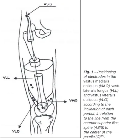
The skin w as previously trichotomized and cleaned w ith alcohol 70% and the electrodes w ere fixed to the skin w ith the aid of mi-cropore surgical tape. For the electrodes fixation on the VM O, VLO and VLL muscles, a line from the anterior-superior iliac spine to the center of the patella w as draw n and used as reference for the measurement of the inclination angles of each portion of the
quad-riceps muscle evaluated(24). For the VM O muscle, the electrode
w as positioned on the muscular core at approximately 2 cm from the femur lateral epicondyle w ith inclination of 50.4o, and for
mus-cle VLL, the electrodes w ere positioned at 10 cm above the patella upper-lateral border w ith approximate inclination of 17o(15) (figure
1). The reference electrode greased w ith electro-conductive gel remained fixed to the tibia anterior tuberosity.
The EM G activity of these muscles w as quantified using the root mean square (RM S) of the three repetitions of each squat exercise.
Fig. 1 – Positioning of electrodes in the
Rev Bras M ed Esporte _ Vol. 11, Nº 3 – M ai/Jun, 2005
157e
The EM G data normalization w as obtained through the average of three repetitions of each w all slide squat isometric exercise (WS
45o and WS 60o) expressed as percentage of the average RM S of
three w all slide squat repetitions at 75o and are presented as
arbi-trary units (A.U.).
RM S average value of the w all slide squat exercise at 45o or 60o
Average value of the w all slide squat exercise at 75o
Statistical analysis
The tw o-w ay ANOVA and Duncan post hoc tests w ere applied (p < 0.05) for the analysis of data.
RESULTS
The results for the control group show ed higher EM G activity in the VLL muscle w hen compared to the VM O (p = 0.022) and VLO
(p = 0.009) muscles during WS 45o; how ever, during WS 60o, no
significant difference w as observed betw een these muscles (ta-ble 2).
With regard to the PPS group, no significant difference betw een
VM O, VLO and VLL muscles w as observed during WS 45o and WS
60o exercises (table 2).
In the intragroup comparison of VM O, VLL and VLO muscles, one could observe for both normal and PPS groups that the WS
60o exercises presented higher EM G activity for all muscles w hen
compared to the WS 45o exercise (table 2).
The intergroup comparison for each exercise analyzed present-ed no statistically significant difference betw een muscles.
increase its EM G activity in order to maintain the patella in its ade-quate alignment.
One observes for group w ith PPS that during WS 45o and WS
60o, no significant differences w ere found betw een VM O, VLO
and VLL muscles. Thus, one believes that WS 45o and WS 60o
exercises provide a balance betw een the medial and lateral por-tions of the patella dynamic stabilizer muscles, and should be indicated during rehabilitation program for individuals w ith PPS. How
-ever, the comparison betw een exercises revealed that the WS 60o
squat exercise presented higher EM G activity of the quadriceps muscle portions.
Similarly, Tang et al.(5) also observed significant differences in
the VM O:VL relation during concentric and eccentric phases of
squat exercise betw een 0-90o knee flexion; how ever, they observed
a better VM O:VL relation during phases evaluated of the squat
exercise at 60o knee flexion, suggesting higher activation of the
VM O muscle both for the control group and for the group w ith PPS.
Despite the methodological differences, our data corroborate those found by Tang et al.(5) and Anderson et al.(22). The results of
this w ork revealed that the quadriceps muscle generally present-ed higher EM G activity as the knee flexion angle increases. The VM O muscle, in turn, presented no higher activation in any of the exercises proposed.
M any authors report that the muscular unbalance may be a pre-ponderant factor of PPS(5,8,11,13,14); how ever, despite not being the
objective of this w ork, one observes that the EM G activity of the medial quadriceps muscles components – VM O and lateral com-ponents – VLL and VLO presented no significant differences be-tw een control and PPS groups, suggesting that the muscular un-balance may not predispose to PPS.
The data found by Cerny(26) also reinforce that hypothesis, once
analyzing the EM G activity of VM O and VL muscles in the w all
slide exercise at 45o in normal individuals and in individuals w ith
PPS, no difference w as observed betw een groups, corroborating results found in this w ork.
According to those data, one may conclude that the w all slide
squat exercise performed at 60o presented higher activation of the
patella stabilizer muscles of normal individuals and individuals w ith symptoms of PPS w hen compared to the w all slide squat exercise
performed at 45o, being able to be indicated during rehabilitation
programs in w hich the objective is the increase on the activation of these muscles.
Different w orks analyzed the squat exercise in different situa-tions associated to adduction(17,26) and to the lateral and medial hip
rotation(27). How ever, no studies on the comparison of VM O, VLL
and VLO muscles betw een squat exercises in different positions and knee angles w ere found in the literature researched.
Thus, studies comparing the EM G activity of VM O, VLL and VLO muscles during different squat exercise modalities are required for a better understanding of the role these muscles play in squat exercises in order to favour the elaboration of exercise protocols aimed at a more effective physiotherapeutic investigation in indi-viduals w ith PPS.
All the authors declared there is not any potential conflict of inter-ests regarding this article.
REFERENCES
1. Biedert RM , Warnke K. Correlation betw een the Q angle and the patella posi-tion: a clinical and axial computed tomography evaluation. Arch Orthop Trauma Surg 2001;121:346-9.
2. Pow ers CM , M affucci R, Hampton S. Rearfoot posture in subjects w ith patel-lofemoral pain. J Orthop Sports Phys Ther 1995;22:155-60.
3. Wilk KE, Reinold M M . Principles of patellofemoral rehabilitation. Sports M edi-cine and Arthroscopy Review 2001;9:325-36.
TABLE 2
Averages (± SD) of the normalized EM G recordings (RM S) (A.U.) of VM O, VLL and VLO muscles in the w all slide squat
exercises (WS) 45o and 60o for control and PPS groups
Control PPS
WS 45o WS 60o* WS 45o WS 60o*
VM O 44.86 (± 14.24)# 75.38 (± 13.65) 49.03 (± 11.84) 76.41 (± 14.23)
VLL 56.57 (± 11.26)# 77.02 (± 14.75) 54.94 (± 10.08) 75.78 (± 11.71)
VLO 44.05 (± 13.43)# 75.55 (± 17.05) 49.54 (± 13.31) 69.76 (± 12.45) * Significant difference in relation to WS 45o (p = 0.0001).
# Significant difference in relation to VM O (p = 0.022) and VLO (p = 0.009) muscles.
DISCUSSION
One know s that the patellofemoral dysfunction presents the conservative treatment as main intervention, in w hich w all slide
squat exercises at 45o and 60o of knee flexion are frequently
per-formed in sportive trainings and in knee rehabilitation programs. Our data show ed higher EM G activity of VLL muscle during WS
45o for the control group; how ever, during the performance of WS
60o, no difference betw een VM O, VLO and VLL muscles w as
ob-served. Therefore, for individuals w ithout PPS, the squat exercise
WS 45o does not seem to be the best alternative for a muscular
strengthening program, once, in this w ork, the VLL muscle is fa-vored at this angle, w hat could result in unbalance on the dynamic
stabilizer muscles, unlike the WS 60o that presented no difference
betw een the portions of the quadriceps muscle, indicating a bal-ance betw een the patella medialis and lateralis dynamic stabilizer muscles.
Despite the methodological differences, these data corroborate
those found by Anderson et al.(22), w ho verified increase on the
158e
Rev Bras M ed Esporte _ Vol. 11, Nº 3 – M ai/Jun, 20054. Baker M M , Juhn M S. Patellofemoral pain syndrome in the female athlete. Clin Sports M ed 2000;19:315-29.
5. Tang SFT, Chen CK, Hsu R, Shih-Wei Chou, Wei-Hsein Hong, Lew HL. Vastus medialis obliquus and vastus lateralis activity in open and closed kinetic chain exercise in patients w ith patellofemoral pain syndrome: an electromyographic study. Arch Phys M ed Rehabil 2001;82:1441-5.
6. Cow an SM , Bennell KL, Hodges PW, Crossley KA, M cConnell J. Delayed onset of electromyographic activity of vastus medialis obliquus relative to vastus later-alis in subjects w ith patellofemoral pain syndrome. Arch Phys M ed Rehabil 2001; 82:183-9.
7. Baker V, Bennell K, Stillman B, Cow an S, Crossley K. Abnormal knee joint posi-tion sense in individuals w ith patellofemoral pain syndrome. J Orthop Res 2002; 20:208-14.
8. Cow an SM , Bennell KL, Crossley KM , Hodges PW, M cConnell J. Physical ther-apy alters recruitment of the vasti in patellofemoral pain syndrome. Arch Phys M ed Rehabil 2002;34:1879-85.
9. Sw enson EJ, Hough DO, M cKeag DB. Patellofemoral dysfunction. How to treat, w hen to refer patients w ith problematic knees. Postgrad M ed 1987;82:125-9. 10. Wilk KE, Davies GJ, M angine RE, M alone TR. Patellofemoral disorders: a
classi-fication system and clinical guidelines for nonoperative rehabilitation. J Orthop Sports Phys Ther 1998;28:307-22.
11. M cGinty G, Irrgang JJ, Pezzullo D. Biomechanical considerations for rehabilita-tion of the knee. Clin Biomech (Bristol, Avon) 2000;15:160-6.
12. W ityrouw E, Sneyers C, Lysens R, Victor J, Bellemans J. Reflex response times of vastus medialis oblique and vastus lateralis in normal subjects and in subjects w ith patellofemoral pain syndrome. J Orthop Sports Phys Ther 1996;24:160-5. 13. Callaghan M J, M cCarthy CJ, Oldham JA. Electromyographic fatigue
characteris-tics of the quadriceps in patellofemoral pain syndrome. M an Ther 2001;6:27-33. 14. Cow an SM , Bennell KL, Hodges PW, Crossley KM , M cConnell J. Simultaneous feedforw ard recruitment of the vasti in untrained postural tasks can be restored by physical therapy. J Orthop Res 2003;21:553-8.
15. Bevilaqua-Grossi D, M onteiro-Pedro V, Bérzin F. Análise funcional dos estabiliza-dores da patela. Acta Ortop Bras 2004;12:99-104.
16. Stiene HA, Brosky T, Reinking M F, Nyland J, M ason M B. A comparison of closed kinetic chain and isokinetic joint isolation exercise in patients w ith patellofemo-ral dysfunction. J Orthop Sports Phys Ther 1996;24:136-41.
17. Earl JE, Schmitz RJ, Arnold BL. Activation of VM O and VL during dynamic mini-squat exercises w ith and w ithout isometric hip adduction. J Electromyogr Kine-siol 2001;11:381-6.
18. Wityrouw E, Lysens R, Bellemans J, Peers K, Vanderstraeten G. Open versus closed kinetic chain exercises for patellofemoral pain. A prospective, random-ized study. Am J Sports M ed 2000;28:687-94.
19. Escamilla RF. Knee biomechanics of the dynamic squat exercise. M ed Sci Sports Exerc 2001;33:127-41.
20. Cabral CM N, M onteiro-Pedro V. Recuperação funcional de indivíduos com dis-função femoropatelar por meio de exercícios em cadeia cinética fechada: revi-são da literatura. Rev Bras Fisioter 2003;7:1-8.
21. Doucette SA, Child DD. The effect of open and closed exercise and knee joint position on patellar tracking in lateral patellar compression syndrome. J Orthop Sports Phys Ther 1996;23:104-10.
22. Anderson R, Courtney C, Carmeli E. EM G analysis of the vastus medialis/vastus lateralis muscles utilizing the unloaded narrow and w ide stance squats. J Sports Rehabil 1998;7:236-47.
23. Pow ers CM . Patellar kinematics, part I: The influence of vastus muscle activity in subjects w ith and w ithout patellofemoral pain. Phys Ther 2000;80:956-64. 24. Bevilaqua-Grossi D, M onteiro-Pedro V, Sousa GC, Silva Z, Bérzin F. Contribution
to the anatomical study of the obliqúe portion of the vastus lateralis muscle. Braz J M orphol Sci 2004;21:47-52.
25. Lieb FJ, Perry J. Quadriceps function. An anatomical and mechanical study us-ing amputed limbs. J Bone Joint Surg 1968;50:1535-48.
26. Cerny K. Vastus medialis oblique/vastus lateralis muscle activity ratios for se-lected exercises in person w ith and w ithout patellofemoral pain syndrome. Phys Ther 1995;75:672-83.