Article
0103 - 5053 $6.00+0.00*e-mail: [email protected]
Palladium(II) Complexes with Thiosemicarbazones. Syntheses, Characterization and
Cytotoxicity against Breast Cancer Cells and Anti-Mycobacterium tuberculosis Activity
Pedro I. da S. Maia,a Angélica Graminha,b Fernando R. Pavan,c Clarice Q. F. Leite,c Alzir A. Batista,b Davi F. Back,d Ernesto S. Lang,d Javier Ellena,e Sebastião de S. Lemos,f
Heloisa S. Salistre-de-Araujog and Victor M. Delon*,a
aInstituto de Química de São Carlos, Universidade de São Paulo, 13566-590 São Carlos-SP, Brazil
bDepartamento de Química, Universidade Federal de São Carlos, 13565-905 São Carlos-SP, Brazil
cFaculdade de Ciências Farmacêuticas, Universidade Estadual Paulista, 14801-902 Araraquara-SP, Brazil
dDepartamento de Química, Universidade Federal de Santa Maria, 97105-900 Santa Maria-RS, Brazil
e nstituto de Física de São Carlos, Universidade de São Paulo, 13560-970 São Carlos-SP, Brazil fInstituto de Química, Universidade de Brasília, 70919-970 Brasília-DF, Brazil
gDepartamento de Ciências Fisiológicas, Universidade Federal de São Carlos, 13565-905 São Carlos-SP, Brazil
Três complexos de PdII com tiossemicarbazonas N(4)-substituídas foram preparados: [Pd(aptsc)
(PPh3)](NO3)•H
2O, 1, [Pd(apmtsc)(PPh3)](NO3), 2, e [Pd(apptsc)(PPh3)](NO3)•H2O, 3, sendo
PPh3 = trifenilfosina; Haptsc = 2-acetilpyridina-tiossemicarbazona; Hapmtsc =
2-acetilpiridina-N(4)-metil-tiossemicarbazona e Happtsc = 2-acetilpiridina-N(4)-fenil-tiossemicarbazona. Os
complexos foram caracterizados por análise elementar, IR, UV-Vis, 1H e 31P{1H} NMR e tiveram
suas estruturas cristalinas determinadas por difratometria de raios X em monocristal. Os ligantes tiossemicarbazonatos monoaniônicos atuam de modo tridentado, ligando-se ao metal pelos átomos de nitrogênio piridínico, nitrogênio azometínico e enxofre. A atividade citotóxica frente à linhagem
de células tumorais MDA-MB231 (tumor de mama) e a atividade anti-Mycobacterium tuberculosis
H37Rv ATCC 27294 dos compostos foram investigadas. Os complexos de PdII mostraram-se
altamente ativos contra as células tumorais, com valores de IC50 em torno de 5 μmol L-1, enquanto
o agente antitumoral em uso clínico cisplatina mostrou-se inativo. Os compostos apresentaram
atividade anti-M. tuberculosis signiicante, com valores de CIM comparáveis ou melhores que
aqueles referentes a alguns fármacos usados clinicamente contra tuberculose.
Three PdII complexes were prepared from N(4)-substituted thiosemicarbazones: [Pd(aptsc)
(PPh3)](NO3)•H
2O, 1, [Pd(apmtsc)(PPh3)](NO3), 2, and [Pd(apptsc)(PPh3)](NO3)•H2O, 3, where
PPh3 = triphenylphosphine; Haptsc = 2-acetylpyridine-thiosemicarbazone; Hapmtsc =
2-acetylpyridine-N(4)-methyl-thiosemicarbazone and Happtsc = 2-acetylpyridine-N
(4)-phenyl-thiosemicarbazone. All complexes were characterized by elemental analysis, IR, UV-Vis, 1H
and 31P{1H} NMR spectroscopies, and had their crystalline structures determined by X-ray
diffractometry from single crystals. The monoanionic thiosemicarbazonate ligands act in a tridentate mode, binding to the metal through the pyridine nitrogen, the azomethine nitrogen and the sulfur atoms. The cytotoxic activity against the breast cancer cell line MDA-MB231 and the
anti-Mycobacteriumtuberculosis H37Rv ATCC 27294 activity were evaluated for the compounds.
All PdII complexes were highly active against the studied cell line, presenting similar values of
IC50, around 5 μmol L-1, while the clinically applied antitumor agent cisplatin was inactive. The
compounds show remarkable anti-M. tuberculosis activities, presenting MIC values comparable
or better than some commercial anti-M tuberculosis drugs.
Keywords: palladium(II) complexes, thiosemicarbazones, cytotoxicity, breast tumor cells,
Introduction
There has been much interest in the development of effective agents against tumor cell lines resistant to cisplatin, as well as of more eficient anti-tuberculosis drugs. Cisplatin has received much attention recently with regard to metal-based chemotherapy due to its disadvantages, such as the occurrence of primary or acquired resistance.1-3
On the other hand, the rising incidence of tuberculosis, especially when associated with AIDS cases, and the occurrence of multidrug-resistant tuberculosis demand the development of new anti-tubercular drugs.4,5 To attenuate
these disadvantages, much effort has been directed toward the development of new antitumor metal coordination compounds with the ability to overcome cisplatin6
resistance, as well as of new drugs addressing the current problems with tuberculosis therapy.
Until some time ago, the great majority of antitumor metal complexes synthesized and characterized were structural analogs of cisplatin.7 However, there has been
a decrease in the number of new compounds of this type because they have not shown improved clinical eficacy, most likely because all cis-[PtX2(amine)2] compounds
show similar DNA-binding modes, and therefore similar biological effects.2,8
Thiosemicarbazone derivatives have received considerable attention due to their pharmacological properties, such as antiviral, antibacterial and antitumor activities.9-13 The compound triapine, which is currently
being tested in a Phase I clinical trial against a variety of tumor cell models,1 and thiacetazone, which has been
widely used in the treatment of tuberculosis,4 are good
examples of the biologic activity of this class of compounds. Structure-activity relationships not only evidence that the complexation with a metal enhances the antimicrobial activity of the ligands against some tested microorganisms, but also indicate that the metal plays a relevant role.14-16 It has already been shown, for example,
that Pd-thiosemicarbazone complexes possess interesting antiproliferative effects on human breast cancer, including tumor cell lines resistant to cisplatin17-20 and have also
presented good antimycobacterial effects.21 The presence
of phosphine ligands in potential anti-cancer complexes is supposed to provide better cytotoxicity by enhancing lipophilicity – and consequently permeability through the cell membrane.22 Thus, our group focused on investigating
both antitumor and antimycobacterial activities of some PdII complexes containing both thiosemicarbazone and
phosphine ligands.
This work reports the preparation, structural characterization, cytotoxicity against breast cancer cells
and anti-M. tuberculosis activity of three thiosemicarbazone
derivatives and their PdII complexes containing
different N(4)-substituted groups:
2-acetylpyridine-thiosemicarbazone (Haptsc), 2-acetylpyridine-N
(4)-methyl-thiosemicarbazone (Hapmtsc) and 2-acetylpyridine-N
(4)-phenyl-thiosemicarbazone (Happtsc).
Experimental
Materials and measurements
[PdCl2(PPh3)2], AgNO3 and other analytical grade reagents and solvents were obtained commercially (Aldrich or Strem Chemicals) and used without further puriication. The complexing agents Haptsc, Hapmtsc and Happtsc were prepared by standard methods described in the literature.23 FTIR spectra were recorded using KBr pellets
on a BOMEM FTIR model BM 100 spectrophotometer in the 4000-400 cm–1 region. The electronic spectra were
recorded with a JASCO V-630 spectrophotometer. NMR spectra were acquired using a Varian MERCURY Plus spectrometer operating at 300.07 and 121.47 MHz for
1H and 31P, respectively. The 1H spectra were internally
referenced to tms (tetramethylsilane). The 31P{1H} spectra
were externally referenced to H3PO4 (85%, d = 0) with positive chemical shifts downield the standard. Elemental analyses (CHNS) were carried out using a FISONS EA-1108 analyzer. A Bruker CCD X8 APEX II diffractometer was used for the X-ray structure analyses of 1 and 2, while
the data collection for 3 was performed on a NONIUS
KAPPA CCD diffractometer. Both equipments were operated using graphite monochromator and Mo-Kα
radiation (λ = 71.073 pm).
Crystal structure determinations
Orange crystals of the complexes 1, 2 and 3 were
obtained by slow evaporation of their methanolic solutions at room temperature. The structures were solved using SHELXS9724 by direct methods. All non-hydrogen atoms
were reined with anisotropic displacement parameters using SHELXL97.25 The hydrogen atom positions were
found in the Fourier map or calculated at idealized positions. More detailed information on the structure determinations is given in Table 1.
Cell culture and cytotoxicity determination
In vitro cytotoxicity assays on cultured human tumor
their pharmacological properties, the new palladium(II) complexes were assayed against the human breast tumor MDA-MB-231 cell line (ATCC No. HTB-26). The cells were routinely maintained at 37 ºC in a humidiied 5% CO2 atmosphere with Dulbecco’s Modified Eagle’s medium (DMEM) supplemented with 10% fetal bovine serum (FBS). After reaching conluence, the cells were detached by tripsinization and counted. For the cytotoxicity assay, 4 × 10⁴ cells well–1 were seeded in 200 μL of
complete medium in 96-well plates (Corning Costar).
The plates were incubated at 37 ºC in 5% CO2 for 24 h to allow cell adhesion prior to drug testing. The complex was dissolved in sterile dimethylsulfoxide (dmso, stock solution with maximum concentration of 20 mmol L-1) and
diluted to 10, 2, 0.2 and 0.02 mmol L-1. Two microliters
of each complex sample were added to 200 μL medium (without FBS), so that the inal concentration of dmso in each well was approximately 1% and the complex was diluted approximately 100 times. The other compounds evaluated (free thiosemicarbazones and cisplatin) were
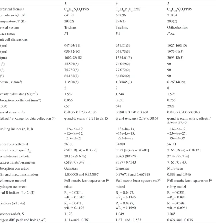
Table 1. X-ray structure data collection and reinement parameters for complexes 1-3
1 2 3
Empirical formula C26H26N5O4PPdS C27H26N5O3PPdS C32H30N5O4PPdS
Formula weight, M 641.95 637.96 718.04
Temperature, T (K) 293(2) 293(2) 293(2)
Crystal system Triclinic Triclinic Orthorhombic
Space group P1– P1– Pbca
Unit cell dimensions
a (pm) 947.95(11) 951.81(3) 1027.160(10)
b (pm) 950.32(10) 968.73(3) 1970.01(3)
c (pm) 1602.98(18) 1584.61(5) 3095.18(5)
α (°) 75.891(6) 74.049(2) 90
β (°) 74.750(6) 77.072(2) 90
γ (°) 84.187(7) 84.664(2) 90
Volume, V (nm3) 1.3501(3) 1.36845(7) 6.26314(15)
Z 2 2 8
Density calculated (Mg/m3) 1.582 1.548 1.523
Absorption coeficient (mm−1) 0.866 0.851 0.756
F(000) 652 648 2928
Crystal size (mm3) 0.410 × 0.170 × 0.130 0.790 × 0.550 × 0.260 0.840 × 0.400 × 0.360
Method / θ Range for data collection (o) ϕ and ω scans / 2.21 to 28.15 ϕ and ω scans / 2.19 to 30.63 ϕ and ω scans with κ offsets /
2.94 to 27.49 Limiting indices (h, k, l) −12←h←12,
−12←k←12, −21←l←21
−13←h←13, −13←k←13, −22←l←22
−13←h←12, −25←k←25, −39←l←39
Relections collected 26183 34380 56101
Relections unique/ Rint 6589 [R(int) = 0.0306] 8357 [R(int) = 0.0682] 7165 [R(int) = 0.0713] Completeness to theta 28.15 (99.6 %) 30.63 (98.9 %) 27.49 (99.7 %) Data/restraints/parameters 6589 / 0 / 349 8357 / 0 / 343 7165 / 0 / 403
Absorption correction Gaussian Gaussian Multi-scan
Min. and max. transmission 1.000000 and 0.835097 0.976719 and 0.667818 0.889 and 0.946
Reinement method Full-matrix least-squares on F2 Full-matrix least-squares on F2 Full-matrix least-squares on F2
Hydrogen treatment mixed mixed riding model
Final R indices [I > 2σ(I)] R1 = 0.0354, wR2 = 0.1010
R1 = 0.0497, wR2 = 0.1345
R1 = 0.0355, wR2 = 0.085 R indices (all data) R1 = 0.0471,
wR2 = 0.1196
R1 = 0.0787, wR2 = 0.1590
R1 = 0.0599, wR2 = 0.0964
Goodness-of-it, S 1.123 1.049 1.045
also dissolved in sterile dmso. The cells were exposed to the complex for 48 h. Cell respiration, as an indicator of cell viability, was determined by the mitochondrial-dependent reduction of MTT (3-(4,5-dimethylthiazol-2-yl)-2,5-diphenyltetrazolium bromide) to formazan.26 MTT
solution (0.5 mg mL-1) was added to the cell cultures and
incubated for 3 h. Thereafter, 100 μL of isopropanol were added in order to dissolve the formazan crystals. The conversion of MTT to formazan by metabolically viable cells was monitored by an automated microplate reader at 570 nm. The percentage of cell viability was calculated by dividing the average absorbance of the cells treated with the compounds by that of the control; % of the cell viability
versus drug concentration (logarithmic scale) was plotted
to determine the IC50 (drug concentration at which 50% of the cells are viable relative to the control), with its estimated error derived from the average of 3 trials.
Anti-Mycobacterium tuberculosis activity assay
The anti-M. tuberculosis activity was determined
applying the Microplate Alamar Blue Assay (MABA) as the analytical method.27 Stock solutions of the tested
compounds were prepared in dmso27 and were diluted in
Middlebrook 7H9 (Difco) broth supplemented with oleic acid, albumin, dextrose and catalase (OADC enrichment - BBL/Becton-Dikinson, Sparks, MD, USA) to obtain inal sample concentration ranges from 1.00 to 100 μg mL-1.
Isoniazid was dissolved in distilled water according to the manufacturers’ recommendations (Difco laboratories, Detroit, MI, USA) and used as a positive control drug. M.
tuberculosis H37Rv ATCC 27294 was grown for 7 to 10
days in Middlebrook 7H9 supplemented with OADC and added of 0.05% Tween 80 to avoid clumps. Suspensions were prepared and their turbidities matched to the optical density of the McFarland No. 1 standard. After further dilution of 1:25 in Middlebrook 7H9 supplemented with OADC, the inoculum was added to each well of the 96-well microtiter plates (Falcon 3072; Becton Dickinson, Lincoln Park, NJ) together with the compounds. Samples were set up in triplicate. Cultures were incubated for 7 days at 37 oC and then Alamar Blue was added for reading. The
minimum inhibitory concentration (MIC) was deined as the lowest concentration of the tested compound necessary to inhibit 90% growth of M. tuberculosis.27 MIC values
were determined by luorescence on a SPECTRAluor Plus microluorimeter (Tecan), with excitation at 530 nm and emission at 590 nm.28 As a control, the MIC value
of isoniazid was determined for each microplate. The acceptable MIC of isoniazid ranged from 0.015 to 0.05 μg mL-1.28
Preparation of [Pd(aptsc)(PPh3)](NO3)•H
2O (1),
[Pd(apmtsc)(PPh3)](NO3) (2) and [Pd(apptsc)(PPh3)] (NO3)•H
2O (3)
Three different solutions of AgNO3 (0.068 g, 0.4 mmol) in MeOH (10 mL) were prepared and mixed with suspensions of [PdCl2(PPh3)2] (0.128 g, 0.2 mmol) in MeOH (20 mL). The resulting mixtures were kept under relux until the formation of red solutions (approximately 30 min). The precipitates formed (AgCl) were iltered off and each iltrate received the addition of 0.2 mmol of one of the free thiosemicarbazones (0.039 g, 0.042 g and 0.054 g for Haptsc, Hapmtsc and Happtsc, respectively) dissolved in MeOH (15 mL). The resulting mixtures were kept under relux for 2 h, forming yellow solutions, which gave orange crystals of complexes 1, 2 and 3 after slow evaporation at
room temperature. The crystals were washed with small portions of cold CH2Cl2 and n-hexane, yielding 0.039
(31.0%), 0.039 (30.3%) and 0.050 g (34.8%) of 1, 2 and 3, respectively. Elemental analysis: Complex 1 (641.98 g
mol-1): Found: C, 48.82; H, 4.27; N, 10.93; S, 5.35. Calc.
for C26H26N5O4PPdS: C, 48.64; H, 4.08; N, 10.91; S, 4.99%. Complex 2 (637.99 g mol-1): Found: C, 50.21;
H, 4.25; N, 10.85; S, 4.80. Calc. for C27H26N5O3PPdS: C, 50.83; H, 4.11; N, 10.98; S, 5.03%. Complex 3 (718.08 g
mol-1): Found: C, 53.44; H, 4.34; N, 10.12; S 4.30. Calc.
for C32H30N5O4PPdS: C, 53.53; H, 4.21; N, 9.75; S, 4.46%. UV-Vis data, CH2Cl2 solution [λmax/nm (log ε)] for 1: 308
(4.47), 369 (4.16), 447 (3.72). Complex 2: 309 (4.41), 380
(4.23), 466 (3.70). Complex 3: 316 (4.29), 420 (4.47).
Results and Discussion
Spectroscopic characterization
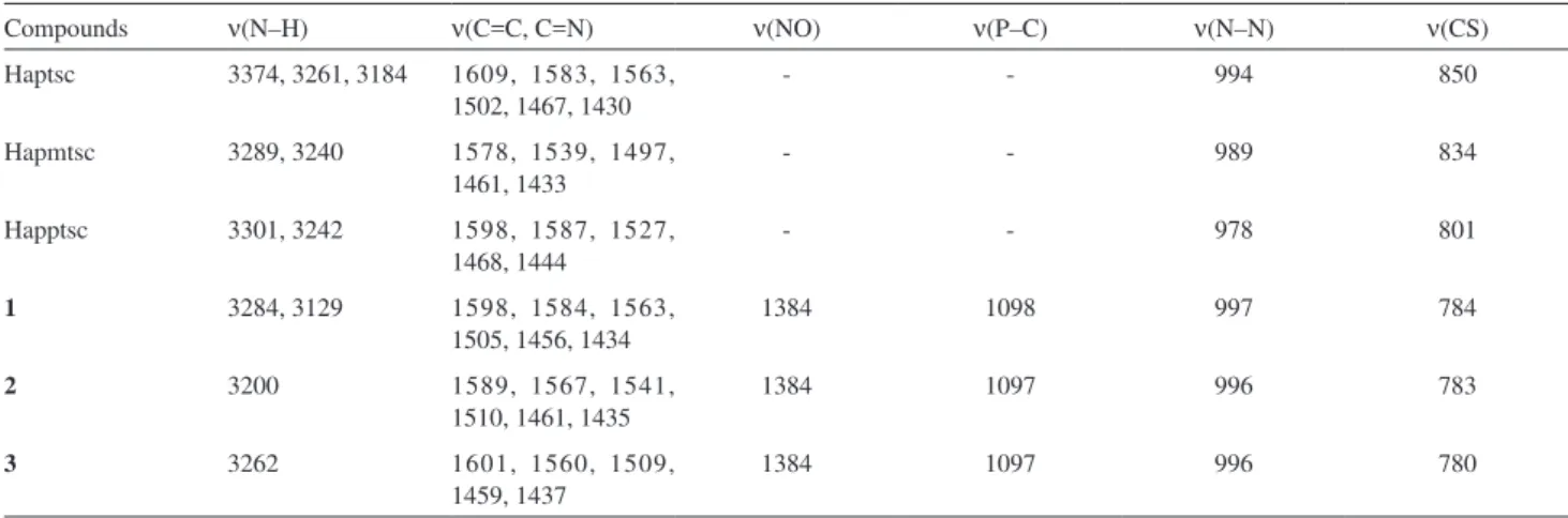
The main IR vibrational bands of the free thiosemicarbazones and their complexes are listed in Table 2. Upon coordination, changes in the ν(C=S),
ν(C=C + C=N) and ν(N–H) wavenumbers, in comparison to the values found for the thiosemicarbazones, were observed for complexes 1-3. They are consistent with the
tridentate coordination of the thiosemicarbazone derivatives through the thiolate sulfur, the azomethine nitrogen and the pyridine nitrogen atoms, respectively.29 The occurrence of
the ν(N−N) band at higher frequencies in the IR spectra of the complexes, compared to those observed for the ligands, conirms coordination through the azomethine nitrogen atom.11 The ν(C=S) bands at 801-850 cm−1 in the spectra
of free thiosemicarbazones shift to the 780-784 cm−1 range
with deprotonation and formation of a C–S single bond.23
A very strong band found at 1384 cm−1 in the spectra of
the complexes is attributed to ν(NO3−).
The presence of the triphenylphosphine ligand is conirmed in the spectra of the complexes by the existence of the characteristic bands around 1097 cm−1 for ν(P–C),
with no signiicant change when compared to the precursor [PdCl2(PPh3)2].
The data obtained from the electronic spectra of the complexes in CH2Cl2 solutions are given in the experimental section. Three d–d spin allowed singlet-singlet and three spin forbidden singlet-triplet transitions are predicted for square-planar complexes of palladium(II). However, strong charge-transfer transitions may interfere and prevent the observation of some of the expected bands, especially for complexes containing sulfur donor atoms.11,17 The
prominent strong bands in the 308-466 nm range are assigned to a combination of intraligand and ligand to metal charge transfer (LMCT) absorptions and d–d bands, which support the idea of a square-planar environment for the metal ions.
The 1H NMR data for the free thiosemicarbazones and
complexes 1, 2 and 3 are given in Table 3. Figure 1 shows
the numbered structures of the free thiosemicarbazones. The three new PdII complexes show similar 1H-chemical
shift behavior. Some hydrogen atom values of d were not observed precisely due to overlapping with the signals of the aromatic hydrogen atoms of triphenylphosphine.
1H NMR integrations and signal multiplicities are in
agreement with the proposed structures.
In the 1H NMR spectra of the complexes 1, 2 and 3,
a high frequency shift of ca. 0.15 ppm for the methyl
hydrogen atoms (C–CH3), compared to the spectra of the thiosemicarbazones, evidences the coordination through the azomethine nitrogen. On the other hand, the signals referred to the hydrogen atoms of the methyl group (N–CH3), observed in the spectrum of 2, remain almost non shifted
in relation to free Hapmtsc. The methyl group bonded to the N4 atom (6 in the numbering scheme) is observed in
the spectrum of Hapmtsc as a doublet, which is consistent with a coupling to the hydrogen atom bonded directly to the N4 atom. In complex 2, however, this signal is observed
as a sharp singlet, similarly to what is reported for a PdII
complex containing the ligands apmtsc− and Cl−.12 The
spectrum of free Haptsc displayed three signals related to N-bonded H atoms, showing non-equivalence for the hydrogens bonded to N4. In complex 1 only one NH signal
is observed, which is consistent with the deprotonation of N3 and with the chemical equivalence of the NH
2 protons.
The 31P{1H} NMR spectrum of the precursor
[PdCl2(PPh3)2] in CD2Cl2 solution shows a singlet peak for the phosphorous donor atoms at d 23.83 ppm. In the spectra of complexes 1, 2 and 3,this singlet peak is shifted
to 28.10, 28.22 and 28.25 ppm, respectively, in conformity with the presence of only one species for each complex in the CD2Cl2 solution.
Table 2. Relevant IR spectral assignments (cm–1) for the free thiosemicarbazones and complexes 1-3
Compounds ν(N–H) ν(C=C, C=N) ν(NO) ν(P–C) ν(N–N) ν(CS)
Haptsc 3374, 3261, 3184 1609, 1583, 1563, 1502, 1467, 1430
- - 994 850
Hapmtsc 3289, 3240 1578, 1539, 1497, 1461, 1433
- - 989 834
Happtsc 3301, 3242 1598, 1587, 1527, 1468, 1444
- - 978 801
1 3284, 3129 1598, 1584, 1563,
1505, 1456, 1434
1384 1098 997 784
2 3200 1589, 1567, 1541,
1510, 1461, 1435
1384 1097 996 783
3 3262 1601, 1560, 1509,
1459, 1437
1384 1097 996 780
Crystal structure of the complexes
Table 4 contains selected interatomic distances and angles for the complexes [Pd(aptsc)(PPh3)](NO3)•H
2O
(1), [Pd(apmtsc)(PPh3)](NO3) (2) and [Pd(apptsc)(PPh3)]
(NO3)•H
2O (3). ORTEP drawings of the products, with an
atomic numbering scheme, are shown in Figures 2, 3 and 4, respectively.
All complexes are cationic, with a nitrate anion as counter ion. Products 1 and 3 also have a water molecule as
solvate. In all complexes, the palladium(II) ion is in a quite similar, nearly planar, fourfold environment. The metal is coordinated to the negatively charged organic molecule through the pyridine nitrogen atom [Pd–N bond distances of 210.2(3) pm for 1, 210.1(3) pm for 2 and 208.8(2) pm
for 3], the azomethine N-atom [at slightly shorter Pd–N
distances of 202.4(3), 203.2(3) and 200.6(2) pm for 1, 2
and 3, respectively] and the sulfur atom [Pd–S distances
of 225.36(9), 225.15(11) and 224.07(7) pm for 1, 2 and 3, respectively]. This forms two ive-membered chelate
rings including the PdII atom and with bond dimensions
comparable to those observed for similar square-planar PdII
complexes.11,29 The remaining binding site is occupied by a
phosphorous atom [Pd–P distances of 229.39(8), 229.23(9) and 227.57(7) pm for 1, 2 and 3, respectively].
The stronger coordination of the metal to the azomethine nitrogen compared to the pyridyl nitrogen is attributed to the higher basicity of N(2).18 The largest deviation in the bond
angles from those expected for square planarity is found for the angle N(1)–Pd–S, in which an average deviation, considering the three complexes, is of 16.7º below the expected value of 180º.
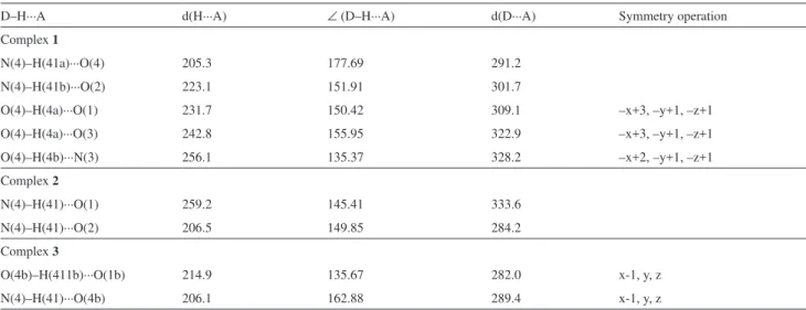
In complex 1, the crystal structure is stabilized by a
net of intermolecular hydrogen bonds toward the direction [001], parallel to the c axis, which is shown in Figure S1
(Supplementary Information, SI). The nitrogen atom N(4), of the NH2 group, is H-bonded through H(41) to the O(2) atom from a nitrate ion, while H(42) interacts with a hydrate water oxygen atom, O(4). Furthermore, the oxygen atom O(4) is H-bonded through H(4a) to the O(1b) and O(3b) from a symmetry-generated nitrate ion and through H(4b) to the
Table 3. 1H NMR data (ppm) for the thiosemicarbazones and complexes 1-3 at room temperature
Compound Assignments Haptsc 1H NMR (dmso-d
6) d 2.39 (s, 3 H, CH3), 7.38 (ddd, J2,3 8 Hz, J2,1 5 Hz, J2,4 1 Hz, 1 H, CH), 7.79 (ddd, J3,4J3,2 8 Hz, J3,1 2 Hz, 1 H, CH),
8.15 (s, 1 H, NH), 8.42 (s, 1 H, NH), 8.44 (ddd, J4,3 8 Hz, J4,2 1 Hz, 1 H, CH), 8.58 (ddd, J1,2 5 Hz, J1,3 2 Hz, 1 H, CH), 10.33 (s, 1 H, NH).
Hapmtsc 1H NMR (dmso-d
6) d 2.38 (s, 3 H, CH3), 3.06 (d, J6,5 5 Hz, 3 H, N–CH3), 7.39 (ddd, J2,3 7 Hz, J2,1 5 Hz, J2,4 1 Hz, 1 H, CH), 7.82 (ddd, J3,4
8 Hz, J3,2 7 Hz, J3,1 1 Hz, 1 H, CH), 8.42 (dq, J4,3 8 Hz, J4,2 1 Hz, 1 H, CH), 8.58 (dq, J1,2 5 Hz, J1,3 1 Hz, 1 H, CH), 8.64 (d, J5,6 5 Hz, 1 H, CH), 10.35 (s, 1 H, NH).
Happtsc 1H NMR (dmso-d
6) d 2.47 (s, 3 H, CH3), 7.24 (ddd, J7,6 7 Hz, J7,5 1 Hz, 1 H, CH), 7.34-7.44 (m, 3 H, CH), 7.55 (dd, J5,6 9 Hz, J5,7 1 Hz, 2
H, CH), 7.82 (ddd, J3,4J3,2 8 Hz, J3,1 2 Hz, 1 H, CH), 8.54 (ddd, J4,3 8 Hz, J4,2 J4,1 1 Hz, 1 H, CH), 8.61 (ddd, J1,2 5 Hz, J1,3 2 Hz, J1,4 1 Hz, 1
H, CH), 10.21 (s, 1 H, NH), 10.67 (s, 1 H, NH).
1 1H NMR (CD
2Cl2) d 2.50 (s, 3 H, CH3), 5.97 (s, 2 H, NH2), 7.02 (ddd, J2,3 8 Hz, J2,1 5 Hz, J2,4 2 Hz, 1 H, CH), 7.19 (d, J1,2 5 Hz, 1 H, CH),
7.49-7.74 (m, 16 H, PPh3 + CH), 8.01 (ddd, J3,2 8 Hz, 1H).
2 1H NMR (CD2Cl2) d 2.52 (s, 3 H, CH3), 3.02 (s, 3 H, CH3), 6.95-8.05 (m, 21 H, PPh3 + CH).
3 1H NMR (CD
2Cl2) d 2.64 (s, 3 H, CH3), 7.04-7.14 (m, 2 H, CH), 7.23 (d, J7,6 8 Hz, 1 H, CH), 7.34 (t, J5,6 8 Hz, 2 H, CH), 7.49-7.73 (m, 18
H, PPh3 + CH), 7.85 (d, J1,2 8 Hz, 1 H, CH), 8.08 (ddd, J3,4J3,2 8 Hz, 1 H, CH).
Table 4. Selected bond lengths (pm) and angles (°) reined from X-ray
data for 1-3
1 2 3
Bond Lengths
Pd–N(1) 210.2(3) 210.1(3) 208.8(2) Pd–N(2) 202.4(3) 203.2(3) 200.6(2) Pd–S 225.36(9) 225.15(11) 224.07(7) Pd–P 229.39(8) 229.23(9) 227.57(7)
S–C(8) 177.6(4) 176.8(4) 176.7(3)
N(3)–N(2) 136.7(4) 136.3(4) 137.0(3) P–C(11) 181.8(3) 182.4(4) 182.3(3) P–C(21) 182.9(3) 181.0(3) 180.9(3) P–C(31) 182.6(3) 182.7(4) 181.5(3) N(5)–O(1) 115.5(6) 114.0(7) 121.6(4) N(5)–O(2) 118.4(6) 121.8(8) 123.9(4) N(5)–O(3) 127.6(7) 119.9(9) 120.2(4) Bond Angles
N(2)–Pd–N(1) 79.54(11) 79.43(12) 79.62(8) N(2)–Pd–S 83.55(9) 83.87(9) 84.22(6) N(1)–Pd–S 162.88(8) 163.29(9) 163.67(6) N(2)–Pd–P 174.95(8) 177.64(9) 175.82(6) N(1)–Pd–P 102.86(7) 102.80(9) 102.13(6)
S–Pd–P 94.20(3) 93.90(4) 93.84(3)
N(3a) atom from a symmetry-related complex molecule. The crystalline structure of complex 2 is stabilized by hydrogen
interactions involving the nitrogen atom N(4) and the oxygen atoms O(2) and O(1), as shown in Figure S2. As observed in 2, the crystal structure of complex 3 is also stabilized by
hydrogen bonds, but in a different mode. The nitrogen atom N(4) is H-bonded through H(41) to a symmetry generated oxygen atom O(4b) from a hydrate water molecule, while the same oxygen O(4) atom is H-bonded through H(411b) to the oxygen atom O(1b) of a nitrate ion, as shown in Figure S3. The distances, angles and symmetry operations related to the hydrogen bonds are given in Table S1.
Cytotoxic activity
The palladium(II) complexes and the respective free thiosemicarbazones were evaluated for their effectiveness against the breast tumor cell line MDA-MB231. For comparison purposes, the cytotoxicity of cisplatin was evaluated under the same experimental conditions. The values of cell viability were calculated after the tested compounds were incubated for 48 h (Figure 5). The IC50 values, calculated from the dose-survival curves from MTT assay, are shown in Table 5.
Comparing only the values of IC50 of the free thiosemicarbazones, the order of cytotoxic activity was Happtsc > Hapmtsc > Haptsc, suggesting that the activity is increased by the presence of bulky groups bonded to
Figure 2. An ORTEP view of [Pd(aptsc)(PPh3)](NO3)•H
2O (1) with the
thermal ellipsoids at the 50% probability level. The labeling scheme used for the phenyl rings is omitted for clarity.
Figure 3. An ORTEP view of [Pd(apmtsc)(PPh3)](NO3) (2) with the
thermal ellipsoids at the 50% probability level. The labeling scheme used for the phenyl rings is omitted for clarity.
Figure 4. An ORTEP view of [Pd(apptsc)(PPh3)](NO3)•H
2O (3) with the
thermal ellipsoids at the 50% probability level. The labeling scheme used for the phenyl rings is omitted for clarity.
Figure 5. The effect of the thiosemicarbazones and their PdII complexes
N(4) of the thiosemicarbazone. On the other hand, all palladium complexes presented values of IC50 around 5 μmol L-1, showing no signiicant variation on the activity
due to the changes in the groups bonded to the N(4) of the thiosemicarbazone ligands. The palladium complexes were more active than the free thiosemicarbazones, except for complex 3, whose thiosemicarbazone precursor Happtsc
had an IC50 value around 3 μmol L-1. The palladium
complex 2 presented an activity ive times higher than
that from Hapmtsc. In addition, the Haptsc and cisplatin were ineffective against the tested cell line, with the IC50 value over 200 μmol L-1. A comparison with the precursor
compound [PdCl2(PPh3)2] was not possible due to the lack of solubility in dmso, even at the concentration of 2 μmol L-1. Alternatively, [PdCl
2(MeCN)2] and PPh3
were tested, showing no activity at the concentration of 200 μmol L-1. The good values of activity found for these
complexes, around 5.0 μmol L-1, show that the complexation
of thiosemicarbazone to PdII may be a good strategy to
obtain antitumor agents, although in this case the free Happtsc presented the best result.
The similarity of the values of IC50 found for the PdII complexes is an evidence in favor of the same
biochemical action mechanism, but different from those of the cisplatin, inactive in this case, and of the free thiosemicarbazones, which varied from inactive (Haptsc) to a signiicant activity (Happtsc). In fact, the literature reports that palladium(II) and platinum(II) complexes of thiosemicarbazone derivatives are able to bind to DNA in vitro, and present enhanced capacity to form interstrand
crosslinks when compared to cisplatin.30,31 Additionally,
the presence of a phosphine ligand may also contribute to higher activity.32
On the other hand, the free thiosemicarbazones could present antitumor effect by inhibiting DNA syntheses through the blockage of the enzyme ribonucleoside diphosphate reductase (RDR), which catalyses the conversion of ribonucleotides into desoxyribonucleotides, as proposed for other α(N)-heterocyclic thiosemicarbazones.33
Anti-Mycobacterium tuberculosis activity
The in vitro antimycobacterial activities of the three
thiosemicarbazones and their PdII complexes were assessed
against the strains of the Mycobacterium tuberculosis H37Rv
ATCC 27294. The results are exposed in Table 5. Comparing the MIC values of the free thiosemicarbazones with the values obtained for the complexes, it can be seen that, except for 2, the activity of the complexes is much
higher than those of the uncoordinated thiosemicarbazones. Complexes 1 and 3 present approximately 8-fold and 6-fold
higher activities, respectively, than the respective free thiosemicarbazones.
The activity found for complex 3 is comparable
to those of some commonly used anti-M. tuberculosis
agents, like cycloserine (MIC = 122.4-489.7μmol L-1),
gentamicin (MIC = 4.19-8.38 μmol L-1), tobramycin
(MIC = 8.56-17.11 μmol L-1) and clarithromycin
(MIC = 10.70-21.40 μmol L-1).27 The precursor complex,
[PdCl2(PPh3)2], as well as the complex [PdCl2(MeCN)2], did not show any inhibition at the concentration of 50 μg mL-1, corresponding to 71.2 and 192.8 μmol L-1,
respectively, suggesting that the thiosemicarbazonate
Figure 6. Comparison of the magnitude of the IC50 values of cisplatin,
free thiosemicarbazones and the respective PdII complexes against breast
tumor MDA-MB-231 cell line.
Table 5. IC50 values of the thiosemicarbazones, PdII complexes and
cisplatin against breast tumor MDA-MB-231 cell line and the MIC values against the strains of the Mycobacterium tuberculosis H37Rv ATCC 27294
IC50 (μmol L-1) MIC (μg mL-1) MIC (μmol L-1)
Haptsc* > 200 31.3 160.9
1 4.9 ± 0.9 12.5 19.5
Hapmtsc 24.6 ± 11.3 7.8 37.4
2 5.2 ± 1.5 25 39.2
Happtsc 2.8 ± 0.5 15.6 57.7
3 5.5 ± 2.8 6.25 8.7
Cisplatin* > 200 Not measured Not measured [PdCl2(MeCN)2] > 200 > 50 -[PdCl2(PPh3)2] Not soluble > 50
ligands are the active species and that their activity may be improved upon complexation to palladium. Previous studies explain this fact as a result of the polarity reduction of the complex molecule as a whole, when compared with the free ions or complexing agents, by partial sharing of their charges within the coordination compounds, favoring their permeation through the lipid layer of the cell membrane, hence resulting in a better cell uptake of the active species.34-37
Conclusions
A series of thiosemicarbazones was used to prepare novel cationic [PdIIL(PPh
3)]+ complexes with nitrate as
counter ion (1-3). The X-ray studies revealed square-planar
structures in which the thiosemicarbazonate ligands bind in a tridentate monoanionic mode (with loss of the N(3) hydrogen atom) by coordination to palladium through the pyridine N, azomethine N and thiolate S atoms. The fourth coordination site is occupied by neutral PPh3. The crystal structure of 1 presents a supramolecular network generated
by intermolecular hydrogen bonds, while in the structures of 2 and 3 only local hydrogen bonds between the cationic
molecule and a nitrate counter ion are observed. Comparing only the values of IC50 of the free thiosemicarbazones, it was observed that the activity is increased by the presence of bulky groups bonded to N(4). All palladium(II) complexes were effective against the studied tumoral cell line, presenting high antiproliferative effect, with IC50 values around 5 μmol L-1, while the clinically applied antitumor
agent cisplatin and the precursor compound [PdCl2(PPh3)2] were ineffective. The similarity of the IC50 values for the PdII complexes indicates that they may have the same
biochemical action mechanism, other than those of the cisplatin and the thiosemicarbazones. The complexes 1 and 3 show higher anti-M. tuberculosis activities, compared
to the free thiosemicarbazones, and their actions are equivalent or greater than those of some commercial anti-M tuberculosis drugs.
Supplementary Information
Crystallographic data have been deposited with the Cambridge Crystallographic Data Centre (deposition numbers CCDC 743883, CCDC 743884 and CCDC 743885 for complexes 1, 2 and 3, respectively). Copies of
available material can be obtained by request to CCDC, 12 Union Road, Cambridge CB2 1EZ, UK (fax 44-1223-336033 or e-mail: [email protected]). Table S1 and Figures S1, S2 and S3 are available free of charge at http://jbcs.sbq.org.br.
Acknowledgments
The authors gratefully acknowledge the financial support of FAPESP, CAPES, CNPq and FINEP.
References
1. Genova, P.; Varadinova, T.; Matesanz, A. I.; Marinova, D.; Souza, P.; Toxicol. Appl. Pharmacol.2004, 197, 107.
2. Kelland, L. R.; Crit. Rev. Oncol. Hematol.1993, 15, 191.
3. Kelland, L. R.; Sharp, S. Y.; ÓNeill, C. F.; Raynaud, F. I.; Beale, P. J.; Judson, I. R.; J. Inorg. Biochem. 1999, 77, 111.
4. Tripathi, R. P.; Tewari, N.; Dwivedi, N.; Tiwari, V. K.; Med. Res. Rev.2005, 25, 93.
5. Sriram, D.; Yogeeswari, P.; Thirumurugan, R.; Bioorg. Med. Chem. Lett.2004, 14, 3923.
6. Lebwohl, D.; Canetta, R.; Eur. J. Cancer1998, 34, 1522.
7. Padhye, S. B.; Kauffman, G. B.; Coord. Chem. Rev.1985, 63,
127.
8. Miernicka, M.; Szulawska, A.; Czyz, M.; Lorenz, I.-P.; Mayer, P.; Karwowski, B.; Budzisz, E.; J. Inorg. Biochem.2008, 102,
157.
9. Pérez, J. M.; Matesanz, A. I.; Martín-Ambite, A.; Navarro, P.; Alonso, C.; Souza, P.; J. Inorg. Biochem. 1999, 75, 255.
10. Nomiya, K.; Sekino, K.; Ishikawa, M.; Honda, A.; Yokoyama, M.; Kasuga, N. C.; Yokoyama, H.; Nakano, S.; Onodera, K.;
J. Inorg. Biochem.2004, 98, 601.
11. Kovala-Demertzi, D.; Domopoulou, A.; Demervzis, M. A.; Valle, G.; Papageorgiou, A.; J. Inorg. Biochem.1997, 68, 147.
12. Maia, P. I. S.; Delon, V. M.; Sousa, G. F.; Batista, A. A.; Nascimento, O. R.; Niquet, E.; Z. Anorg. Allg. Chem.2007, 633, 783.
13. Cocco, M. T.; Congiu, C.; Onnis, V.; Pellerano, M. L.; Logu, A. D.; Bioorg. Med. Chem. 2002, 10, 501.
14. do Nascimento, F. B.; Poelhsitz, G. V.; Pavan, F. R.; Sato, D. N.; Leite, C. Q. F.; Selistre-de-Araújo, H. S.; Ellena, J.; Castellano, E. E.; Delon, V. M.; Batista, A. A.; J. Inorg. Biochem.2008, 102, 1783.
15. Rodríguez-Argüelles, M. C.; López-Silva, E. C.; Sanmartín, J.; Pelagatti, P.; Zani, F.; J. Inorg. Biochem. 2005, 99, 2231.
16. Sau, D. K.; Butcher, R. J.; Chaudhuri, S.; Saha, N.; Mol. Cell. Biochem. 2003, 253, 21.
17. Matesanz, A. I.; Perez, J. M.; Navarro, P.; Moreno, J. M.; Colacio, E.; Souza, P.; J. Inorg. Biochem.1999, 76, 29.
18. Kovala-Demertzi, D.; Demertzis, M. A.; Miller, J. R.; Papadopoulou, C.; Dodorou, C.; Filousis, G.; J. Inorg. Biochem. 2001, 86, 555.
19. Jalilian, A. R.; Sadeghi, M.; Kamrani, Y. Y.; Radiochim. Acta 2006, 94, 865.
21. Bharti, N.; Athar, F.; Maurya, M. R.; Azam, A.; Bioorg. Med. Chem.2004, 12, 4679.
22. Queiroz, S. L.; Batista, A. A.; Quim. Nova 1996, 19, 651.
23. Klayman, D. L.; Bartosevich, J. F.; Grifin, T. S.; Mason, C. J.; Scovill, J. P.; J. Med. Chem. 1979, 22,855.
24. Sheldrick, G. M.; SHELXS97, Program for the Solution of Crystal Structures, University of Göttingen, Germany, 1997.
25. Sheldrick, G. M.; SHELXL97, Program for the Reinement of Crystal Structures, University of Göttingen, Germany, 1997.
26. Mosmann, T.; J. Immunol. Methods1983, 65, 55.
27. Franzblau, S. G.; Witzig, R. S.; McLaughlin, J. C.; Torres, P.; Madico, G.; Hernandez, A.; Degnan, M. T.; Cook, M. B.; Quenzer, V. K.; Ferguson, R. M.; Gilman, R. H.; J. Clin. Microbiol. 1998, 36, 362.
28. Collins, L. A.; Franzblau, S. G.; Antimicrob. Agents Chemother. 1997, 41, 1004.
29. Rebolledo, A. P.; Vieites, M.; Gambino, D.; Piro, O. E.; Castellano, E. E.; Zani, C. L.; Souza-Fagundes, E. M.; Teixeira, L. R.; Batista, A. A.; Beraldo, H.; J. Inorg. Biochem. 2005, 99,
698.
30. Quiroga, A. G.; Navarro-Ranninger, C.; Coord. Chem. Rev. 2004, 248, 119.
31. Quiroga, A. G.; Perez, J. M.; Montero, E. I.; Masaguer, J. R.; Alonso, C.; Navarro-Ranninger, C.; J. Inorg. Biochem.1998, 70, 117.
32. da Rocha, M. C.; Santana, A. M.; Ananias, S. R.; de Almeida, E. T.; Mauro, A. E.; Placeresa, M. C. P.; Carlos, I. Z.; J. Braz. Chem. Soc. 2007, 18, 1473.
33. Cory, J. G.; Cory, A. H.; Rappa, G.; Lorico, A.; Liu, M.; Lin, T.; Sartorelli, A. C.; Biochem. Pharmacol.1994, 48, 335.
34. Maia, P. I. S.; Pavan, F. R.; Leite, C. Q. F.; Lemos, S. S.; de Sousa, G. F.; Batista, A. A.; Nascimento, O. R.; Ellena, J.; Castellano, E. E.; Niquet, E.; Delon, V. M.; Polyhedron 2009, 28, 398.
35. Maurya, M. R.; Kumar, A.; Abid, M.; Azam, A.; Inorg. Chim. Acta 2006, 359, 2439.
36. Mendes, I. C.; Moreira, J. P.; Speziali, N. L.; Mangrich, A. S.; Takahashia, J. A.; Beraldo, H.; J. Braz. Chem. Soc. 2006, 17,
1571.
37. Maurya, M. R.; Kumar, A.; Bhat, A. R.; Azam, A.; Bader, C.; Rehder, D. Inorg. Chem.2006, 45, 1260.
Received: August 14, 2009 Web Release Date: February 25, 2010
Supplementary Information
0103 - 5053 $6.00+0.00*e-mail: [email protected]
Palladium(II) Complexes with Thiosemicarbazones. Syntheses, Characterization,
Cytotoxicity against Breast Cancer Cells and Anti-Mycobacterium tuberculosis Activity
Pedro I. da S. Maia,a Angélica Graminha,b Fernando R. Pavan,c Clarice Q. F. Leite,c Alzir A. Batista,b Davi F. Back,d Ernesto S. Lang,d Javier Ellena,e Sebastião de S. Lemos,f
Heloisa S. Salistre-de-Araujog and Victor M. Delon*,a
a Instituto de Química de São Carlos, Universidade de São Paulo, 13566-590 São Carlos - SP, Brazil
b Departamento de Química, Universidade Federal de São Carlos, 13565-905 São Carlos - SP, Brazil
c Faculdade de Ciências Farmacêuticas, Universidade Estadual Paulista, 14801-902 Araraquara - SP, Brazil
d Departamento de Química, Universidade Federal de Santa Maria, 97105-900 Santa Maria - RS, Brazil
e Instituto de Física de São Carlos, Universidade de São Paulo, 13560-970 São Carlos - SP, Brazil f Instituto de Química, Universidade de Brasília, 70919-970 Brasília - DF, Brazil
g Departamento de Ciências Fisiológicas, Universidade Federal de São Carlos, 13565-905 São Carlos - SP, Brazil
Table S1.Hydrogen bonds (distances in pm and angles in °) for complexes 1-3, calculated for distances d(H···A) < r(A) + 200.0 pm and angles ∠ (D–H···A) > 110° (D = H–donor, A = H–acceptor)
D–H···A d(H···A) ∠ (D–H···A) d(D···A) Symmetry operation
Complex 1
N(4)–H(41a)···O(4) 205.3 177.69 291.2
N(4)–H(41b)···O(2) 223.1 151.91 301.7
O(4)–H(4a)···O(1) 231.7 150.42 309.1 –x+3, –y+1, –z+1
O(4)–H(4a)···O(3) 242.8 155.95 322.9 –x+3, –y+1, –z+1
O(4)–H(4b)···N(3) 256.1 135.37 328.2 –x+2, –y+1, –z+1
Complex 2
N(4)–H(41)···O(1) 259.2 145.41 333.6
N(4)–H(41)···O(2) 206.5 149.85 284.2
Complex 3
O(4b)–H(411b)···O(1b) 214.9 135.67 282.0 x-1, y, z
Figure S1. View of the crystal structure of [Pd(aptsc)(PPh3)](NO3)•H
2O (1) toward the direction [001], showing the hydrogen bonds as dashed lines.
Figure S3. View of the crystal structure of [Pd(apptsc)(PPh3)](NO3)•H
 • H 2 O (1), [Pd(apmtsc)(PPh 3 )](NO 3 ) (2) and [Pd(apptsc)(PPh 3 )]](https://thumb-eu.123doks.com/thumbv2/123dok_br/18994071.461638/6.892.67.805.139.399/table-contains-selected-interatomic-distances-angles-complexes-apmtsc.webp)
 • H 2 O (3) with the thermal ellipsoids at the 50% probability level](https://thumb-eu.123doks.com/thumbv2/123dok_br/18994071.461638/7.892.479.821.680.1009/figure-ortep-view-apptsc-thermal-ellipsoids-probability-level.webp)

 • H 2 O (1) toward the direction [001], showing the hydrogen bonds as dashed lines](https://thumb-eu.123doks.com/thumbv2/123dok_br/18994071.461638/12.892.178.670.113.488/figure-view-crystal-structure-direction-showing-hydrogen-dashed.webp)
 • H 2 O (3) toward the direction [010], showing the hydrogen bonds as dashed lines.](https://thumb-eu.123doks.com/thumbv2/123dok_br/18994071.461638/13.892.199.724.112.491/figure-crystal-structure-apptsc-direction-showing-hydrogen-dashed.webp)
